第28章無脊椎動物
28.7 Superphylum Deuterostomia28.7 後口動物上門
Learning Objectives学習目標
By the end of this section, you will be able to do the following:
この節を学ぶと、次のことができるようになる。
- Describe the distinguishing characteristics of echinoderms棘皮動物を特徴づける性質を説明する。
- Describe the distinguishing characteristics of chordates脊索動物を特徴づける性質を説明する。
The phyla Echinodermata and Chordata (the phylum that includes humans) both belong to the superphylum Deuterostomia. Recall that protostomes and deuterostomes differ in certain aspects of their embryonic development, and they are named based on which opening of the archenteron (primitive gut tube) develops first. The word deuterostome comes from the Greek word meaning “mouth second,” indicating that the mouth develops as a secondary structure opposite the location of the blastopore, which becomes the anus. In protostomes (“mouth first”), the first embryonic opening becomes the mouth, and the second opening becomes the anus.
英語のヒント
棘皮動物門と、人を含む脊索動物門は、どちらも後口動物上門に属する。前口動物と後口動物は、胚発生のいくつかの点で異なり、原腸、すなわち原始的な腸管のどの開口部が先に発達するかに基づいて名付けられていることを思い出そう。後口動物という語は、「口が二番目」を意味するギリシャ語に由来する。肛門となる原口の反対側に、二次的な構造として口ができることを示している。前口動物(「口が最初」)では、胚の最初の開口部が口となり、二番目の開口部が肛門となる。
There are a series of other developmental characteristics that differ between protostomes and deuterostomes, including the type of early cleavage (embryonic cell division) and the mode of formation of the coelom of the embryo: Protosomes typically exhibit spiral mosaic cleavage whereas deuterostomes exhibit radial regulative cleavage. In deuterostomes, the endodermal lining of the archenteron usually forms buds called coelomic pouches that expand and ultimately obliterate the embryonic blastocoel (the cavity within the blastula and early gastrula) to become the embryonic mesoderm, the third germ layer. This happens when the mesodermal pouches become separated from the invaginating endodermal layer forming the archenteron, then expand and fuse to form the coelomic cavity. The resulting coelom is termed an enterocoelom. The archenteron develops into the alimentary canal, and a mouth opening is formed by invagination of ectoderm at the pole opposite the blastopore of the gastrula. The blastopore forms the anus of the alimentary system in the juvenile and adult forms. Cleavage in most deuterostomes is also indeterminant, meaning that the developmental fates of early embryonic cells are not decided at that point of embryonic development (this is why we could potentially clone most deuterostomes, including ourselves).
英語のヒント
前口動物と後口動物は、初期卵割(胚の細胞分裂)の型や胚の体腔形成の様式など、ほかの発生上の特徴にも一連の違いがある。前口動物は通常、らせん状のモザイク卵割を示し、後口動物は放射状の調節卵割を示す。後口動物では、原腸を裏打ちする内胚葉が通常、体腔嚢という芽をつくる。これが広がり、最終的に胞胚腔(胞胚や初期原腸胚の内部の空洞)を消失させ、第三の胚葉である中胚葉になる。これは、原腸を形成する陥入した内胚葉の層から中胚葉の袋が分離し、拡大して融合し、体腔をつくることで起こる。こうして生じる体腔を腸体腔という。原腸は消化管へ発達する。原腸胚の原口と反対の極で外胚葉が陥入し、口の開口部ができる。原口は幼体と成体の消化系の肛門を形成する。多くの後口動物の卵割は不定的でもある。つまり、初期胚細胞の発生運命は、その時点ではまだ決まっていない。このため、私たち自身を含む多くの後口動物で、クローン作製が可能になりうるのである。
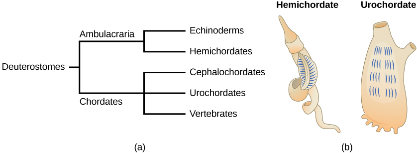
The deuterostomes consist of two major clades—the Chordata and the Ambulacraria. The Chordata include the vertebrates and two invertebrate subphyla, the urochordates and the cephalochordates. The Ambulacraria include the echinoderms and the hemichordates, which were once considered to be a chordate subphylum (Figure 28.46). The two clades, in addition to being deuterostomes, have some other interesting features in common. As we have seen, the vast majority of invertebrate animals do not possess a defined bony vertebral endoskeleton, or a bony cranium. However, one of the most ancestral groups of deuterostome invertebrates, the Echinodermata, do produce tiny skeletal “bones” called ossicles that make up a true endoskeleton, or internal skeleton, covered by an epidermis. The Hemichordata (acorn worms and pterobranchs) will not be covered here, but share with the echinoderms a three-part (tripartite) coelom, similar larval forms, and a derived metanephridium that rids the animals of nitrogenous wastes. They also share pharyngeal slits with the chordates (Figure 28.46). In addition, hemichordates have a dorsal nerve cord in the midline of the epidermis, but lack a neural tube, a true notochord and the endostyle and post-anal tail characteristic of chordates.
英語のヒント
後口動物は、脊索動物と歩帯動物という二つの主要クレードからなる。脊索動物には、脊椎動物と、無脊椎の二亜門である尾索動物・頭索動物が含まれる。歩帯動物には棘皮動物と、かつて脊索動物の亜門と考えられていた半索動物が含まれる(図28.46)。この二つのクレードは、後口動物であることに加え、いくつかの興味深い特徴を共有する。すでに見たように、無脊椎動物の大多数には、明確な骨性の脊椎内骨格や骨性の頭蓋がない。しかし、後口動物の無脊椎動物の中で最も祖先的な群の一つである棘皮動物は、骨片と呼ばれる小さな骨格性の「骨」をつくる。これは、表皮で覆われた真の内骨格を構成する。ここでは半索動物(ギボシムシとフサカツギ)を詳しく扱わないが、棘皮動物と共通して、三部に分かれた体腔、似た幼生型、窒素性老廃物を除く派生的な後腎管をもつ。脊索動物と共通する咽頭裂もある(図28.46)。また、半索動物は表皮の背側正中線に神経索をもつが、脊索動物の特徴である神経管、真の脊索、内柱、肛門後方の尾を欠く。
OpenStax, Rice University; Biology 2e. (credit a MAC; credit b modification of Gill Slits By Own work by Zebra.element [Public domain], via Wikimedia Commons) · CC BY 4.0 · Source出典
Figure 28.46図 28.46
Ambulacraria and Chordata. (a) The major deuterostome taxa. (b) pharyngeal slits in hemichordates and urochordates. (credit a MAC; credit b modification of Gill Slits By Own work by Zebra.element [Public domain], via Wikimedia Commons)
歩帯動物と脊索動物。(a)後口動物の主要分類群。(b)半索動物と尾索動物の咽頭裂。(a:MAC。b:Zebra.element自身による作品「Gill Slits」(パブリックドメイン)、Wikimedia Commons経由の図を改変)
Phylum Echinodermata棘皮動物門
Echinodermata are named after their “prickly skin” (from the Greek “echinos” meaning “prickly” and “dermos” meaning “skin”). This phylum is a collection of about 7,000 described living species of exclusively marine, bottom-dwelling organisms. Sea stars (Figure 28.47), sea cucumbers, sea urchins, sand dollars, and brittle stars are all examples of echinoderms.
英語のヒント
棘皮動物は「とげのある皮膚」にちなんで名付けられた。ギリシャ語の echinos は「とげのある」、dermos は「皮膚」を意味する。この門は、海産の底生生物だけからなり、現生の記載種は約7,000種である。ヒトデ(図28.47)、ナマコ、ウニ、タコノマクラ類、クモヒトデは、すべて棘皮動物の例である。
Morphology and Anatomy形態と体の構造
Despite the adaptive value of bilaterality for most free-living cephalized animals, adult echinoderms exhibit pentaradial symmetry (with “arms” typically arrayed in multiples of five around a central axis). Echinoderms have an endoskeleton made of calcareous ossicles (small bony plates), covered by the epidermis. For this reason, it is an endoskeleton like our own, not an exoskeleton like that of arthropods. The ossicles may be fused together, embedded separately in the connective tissue of the dermis, or be reduced to minute spicules of bone as in sea cucumbers. The spines for which the echinoderms are named are connected to some of the plates. The spines may be moved by small muscles, but they can also be locked into place for defense. In some species, the spines are surrounded by tiny stalked claws called pedicellaria, which help keep the animal's surface clean of debris, protect papulae used in respiration, and sometimes aid in food capture.
英語のヒント
頭化した自由生活性動物の多くにとって、左右相称には適応上の価値がある。それにもかかわらず、棘皮動物の成体は五放射相称を示し、通常、中央の軸の周囲に5の倍数の「腕」が並ぶ。棘皮動物の内骨格は、表皮に覆われた石灰質の骨片(小さな骨の板)からなる。このため、節足動物のような外骨格ではなく、私たちと同じく内骨格である。骨片は互いに融合することも、真皮の結合組織中に個別に埋まることも、ナマコのように微小な骨の棘へ縮小することもある。棘皮動物の名の由来となる棘は、板の一部につながっている。小さな筋肉で動かせるが、防御のために固定することもできる。一部の種では、叉棘という小さな柄付きの爪が棘の周囲にある。叉棘は体表からごみを取り除き、呼吸に使われる皮鰓を守り、ときには食物の捕獲も助ける。
The endoskeleton is produced by dermal cells, which also produce several kinds of pigments, imparting vivid colors to these animals. In sea stars, fingerlike projections (papillae) of dermal tissue extend through the endoskeleton and function as gills. Some cells are glandular, and may produce toxins. Each arm or section of the animal contains several different structures: for example, digestive glands, gonads, and the tube feet that are unique to the echinoderms. In echinoderms like sea stars, every arm bears two rows of tube feet on the oral side, running along an external ambulacral groove. These tube feet assist in locomotion, feeding, and chemical sensations, as well as serve to attach some species to the substratum.
英語のヒント
内骨格は真皮の細胞によってつくられる。これらの細胞は複数種の色素もつくり、体に鮮やかな色を与える。ヒトデでは、真皮組織の指状の突起(乳頭)が内骨格を通って外へ伸び、鰓として働く。一部の細胞は腺性で、毒素をつくることがある。各腕や区画には、消化腺、生殖腺、そして棘皮動物に固有の管足など、さまざまな構造が含まれる。ヒトデのような棘皮動物では、各腕の口側に、外部の歩帯溝に沿って2列の管足が並ぶ。管足は移動、摂食、化学感覚を助け、一部の種では基質への付着にも使われる。
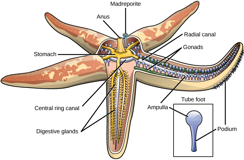


Figure 28.47図 28.47
Anatomy of a sea star. This diagram of a sea star shows the pentaradial pattern typical of adult echinoderms, and the water vascular system that is their defining characteristic.
ヒトデの体の構造。棘皮動物の成体に典型的な五放射の配置と、この動物群を特徴づける水管系を示す。