第28章無脊椎動物
28.1 Phylum Porifera28.1 海綿動物門
Learning Objectives学習目標
By the end of this section, you will be able to do the following:
この節を学ぶと、次のことができるようになる。
- Describe the organizational features of the simplest multicellular organisms最も単純な多細胞生物の構成上の特徴を説明する。
- Explain the various body forms and bodily functions of sponges海綿の多様な体形と体の機能を説明する。
As we have seen, the vast majority of invertebrate animals do not possess a defined bony vertebral endoskeleton, or a bony cranium. However, one of the most ancestral groups of deuterostome invertebrates, the Echinodermata, do produce tiny skeletal “bones” called ossicles that make up a true endoskeleton, or internal skeleton, covered by an epidermis.
英語のヒント
すでに見たように、無脊椎動物の大多数は、明確な骨性の脊椎内骨格も骨性の頭蓋ももたない。しかし、後口動物に属する無脊椎動物の中でも祖先的な群の一つである棘皮動物は、骨片と呼ばれる小さな骨格性の「骨」を形成する。それらが、表皮に覆われた真の内骨格を構成する。
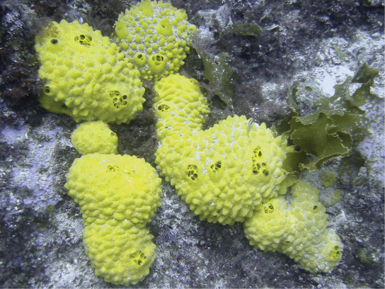
We will start our investigation with the simplest of all the invertebrates—animals sometimes classified within the clade Parazoa (“beside the animals”). This clade currently includes only the phylum Placozoa (containing a single species, Trichoplax adhaerens), and the phylum Porifera, containing the more familiar sponges (Figure 28.2). The split between the Parazoa and the Eumetazoa (all animal clades above Parazoa) likely took place over a billion years ago.
英語のヒント
無脊椎動物の中で最も単純な動物から調べ始めよう。これらは、側生動物(Parazoa、「動物のそばに」の意)というクレードに分類されることがある。このクレードに現在含まれるのは、1種の Trichoplax adhaerens からなる平板動物門と、よりなじみ深い海綿を含む海綿動物門だけである(図28.2)。側生動物と真正後生動物(側生動物より上位にあるすべての動物クレード)の分岐は、10億年以上前に起こったと考えられる。
We should reiterate here that the Porifera do not possess “true” tissues that are embryologically homologous to those of all other derived animal groups such as the insects and mammals. This is because they do not create a true gastrula during embryogenesis, and as a result do not produce a true endoderm or ectoderm. But even though they are not considered to have true tissues, they do have specialized cells that perform specific functions like tissues (for example, the external “pinacoderm” of a sponge acts like our epidermis). Thus, functionally, the poriferans can be said to have tissues; however, these tissues are likely not embryologically homologous to our own.
英語のヒント
ここで繰り返しておくと、海綿動物は、昆虫や哺乳類など、ほかのより派生的な動物群すべての組織と発生学的に相同な「真の」組織をもたない。これは、胚発生の過程で真の原腸胚を形成せず、その結果として真の内胚葉や外胚葉を形成しないためである。しかし、真の組織をもつとはみなされないものの、組織のように特定の機能を果たす特殊化した細胞はもっている。例えば、海綿の外側の「扁平上皮」は、私たちの表皮のように働く。したがって、機能の面では海綿動物にも組織があるといえるが、それらは私たちの組織と発生学的に相同ではない可能性が高い。
Sponge larvae (e.g., parenchymula and amphiblastula) are flagellated and able to swim; however, adults are non-motile and spend their life attached to a substratum. Since water is vital to sponges for feeding, excretion, and gas exchange, their body structure facilitates the movement of water through the sponge. Various canals, chambers, and cavities enable water to move through the sponge to allow the exchange of food and waste as well as the exchange of gases to nearly all body cells.
英語のヒント
海綿の幼生(例えばパレンキムラ幼生やアンフィブラストラ幼生)は鞭毛をもち、泳ぐことができる。一方、成体は移動せず、基質に付着して一生を過ごす。海綿では摂食、排泄、ガス交換に水が不可欠なため、その体の構造は、水が内部を通り抜けやすくできている。さまざまな水路、室、空洞を水が通ることで、ほぼすべての体細胞において、栄養と老廃物のやりとりやガス交換が可能になる。
OpenStax, Rice University; Biology 2e. (credit: Andrew Turner) Embedded rights notice: Andrew Turner · CC BY 4.0 · Source出典

Figure 28.2図 28.2
Sponges. Sponges are members of the phylum Porifera, which contains the simplest invertebrates. (credit: Andrew Turner)
海綿。海綿は、最も単純な無脊椎動物を含む海綿動物門の一員である。(提供:Andrew Turner)
Morphology of Sponges海綿の形態
There are at least 5,000 named species of sponges, likely with thousands more yet to be classified. The morphology of the simplest sponges takes the shape of an irregular cylinder with a large central cavity, the spongocoel, occupying the inside of the cylinder (Figure 28.3). Water enters into the spongocoel through numerous pores, or ostia, that create openings in the body wall. Water entering the spongocoel is expelled via a large common opening called the osculum. However, we should note that sponges exhibit a range of diversity in body forms, including variations in the size and shape of the spongocoel, as well as the number and arrangement of feeding chambers within the body wall. In some sponges, multiple feeding chambers open off of a central spongocoel and in others, several feeding chambers connecting to one another may lie between the entry pores and the spongocoel.
英語のヒント
海綿には少なくとも5,000種の命名済みの種があり、未分類の種もさらに数千種あると考えられる。最も単純な海綿は不規則な円筒形をしており、その内部を囲胃腔と呼ばれる大きな中央の空洞が占める(図28.3)。体壁に開く無数の小孔から水が囲胃腔に入り、大孔と呼ばれる一つの大きな共通の開口部から排出される。ただし、海綿の体形は多様で、囲胃腔の大きさや形、体壁内の摂食室の数や配置にもさまざまな違いがある。中央の囲胃腔につながって複数の摂食室が開く海綿もあれば、入口となる小孔と囲胃腔の間に、互いにつながった複数の摂食室がある海綿もある。
While sponges do not exhibit true tissue-layer organization, they do have a number of functional “tissues” composed of different cell types specialized for distinct functions. For example, epithelial-like cells called pinacocytes form the outermost body, called a pinacoderm, that serves a protective function similar that of our epidermis. Scattered among the pinacoderm are the ostia that allow entry of water into the body of the sponge. These pores have given the sponges their phylum name Porifera—pore-bearers. In some sponges, ostia are formed by porocytes, single tube-shaped cells that act as valves to regulate the flow of water into the spongocoel. In other sponges, ostia are formed by folds in the body wall of the sponge. Between the outer layer and the feeding chambers of the sponge is a jelly-like substance called the mesohyl, which contains collagenous fibers. Various cell types reside within the mesohyl, including amoebocytes, the “stem cells” of sponges, and sclerocytes, which produce skeletal materials. The gel-like consistency of mesohyl acts like an endoskeleton and maintains the tubular morphology of sponges.
英語のヒント
海綿には真の組織層による構成は見られないが、それぞれ異なる機能に特殊化した細胞型からなる、機能上の「組織」がいくつもある。例えば、扁平細胞と呼ばれる上皮に似た細胞は、体の最外層である扁平上皮を形成し、私たちの表皮に似た保護機能を果たす。扁平上皮の各所には、水を海綿の体内へ取り込む小孔が散在する。この孔にちなみ、海綿には「孔をもつもの」を意味する Porifera という門名が付けられている。一部の海綿では、小孔は孔細胞という単一の管状細胞によって形成される。この細胞は弁として働き、囲胃腔へ入る水の流れを調節する。ほかの海綿では、体壁の折れ込みによって小孔が形成される。海綿の外層と摂食室の間には、コラーゲン線維を含む、メソヒルと呼ばれるゼリー状の物質がある。メソヒルの内部には、海綿の「幹細胞」であるアメーバ細胞や、骨格物質を産生する骨片形成細胞など、さまざまな細胞型が存在する。メソヒルはゲル状の硬さをもち、内骨格のように働いて、海綿の管状の形態を保つ。
The feeding chambers inside the sponge are lined by choanocytes ("collar cells"). The structure of a choanocyte is critical to its function, which is to generate a directed water current through the sponge and to trap and ingest microscopic food particles by phagocytosis. These feeding cells are similar in appearance to unicellular choanoflagellates (Protista). This similarity suggests that sponges and choanoflagellates are closely related and likely share common ancestry. The body of the choanocyte is embedded in mesohyl and contains all the organelles required for normal cell function. Protruding into the “open space” inside the feeding chamber is a mesh-like collar composed of microvilli with a single flagellum in the center of the column. The beating of the flagella from all choanocytes draws water into the sponge through the numerous ostia, into the spaces lined by choanocytes, and eventually out through the osculum (or osculi, if the sponge consists of a colony of attached sponges). Food particles, including waterborne bacteria and unicellular organisms such as algae and various animal-like protists, are trapped by the sieve-like collar of the choanocytes, slide down toward the body of the cell, and are ingested by phagocytosis. Choanocytes also serve another surprising function: They can differentiate into sperm for sexual reproduction, at which time they become dislodged from the mesohyl and leave the sponge with expelled water through the osculum.
英語のヒント
海綿内部の摂食室は、襟細胞で裏打ちされている。襟細胞の構造は、その機能にとって決定的に重要である。その機能とは、海綿を通り抜ける一定方向の水流をつくり、微小な食物粒子を捕らえて食作用で取り込むことである。この摂食細胞の外見は、単細胞の襟鞭毛虫(原生生物)に似ている。この類似は、海綿と襟鞭毛虫が近縁であり、共通の祖先をもつ可能性が高いことを示唆する。襟細胞の細胞体はメソヒルに埋まっており、通常の細胞機能に必要な細胞小器官をすべて含んでいる。摂食室内の「開いた空間」へ突き出しているのは、微絨毛でできた網状の襟であり、その柱状構造の中央には1本の鞭毛がある。すべての襟細胞の鞭毛が打つことによって、水が多数の小孔から海綿内へ入り、襟細胞に囲まれた空間を通り、最終的に大孔から出ていく。海綿が互いに付着した個体の群体からなる場合には、大孔は複数ある。水中の細菌、藻類などの単細胞生物、さまざまな動物様の原生生物を含む食物粒子は、襟細胞のふるい状の襟に捕らえられ、細胞体のほうへ滑り落ち、食作用で取り込まれる。襟細胞には、もう一つ驚くべき機能もある。有性生殖のための精子に分化できるのである。その際にはメソヒルから離れ、排出される水とともに大孔を通って海綿の外へ出る。
The amoebocytes (derived from stem-cell-like archaeocytes), are so named because they move throughout the mesohyl in an amoeba-like fashion. They have a variety of functions: In addition to delivering nutrients from choanocytes to other cells within the sponge, they also give rise to eggs for sexual reproduction. (The eggs remain in the mesohyl, whereas the sperm cells are released into the water.) The amoebocytes can differentiate into other cell types of the sponge, such as collenocytes and lophocytes, which produce the collagen-like protein that support the mesohyl. Amoebocytes can also give rise to sclerocytes, which produce spicules (skeletal spikes of silica or calcium carbonate) in some sponges, and spongocytes, which produce the protein spongin in the majority of sponges. These different cell types in sponges are shown in Figure 28.3.
英語のヒント
アメーバ細胞は、幹細胞に似た原始細胞に由来し、メソヒル全体をアメーバのように移動することからその名が付いている。機能は多様である。襟細胞から海綿内のほかの細胞へ栄養を運ぶほか、有性生殖のための卵も生じさせる。卵はメソヒル内にとどまる一方、精子は水中へ放出される。アメーバ細胞は、膠細胞やロフォサイトなど、海綿のほかの細胞型へ分化できる。これらはメソヒルを支えるコラーゲン様タンパク質を産生する。またアメーバ細胞からは、一部の海綿で骨片(シリカや炭酸カルシウムからなる骨格の棘)をつくる骨片形成細胞や、大多数の海綿で海綿質というタンパク質をつくる海綿質形成細胞も生じる。海綿のこれらの異なる細胞型を図28.3に示す。
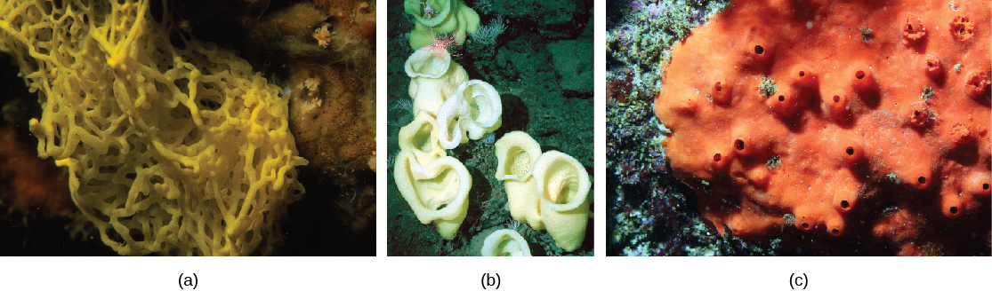
As we’ve seen, most sponges are supported by small bone-like spicules (usually tiny pointed structures made of calcium carbonate or silica) in the mesohyl. Spicules provide support for the body of the sponge, and may also deter predation. The presence and composition of spicules form the basis for differentiating three of the four classes of sponges (Figure 28.4). Sponges in class Calcarea produce calcium carbonate spicules and no spongin; those in class Hexactinellida produce six-rayed siliceous (glassy) spicules and no spongin; and those in class Demospongia contain spongin and may or may not have spicules; if present, those spicules are siliceous. Sponges in this last class have been used as bath sponges. Spicules are most conspicuously present in the glass sponges, class Hexactinellida. Some of the spicules may attain gigantic proportions. For example, relative to typical glass sponge spicules, whose size generally ranges from 3 to 10 mm, some of the basal spicules of the hexactinellid Monorhaphis chuni are enormous and grow up to 3 meters long! The glass sponges are also unusual in that most of their body cells are fused together to form a multinucleate syncytium. Because their cells are interconnected in this way, the hexactinellid sponges have no mesohyl. A fourth class of sponges, the Sclerospongiae, was described from species discovered in underwater tunnels. These are also called coralline sponges after their multilayered calcium carbonate skeletons. Dating based on the rate of deposition of the skeletal layers suggests that some of these sponges are hundreds of years old.
英語のヒント
すでに見たように、ほとんどの海綿は、メソヒル内にある小さな骨のような骨片によって支えられている。骨片は通常、炭酸カルシウムやシリカでできた微小な尖った構造である。骨片は海綿の体を支え、捕食を抑える働きもあると考えられる。骨片の有無と組成は、海綿の四つの綱のうち三つを区別する基準となる(図28.4)。石灰海綿綱の海綿は炭酸カルシウムの骨片をつくるが、海綿質はつくらない。六放海綿綱は6本の放射枝をもつシリカ質(ガラス質)の骨片をつくるが、海綿質はつくらない。普通海綿綱は海綿質を含み、骨片をもつものともたないものがある。骨片がある場合はシリカ質である。この最後の綱に属する海綿は、入浴用のスポンジとして使われてきた。骨片が特に目立つのは、六放海綿綱のガラス海綿である。骨片の中には巨大になるものもある。例えば、ガラス海綿の典型的な骨片の大きさは一般に3〜10 mmだが、六放海綿 Monorhaphis chuni の基部の骨片の一部は、長さ3 mにも達する。ガラス海綿は、体の細胞の大部分が融合して、多核の合胞体を形成する点でも独特である。このように細胞が互いにつながっているため、六放海綿にはメソヒルがない。第四の綱である硬骨海綿綱は、水中のトンネルで発見された種から記載された。炭酸カルシウムの骨格が何層にも重なることから、サンゴ海綿とも呼ばれる。骨格層が堆積する速さに基づく年代推定から、その一部は数百年生きていると考えられる。
OpenStax, Rice University; Biology 2e. (credit a: modification of work by Parent Géry; credit b: modification of work by Monterey Bay Aquarium Research Institute, NOAA; credit c: modification of work by Sanctuary Integrated Monitoring Network, Monterey Bay National Marine Sanctuary, NOAA) · CC BY 4.0 · Source出典

Figure 28.4図 28.4
Several classes of sponges. (a) Clathrina clathrus belongs to class Calcarea, (b) Staurocalyptus spp. (common name: yellow Picasso sponge) belongs to class Hexactinellida, and (c) Acarnus erithacus belongs to class Demospongia. (credit a: modification of work by Parent Géry; credit b: modification of work by Monterey Bay Aquarium Research Institute, NOAA; credit c: modification of work by Sanctuary Integrated Monitoring Network, Monterey Bay National Marine Sanctuary, NOAA)
海綿のいくつかの綱。(a)Clathrina clathrus は石灰海綿綱、(b)Staurocalyptus 属の種(通称:イエロー・ピカソ・スポンジ)は六放海綿綱、(c)Acarnus erithacus は普通海綿綱に属する。(a:Parent Géryの作品を改変。b:Monterey Bay Aquarium Research Institute、NOAAの作品を改変。c:Sanctuary Integrated Monitoring Network、Monterey Bay National Marine Sanctuary、NOAAの作品を改変)