第35章神経系
35.3 The Central Nervous System35.3 中枢神経系
Learning Objectives学習目標
By the end of this section, you will be able to do the following:
この節を学び終えると、次のことができるようになる。
- Identify the spinal cord, cerebral lobes, and other brain areas on a diagram of the brain脳の図で、脊髄、大脳の各葉、その他の脳領域を見分ける。
- Describe the basic functions of the spinal cord, cerebral lobes, and other brain areas脊髄、大脳の各葉、その他の脳領域の基本的な機能を説明する。
The central nervous system (CNS) is made up of the brain, a part of which is shown in Figure 35.19 and spinal cord and is covered with three layers of protective coverings called meninges (from the Greek word for membrane). The outermost layer is the dura mater (Latin for “hard mother”). As the Latin suggests, the primary function for this thick layer is to protect the brain and spinal cord. The dura mater also contains vein-like structures that carry blood from the brain back to the heart. The middle layer is the web-like arachnoid mater. The last layer is the pia mater (Latin for “soft mother”), which directly contacts and covers the brain and spinal cord like plastic wrap. The space between the arachnoid and pia maters is filled with cerebrospinal fluid (CSF). CSF is produced by a tissue called choroid plexus in fluid-filled compartments in the CNS called ventricles. The brain floats in CSF, which acts as a cushion and shock absorber and makes the brain neutrally buoyant. CSF also functions to circulate chemical substances throughout the brain and into the spinal cord.
英語のヒント
中枢神経系(CNS)は、図35.19に一部を示す脳と脊髄からなり、髄膜(ギリシャ語の「膜」に由来)という三層の保護膜で覆われている。最外層は硬膜(ラテン語で「硬い母」)である。その名称が示すように、この厚い層の主な働きは脳と脊髄の保護である。硬膜には、脳から心臓へ血液を戻す静脈状の構造もある。中間層は網状のくも膜である。最内層は軟膜(ラテン語で「柔らかい母」)で、食品用ラップのように脳と脊髄に直接接して覆う。くも膜と軟膜の間は脳脊髄液(CSF)で満たされる。CSFは、中枢神経系の脳室という液体で満たされた区画にある、脈絡叢という組織によってつくられる。脳はCSFに浮かび、CSFはクッションと衝撃吸収材として働き、脳を中性浮力の状態にする。また、脳全体から脊髄へ化学物質を循環させる働きもある。
The entire brain contains only about 8.5 tablespoons of CSF, but CSF is constantly produced in the ventricles. This creates a problem when a ventricle is blocked—the CSF builds up and creates swelling and the brain is pushed against the skull. This swelling condition is called hydrocephalus (“water head”) and can cause seizures, cognitive problems, and even death if a shunt is not inserted to remove the fluid and pressure.
英語のヒント
脳全体にあるCSFは大さじ約8.5杯にすぎないが、脳室では常につくられている。脳室が閉塞すると、CSFがたまって腫れ、脳が頭蓋骨に押しつけられるため問題となる。この腫脹状態を水頭症(「水の頭」)という。液体と圧力を逃がすシャントが挿入されなければ、けいれん発作、認知機能の問題、さらには死を招くことがある。
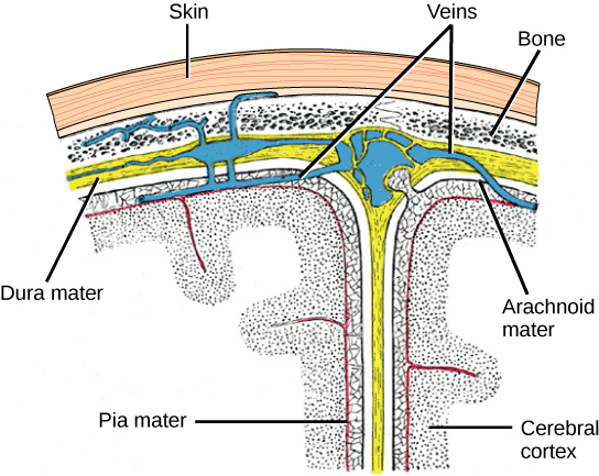
OpenStax, Rice University; Biology 2e. (credit: modification of work by Gray’s Anatomy) · CC BY 4.0 · Source出典
Figure 35.19図 35.19
The cerebral cortex is covered by three layers of meninges: the dura, arachnoid, and pia maters. (credit: modification of work by Gray’s Anatomy)
大脳皮質は、硬膜、くも膜、軟膜という三層の髄膜で覆われる。(クレジット:Gray’s Anatomyの作品を改変)
Brain脳
The brain is the part of the central nervous system that is contained in the cranial cavity of the skull. It includes the cerebral cortex, limbic system, basal ganglia, thalamus, hypothalamus, and cerebellum. There are three different ways that a brain can be sectioned in order to view internal structures: a sagittal section cuts the brain left to right, as shown in Figure 35.20b, a coronal section cuts the brain front to back, as shown in These illustrations show the (a) coronal and (b) sagittal sections of the human brain.a, and a horizontal section cuts the brain top to bottom.
脳は、頭蓋骨の頭蓋腔内にある中枢神経系の部分である。大脳皮質、大脳辺縁系、大脳基底核、視床、視床下部、小脳を含む。内部構造を見るための切り方には三つある。図35.20bに示す矢状断は脳を左右に分け、冠状断は「ヒトの脳の(a)冠状断と(b)矢状断を示す図」aのように前後に分け、水平断は上下に分ける。
Cerebral Cortex大脳皮質
The outermost part of the brain is a thick piece of nervous system tissue called the cerebral cortex, which is folded into hills called gyri (singular: gyrus) and valleys called sulci (singular: sulcus). The cortex is made up of two hemispheres—right and left—which are separated by a large sulcus. A thick fiber bundle called the corpus callosum (Latin: “tough body”) connects the two hemispheres and allows information to be passed from one side to the other. Although there are some brain functions that are localized more to one hemisphere than the other, the functions of the two hemispheres are largely redundant. In fact, sometimes (very rarely) an entire hemisphere is removed to treat severe epilepsy. While patients do suffer some deficits following the surgery, they can have surprisingly few problems, especially when the surgery is performed on children who have very immature nervous systems.
英語のヒント
脳の最外側には、大脳皮質という厚い神経組織があり、脳回(単数gyrus、複数gyri)という山と、脳溝(単数sulcus、複数sulci)という谷に折りたたまれている。皮質は左右二つの半球からなり、大きな溝で隔てられる。脳梁(ラテン語で「丈夫な体」)という太い線維束が両半球をつなぎ、一方から他方へ情報を渡せるようにする。一部の機能は一方の半球により強く局在するが、左右の機能は大部分が重複している。実際、重いてんかんの治療で、ごくまれに半球全体を除去することがある。手術後には多少の機能障害が生じるものの、特に神経系がまだ非常に未熟な子どもに手術した場合、驚くほど問題が少ないことがある。
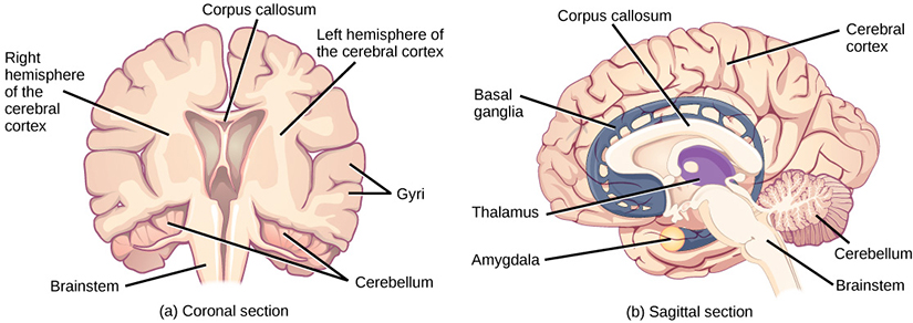
These illustrations show the (a) coronal and (b) sagittal sections of the human brain.
ヒトの脳の(a)冠状断と(b)矢状断を示す図。
In other surgeries to treat severe epilepsy, the corpus callosum is cut instead of removing an entire hemisphere. This causes a condition called split-brain, which gives insights into unique functions of the two hemispheres. For example, when an object is presented to patients’ left visual field, they may be unable to verbally name the object (and may claim to not have seen an object at all). This is because the visual input from the left visual field crosses and enters the right hemisphere and cannot then signal to the speech center, which generally is found in the left side of the brain. Remarkably, if a split-brain patient is asked to pick up a specific object out of a group of objects with the left hand, the patient will be able to do so but will still be unable to vocally identify it.
英語のヒント
重いてんかんを治療する別の手術では、半球全体を除く代わりに脳梁を切断する。これによって分離脳と呼ばれる状態が生じ、両半球の固有の機能を知る手掛かりが得られる。例えば患者の左視野に物体を提示すると、物体の名前を言えないことがあり、何も見なかったと主張することさえある。これは左視野からの視覚入力が交差して右半球へ入り、その後、通常は左半球にある言語中枢へ信号を送れないためである。注目すべきことに、分離脳患者に、複数の物体から特定のものを左手で取り上げるよう求めると、それはできる。しかし、その物体を声で言い当てることは依然としてできない。
Each cortical hemisphere contains regions called lobes that are involved in different functions. Scientists use various techniques to determine what brain areas are involved in different functions: they examine patients who have had injuries or diseases that affect specific areas and see how those areas are related to functional deficits. They also conduct animal studies where they stimulate brain areas and see if there are any behavioral changes. They use a technique called transcranial magnetic stimulation (TMS) to temporarily deactivate specific parts of the cortex using strong magnets placed outside the head; and they use functional magnetic resonance imaging (fMRI) to look at changes in oxygenated blood flow in particular brain regions that correlate with specific behavioral tasks. These techniques, and others, have given great insight into the functions of different brain regions but have also showed that any given brain area can be involved in more than one behavior or process, and any given behavior or process generally involves neurons in multiple brain areas. That being said, each hemisphere of the mammalian cerebral cortex can be broken down into four functionally and spatially defined lobes: frontal, parietal, temporal, and occipital. Figure 35.20 illustrates these four lobes of the human cerebral cortex.
英語のヒント
各大脳皮質半球には、異なる機能に関わる葉という領域がある。科学者は、どの領域がどの機能に関わるかを調べるため、さまざまな方法を使う。特定部位を損なうけがや病気の患者を調べ、その部位と機能障害の関係を確かめる。また動物研究では、脳の領域を刺激し、行動が変わるかを調べる。経頭蓋磁気刺激(TMS)では、頭の外に置いた強い磁石を使い、皮質の特定の部分を一時的に不活性化する。機能的磁気共鳴画像法(fMRI)では、特定の行動課題と相関する、脳の各領域の酸素化血流の変化を見る。これらや他の手法によって各領域の機能の理解は進んだが、ある領域が複数の行動や過程に関わり、ある行動や過程にも通常は複数領域のニューロンが関わることが分かった。それでも、哺乳類の大脳皮質の各半球は、機能と空間的位置によって定義される四つの葉、前頭葉、頭頂葉、側頭葉、後頭葉に分けられる。図35.20は、ヒトの大脳皮質の四葉を示す。
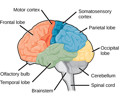
Figure 35.20図 35.20
The human cerebral cortex includes the frontal, parietal, temporal, and occipital lobes.
ヒトの大脳皮質には、前頭葉、頭頂葉、側頭葉、後頭葉がある。
The frontal lobe is located at the front of the brain, over the eyes. This lobe contains the olfactory bulb, which processes smells. The frontal lobe also contains the motor cortex, which is important for planning and implementing movement. Areas within the motor cortex map to different muscle groups, and there is some organization to this map, as shown in Figure 35.21. For example, the neurons that control movement of the fingers are next to the neurons that control movement of the hand. Neurons in the frontal lobe also control cognitive functions like maintaining attention, speech, and decision-making. Studies of humans who have damaged their frontal lobes show that parts of this area are involved in personality, socialization, and assessing risk.
前頭葉は脳の前部、眼の上にある。匂いを処理する嗅球を含み、運動の計画と実行に重要な運動皮質も含む。運動皮質の領域は各筋群と対応し、図35.21のように、一定の配置を持つ。例えば指の運動を制御するニューロンは、手の運動を制御するニューロンの隣にある。前頭葉のニューロンは、注意の維持、発話、意思決定などの認知機能も制御する。前頭葉に損傷を受けた人々の研究は、この領域の一部が人格、社会化、危険の評価に関わることを示している。
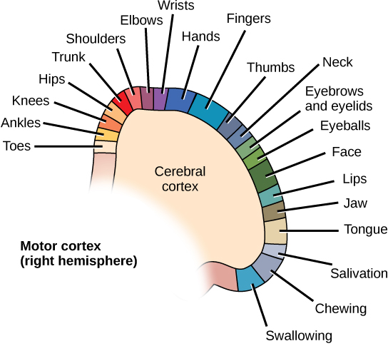


Figure 35.21図 35.21
Different parts of the motor cortex control different muscle groups. Muscle groups that are neighbors in the body are generally controlled by neighboring regions of the motor cortex as well. For example, the neurons that control finger movement are near the neurons that control hand movement.
運動皮質の異なる部分が、異なる筋群を制御する。体内で隣り合う筋群は、一般に運動皮質でも隣り合う領域によって制御される。例えば指の運動を制御するニューロンは、手の運動を制御するニューロンの近くにある。
The parietal lobe is located at the top of the brain. Neurons in the parietal lobe are involved in speech and also reading. Two of the parietal lobe’s main functions are processing somatosensation—touch sensations like pressure, pain, heat, cold—and processing proprioception—the sense of how parts of the body are oriented in space. The parietal lobe contains a somatosensory map of the body similar to the motor cortex.
英語のヒント
頭頂葉は脳の上部にある。そのニューロンは発話や読字に関与する。主な機能のうち二つは、圧力、痛み、熱、冷たさなどの触覚的な体性感覚の処理と、体の各部が空間内でどう向いているかを知る固有感覚の処理である。頭頂葉には、運動皮質と同様に、体の体性感覚地図がある。
The occipital lobe is located at the back of the brain. It is primarily involved in vision—seeing, recognizing, and identifying the visual world.
後頭葉は脳の後部にある。主に視覚、すなわち視覚世界を見る、認識する、識別することに関与する。
The temporal lobe is located at the base of the brain by your ears and is primarily involved in processing and interpreting sounds. It also contains the hippocampus (Greek for “seahorse”)—a structure that processes memory formation. The hippocampus is illustrated in Figure 35.23. The role of the hippocampus in memory was partially determined by studying one famous epileptic patient, HM, who had both sides of his hippocampus removed in an attempt to cure his epilepsy. His seizures went away, but he could no longer form new memories (although he could remember some facts from before his surgery and could learn new motor tasks).
側頭葉は耳のそば、脳の下部にあり、主に音の処理と解釈に関与する。また、記憶形成を処理する構造である海馬(ギリシャ語で「タツノオトシゴ」)を含む。図35.23に海馬を示す。その記憶における役割は、てんかんを治そうとして両側の海馬を除去された有名な患者HMの研究から、一部が分かった。発作はなくなったが、新しい記憶をつくれなくなった。ただし、手術前の事実の一部を思い出したり、新しい運動課題を学んだりすることはできた。