第35章神経系
Basal Ganglia大脳基底核
Interconnected brain areas called the basal ganglia (or basal nuclei), shown in These illustrations show the (a) coronal and (b) sagittal sections of the human brain.b, play important roles in movement control and posture. Damage to the basal ganglia, as in Parkinson’s disease, leads to motor impairments like a shuffling gait when walking. The basal ganglia also regulate motivation. For example, when a wasp sting led to bilateral basal ganglia damage in a 25-year-old businessman, he began to spend all his days in bed and showed no interest in anything or anybody. But when he was externally stimulated—as when someone asked to play a card game with him—he was able to function normally. Interestingly, he and other similar patients do not report feeling bored or frustrated by their state.
英語のヒント
「ヒトの脳の(a)冠状断と(b)矢状断を示す図」bの大脳基底核は、相互につながった脳領域で、運動制御と姿勢に重要な役割を果たす。パーキンソン病のように大脳基底核が損傷すると、すり足歩行などの運動障害が生じる。大脳基底核は意欲も調節する。例えば、ハチに刺されて両側の大脳基底核を損傷した25歳の会社員は、一日中ベッドで過ごすようになり、何にも、誰にも関心を示さなくなった。しかし、誰かがカードゲームに誘うなど、外から刺激されると、正常に行動できた。興味深いことに、本人や同様の患者は、自分の状態に退屈や不満を感じるとは報告していない。
Thalamus視床
The thalamus (Greek for “inner chamber”), illustrated in Figure 35.23, acts as a gateway to and from the cortex. It receives sensory and motor inputs from the body and also receives feedback from the cortex. This feedback mechanism can modulate conscious awareness of sensory and motor inputs depending on the attention and arousal state of the animal. The thalamus helps regulate consciousness, arousal, and sleep states. A rare genetic disorder called fatal familial insomnia causes the degeneration of thalamic neurons and glia. This disorder prevents affected patients from being able to sleep, among other symptoms, and is eventually fatal.
英語のヒント
図35.23の視床(ギリシャ語で「内なる部屋」)は、皮質への出入り口として働く。体から感覚入力と運動入力を受け、皮質からのフィードバックも受ける。このフィードバック機構は、動物の注意や覚醒の状態に応じて、感覚・運動入力が意識に上る程度を調節できる。視床は意識、覚醒、睡眠状態の調節を助ける。致死性家族性不眠症というまれな遺伝性疾患は、視床のニューロンとグリアを変性させる。この病気には、患者が眠れなくなるなどの症状があり、最終的には致死的となる。
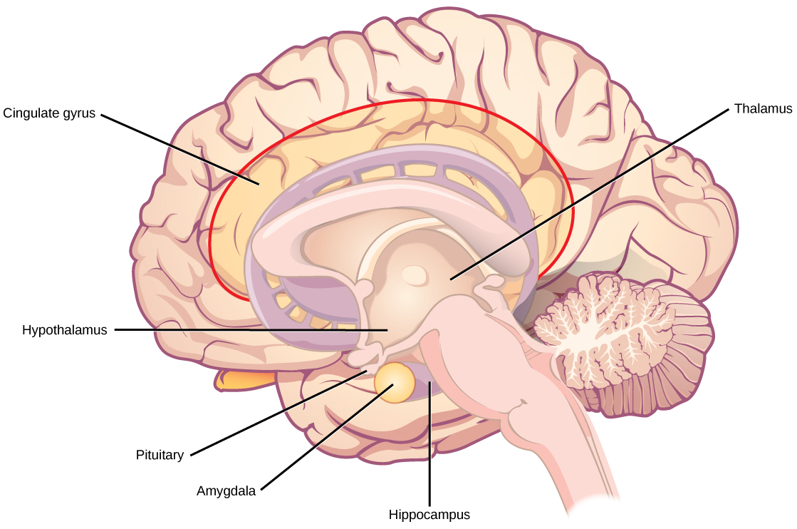
Figure 35.23図 35.23
The limbic system regulates emotion and other behaviors. It includes parts of the cerebral cortex located near the center of the brain, including the cingulate gyrus and the hippocampus as well as the thalamus, hypothalamus, and amygdala.
大脳辺縁系は感情やその他の行動を調節する。脳の中央近くの大脳皮質の一部である帯状回と海馬に加え、視床、視床下部、扁桃体を含む。
Hypothalamus視床下部
Below the thalamus is the hypothalamus, shown in Figure 35.23. The hypothalamus controls the endocrine system by sending signals to the pituitary gland, a pea-sized endocrine gland that releases several different hormones that affect other glands as well as other cells. This relationship means that the hypothalamus regulates important behaviors that are controlled by these hormones. The hypothalamus is the body’s thermostat—it makes sure key functions like food and water intake, energy expenditure, and body temperature are kept at appropriate levels. Neurons within the hypothalamus also regulate circadian rhythms, sometimes called sleep cycles.
英語のヒント
図35.23の視床下部は視床の下にある。エンドウ豆ほどの大きさの内分泌腺である下垂体へ信号を送り、内分泌系を制御する。下垂体は、他の腺や細胞に影響する複数のホルモンを放出する。この関係により、視床下部は、それらのホルモンに制御される重要な行動を調節する。視床下部は体の温度調節器であり、食物や水の摂取、エネルギー消費、体温といった重要な機能が適切な水準に保たれるようにする。そのニューロンは、睡眠周期とも呼ばれる概日リズムも調節する。
Limbic System大脳辺縁系
The limbic system is a connected set of structures that regulates emotion, as well as behaviors related to fear and motivation. It plays a role in memory formation and includes parts of the thalamus and hypothalamus as well as the hippocampus. One important structure within the limbic system is a temporal lobe structure called the amygdala (Greek for “almond”), illustrated in Figure 35.23. The two amygdala are important both for the sensation of fear and for recognizing fearful faces. The cingulate gyrus helps regulate emotions and pain.
大脳辺縁系は、感情と、恐怖や意欲に関する行動を調節する、互いにつながった構造群である。記憶形成にも役割を果たし、海馬に加えて視床と視床下部の一部を含む。その重要な構造の一つが、図35.23に示す、側頭葉の扁桃体(ギリシャ語で「アーモンド」)である。左右二つの扁桃体は、恐怖を感じることと、恐怖を示す顔を認識することの両方に重要である。帯状回は感情と痛みの調節を助ける。
Cerebellum小脳
The cerebellum (Latin for “little brain”), shown in Figure 35.20, sits at the base of the brain on top of the brainstem. The cerebellum controls balance and aids in coordinating movement and learning new motor tasks.
図35.20の小脳(ラテン語で「小さな脳」)は、脳の下部、脳幹の上に位置する。平衡を制御し、運動の協調や新しい運動課題の学習を助ける。
Brainstem脳幹
The brainstem, illustrated in Figure 35.20, connects the rest of the brain with the spinal cord. It consists of the midbrain, medulla oblongata, and the pons. Motor and sensory neurons extend through the brainstem allowing for the relay of signals between the brain and spinal cord. Ascending neural pathways cross in this section of the brain allowing the left hemisphere of the cerebrum to control the right side of the body and vice versa. The brainstem coordinates motor control signals sent from the brain to the body. The brainstem controls several important functions of the body including alertness, arousal, breathing, blood pressure, digestion, heart rate, swallowing, walking, and sensory and motor information integration.
英語のヒント
図35.20の脳幹は、脳の他の部分と脊髄をつなぐ。中脳、延髄、橋からなる。運動ニューロンと感覚ニューロンが脳幹を通り、脳と脊髄の間で信号を中継する。上行神経路は脳のこの部分で交差し、大脳の左半球が体の右側を制御できるようにする。逆も同様である。脳幹は、脳から体へ送られる運動制御信号を協調させる。注意の維持、覚醒、呼吸、血圧、消化、心拍数、嚥下、歩行、感覚情報と運動情報の統合など、体の重要な機能をいくつも制御する。
Spinal Cord脊髄
Connecting to the brainstem and extending down the body through the spinal column is the spinal cord, shown in Figure 35.20. The spinal cord is a thick bundle of nerve tissue that carries information about the body to the brain and from the brain to the body. The spinal cord is contained within the bones of the vertebrate column but is able to communicate signals to and from the body through its connections with spinal nerves (part of the peripheral nervous system). A cross-section of the spinal cord looks like a white oval containing a gray butterfly-shape, as illustrated in Figure 35.24. Myelinated axons make up the “white matter” and neuron and glial cell bodies make up the “gray matter.” Gray matter is also composed of interneurons, which connect two neurons each located in different parts of the body. Axons and cell bodies in the dorsal (facing the back of the animal) spinal cord convey mostly sensory information from the body to the brain. Axons and cell bodies in the ventral (facing the front of the animal) spinal cord primarily transmit signals controlling movement from the brain to the body.
英語のヒント
図35.20の脊髄は、脳幹につながり、脊柱を通って体の下方へ伸びる。体に関する情報を脳へ、また脳から体へ運ぶ、太い神経組織の束である。脊柱の骨の内部に収まるが、末梢神経系の一部である脊髄神経との結合を通じ、体との間で信号をやり取りできる。脊髄の断面は、図35.24のように、灰色のチョウ形を含む白い楕円に見える。髄鞘化された軸索が「白質」を、ニューロンとグリアの細胞体が「灰白質」をつくる。灰白質には、体の異なる部分にある二つのニューロンをつなぐ介在ニューロンもある。脊髄の背側(動物の背中側)の軸索と細胞体は、主に体から脳へ感覚情報を伝える。腹側(動物の前面側)の軸索と細胞体は、主に脳から体へ、運動を制御する信号を送る。
The spinal cord also controls motor reflexes. These reflexes are quick, unconscious movements—like automatically removing a hand from a hot object. Reflexes are so fast because they involve local synaptic connections. For example, the knee reflex that a doctor tests during a routine physical is controlled by a single synapse between a sensory neuron and a motor neuron. While a reflex may only require the involvement of one or two synapses, synapses with interneurons in the spinal column transmit information to the brain to convey what happened (the knee jerked, or the hand was hot).
英語のヒント
脊髄は運動反射も制御する。熱い物に触れた手を自動的に引っ込めるような、速く無意識的な動作である。反射が速いのは、局所的なシナプス結合によって行われるからである。例えば、通常の診察で医師が調べる膝の反射は、感覚ニューロンと運動ニューロンの間の一つのシナプスで制御される。反射自体には一つか二つのシナプスが関わるだけでよい場合もあるが、脊柱内の介在ニューロンとのシナプスは、膝が跳ねた、手が熱かったという出来事を脳へ伝える。
In the United States, there around 10,000 spinal cord injuries each year. Because the spinal cord is the information superhighway connecting the brain with the body, damage to the spinal cord can lead to paralysis. The extent of the paralysis depends on the location of the injury along the spinal cord and whether the spinal cord was completely severed. For example, if the spinal cord is damaged at the level of the neck, it can cause paralysis from the neck down, whereas damage to the spinal column further down may limit paralysis to the legs. Spinal cord injuries are notoriously difficult to treat because spinal nerves do not regenerate, although ongoing research suggests that stem cell transplants may be able to act as a bridge to reconnect severed nerves. Researchers are also looking at ways to prevent the inflammation that worsens nerve damage after injury. One such treatment is to pump the body with cold saline to induce hypothermia. This cooling can prevent swelling and other processes that are thought to worsen spinal cord injuries.
英語のヒント
米国では、年間約1万件の脊髄損傷が起こる。脊髄は脳と体を結ぶ情報の幹線であるため、損傷は麻痺につながりうる。その範囲は、脊髄のどの位置が傷ついたかと、完全に切断されたかどうかによる。例えば頸部の高さで損傷すると首から下が麻痺しうるが、さらに下方の脊柱の損傷では、麻痺が脚だけに限られることがある。脊髄神経は再生しないため、脊髄損傷の治療は非常に難しいことで知られる。ただし現在の研究は、幹細胞移植が切断された神経を再接続する橋として働く可能性を示している。研究者は、損傷後に神経障害を悪化させる炎症を防ぐ方法も探っている。その一つは、冷たい生理食塩水を体内へ送り、低体温を誘発する治療である。冷却により、脊髄損傷を悪化させると考えられる腫れやその他の過程を防げることがある。
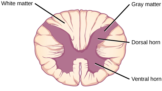


Figure 35.24図 35.24
A cross-section of the spinal cord shows gray matter (containing cell bodies and interneurons) and white matter (containing axons).
脊髄の断面には、細胞体と介在ニューロンを含む灰白質と、軸索を含む白質が見られる。