第35章神経系
Synaptic Transmissionシナプス伝達
The synapse or “gap” is the place where information is transmitted from one neuron to another. Synapses usually form between axon terminals and dendritic spines, but this is not universally true. There are also axon-to-axon, dendrite-to-dendrite, and axon-to-cell body synapses. The neuron transmitting the signal is called the presynaptic neuron, and the neuron receiving the signal is called the postsynaptic neuron. Note that these designations are relative to a particular synapse—most neurons are both presynaptic and postsynaptic. There are two types of synapses: chemical and electrical.
英語のヒント
シナプス、すなわち「隙間」は、あるニューロンから別のニューロンへ情報が伝えられる場所である。通常は軸索終末と樹状突起スパインの間にできるが、常にそうとは限らない。軸索どうし、樹状突起どうし、軸索と細胞体の間のシナプスもある。信号を伝えるニューロンをシナプス前ニューロン、受け取るニューロンをシナプス後ニューロンという。これらの呼称は、特定のシナプスに対して決まる。ほとんどのニューロンは、シナプス前でもシナプス後でもある。シナプスには化学シナプスと電気シナプスの二種類がある。
Chemical Synapse化学シナプス
When an action potential reaches the axon terminal it depolarizes the membrane and opens voltage-gated Na+ channels. Na+ ions enter the cell, further depolarizing the presynaptic membrane. This depolarization causes voltage-gated Ca2+ channels to open. Calcium ions entering the cell initiate a signaling cascade that causes small membrane-bound vesicles, called synaptic vesicles, containing neurotransmitter molecules to fuse with the presynaptic membrane. Synaptic vesicles are shown in Figure 35.14, which is an image from a scanning electron microscope.
英語のヒント
活動電位が軸索終末に達すると、膜が脱分極し、電位依存性Na+チャネルが開く。Na+が細胞へ入り、シナプス前膜をさらに脱分極させる。この脱分極によって、電位依存性Ca2+チャネルが開く。流入したカルシウムイオンはシグナル伝達の連鎖を始動させる。それにより、神経伝達物質分子を含み、膜で囲まれたシナプス小胞という小さな袋が、シナプス前膜と融合する。走査電子顕微鏡像である図35.14にシナプス小胞を示す。
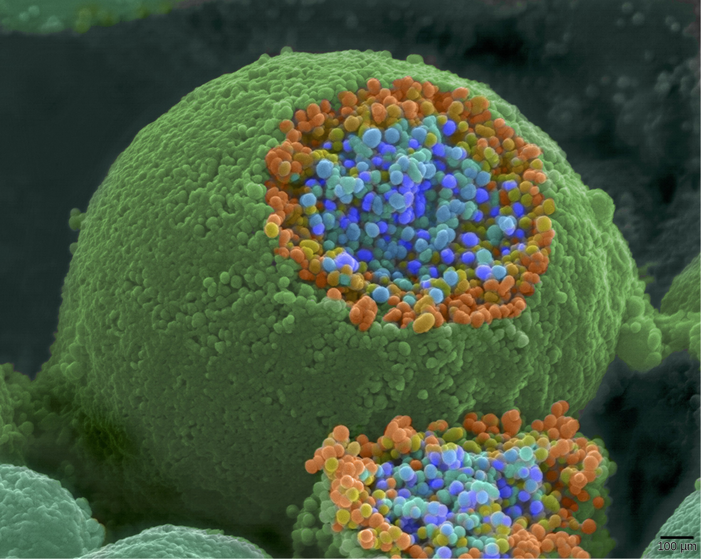
OpenStax, Rice University; Biology 2e. (credit: modification of work by Tina Carvalho, NIH-NIGMS; scale-bar data from Matt Russell) · CC BY 4.0 · Source出典
Figure 35.14図 35.14
This pseudocolored image taken with a scanning electron microscope shows an axon terminal that was broken open to reveal synaptic vesicles (blue and orange) inside the neuron. (credit: modification of work by Tina Carvalho, NIH-NIGMS; scale-bar data from Matt Russell)
走査電子顕微鏡で撮影し、擬似着色した像。軸索終末を割って開き、ニューロン内部のシナプス小胞(青と橙)を見えるようにしている。(クレジット:Tina Carvalho、NIH-NIGMSの作品を改変、スケールバーのデータ:Matt Russell)
Fusion of a vesicle with the presynaptic membrane causes neurotransmitter to be released into the synaptic cleft, the extracellular space between the presynaptic and postsynaptic membranes, as illustrated in Figure 35.15. The neurotransmitter diffuses across the synaptic cleft and binds to receptor proteins on the postsynaptic membrane.
図35.15に示すように、小胞がシナプス前膜と融合すると、シナプス前膜と後膜の間の細胞外空間であるシナプス間隙へ、神経伝達物質が放出される。神経伝達物質は間隙を拡散して渡り、シナプス後膜の受容体タンパク質に結合する。
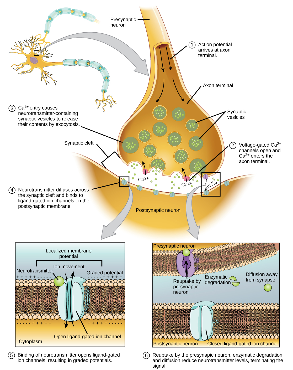


Figure 35.15図 35.15
Communication at chemical synapses requires release of neurotransmitters. When the presynaptic membrane is depolarized, voltage-gated Ca2+ channels open and allow Ca2+ to enter the cell. The calcium entry causes synaptic vesicles to fuse with the membrane and release neurotransmitter molecules into the synaptic cleft. The neurotransmitter diffuses across the synaptic cleft and binds to ligand-gated ion channels in the postsynaptic membrane, resulting in a localized depolarization or hyperpolarization of the postsynaptic neuron.
化学シナプスでの情報伝達には、神経伝達物質の放出が必要である。シナプス前膜が脱分極すると、電位依存性Ca2+チャネルが開き、Ca2+が細胞内へ入る。カルシウムの流入により、シナプス小胞が膜と融合し、神経伝達物質分子をシナプス間隙へ放出する。神経伝達物質は間隙を拡散して渡り、シナプス後膜のリガンド依存性イオンチャネルに結合する。その結果、シナプス後ニューロンに局所的な脱分極または過分極が生じる。
The binding of a specific neurotransmitter causes particular ion channels, in this case ligand-gated channels, on the postsynaptic membrane to open. Neurotransmitters can either have excitatory or inhibitory effects on the postsynaptic membrane. For example, when acetylcholine is released at the synapse between a nerve and muscle (called the neuromuscular junction) by a presynaptic neuron, it causes postsynaptic Na+ channels to open. Na+ enters the postsynaptic cell and causes the postsynaptic membrane to depolarize. This depolarization is called an excitatory postsynaptic potential (EPSP) and makes the postsynaptic neuron more likely to fire an action potential. Release of neurotransmitter at inhibitory synapses causes inhibitory postsynaptic potentials (IPSPs), a hyperpolarization of the presynaptic membrane. For example, when the neurotransmitter GABA (gamma-aminobutyric acid) is released from a presynaptic neuron, it binds to and opens Cl- channels. Cl- ions enter the cell and hyperpolarizes the membrane, making the neuron less likely to fire an action potential.
英語のヒント
特定の神経伝達物質が結合すると、シナプス後膜の特定のイオンチャネル、この場合はリガンド依存性チャネルが開く。神経伝達物質は、シナプス後膜に興奮性または抑制性の効果を及ぼしうる。例えば、神経と筋の間のシナプス(神経筋接合部)でシナプス前ニューロンがアセチルコリンを放出すると、シナプス後のNa+チャネルが開く。Na+がシナプス後細胞に入り、後膜を脱分極させる。この脱分極を興奮性シナプス後電位(EPSP)といい、シナプス後ニューロンが活動電位を発火しやすくなる。抑制性シナプスでの神経伝達物質の放出は、シナプス前膜の過分極である抑制性シナプス後電位(IPSP)を起こす。例えば、シナプス前ニューロンから放出されたGABA(γ-アミノ酪酸)は、Cl-チャネルに結合して開かせる。Cl-が細胞内へ入り、膜を過分極させるので、ニューロンは活動電位を発火しにくくなる。
Once neurotransmission has occurred, the neurotransmitter must be removed from the synaptic cleft so the postsynaptic membrane can “reset” and be ready to receive another signal. This can be accomplished in three ways: the neurotransmitter can diffuse away from the synaptic cleft, it can be degraded by enzymes in the synaptic cleft, or it can be recycled (sometimes called reuptake) by the presynaptic neuron. Several drugs act at this step of neurotransmission. For example, some drugs that are given to Alzheimer’s patients work by inhibiting acetylcholinesterase, the enzyme that degrades acetylcholine. This inhibition of the enzyme essentially increases neurotransmission at synapses that release acetylcholine. Once released, the acetylcholine stays in the cleft and can continually bind and unbind to postsynaptic receptors.
英語のヒント
神経伝達が起こった後は、シナプス後膜が「リセット」して次の信号を受け取る準備をできるように、神経伝達物質をシナプス間隙から除く必要がある。これには三つの方法がある。間隙から拡散して離れる、間隙内の酵素で分解される、シナプス前ニューロンによって回収・再利用される(再取込みともいう)ことである。神経伝達のこの段階に作用する薬物もある。例えば、アルツハイマー病患者に投与される一部の薬は、アセチルコリンを分解する酵素アセチルコリンエステラーゼを阻害して働く。酵素の阻害によって、実質的にアセチルコリンを放出するシナプスの神経伝達が増強される。放出されたアセチルコリンは間隙にとどまり、シナプス後受容体への結合と解離を繰り返せる。
Neurotransmitter Function and Location 神経伝達物質の機能と分布 |
||
|---|---|---|
Neurotransmitter 神経伝達物質 |
Example 例 |
Location 分布 |
Acetylcholine アセチルコリン |
— |
CNS and/or PNS 中枢神経系および/または末梢神経系 |
Biogenic amine 生体アミン |
Dopamine, serotonin, norepinephrine ドーパミン、セロトニン、ノルアドレナリン |
CNS and/or PNS 中枢神経系および/または末梢神経系 |
Amino acid アミノ酸 |
Glycine, glutamate, aspartate, gamma aminobutyric acid グリシン、グルタミン酸、アスパラギン酸、γ-アミノ酪酸 |
CNS 中枢神経系 |
Neuropeptide 神経ペプチド |
Substance P, endorphins サブスタンスP、エンドルフィン |
CNS and/or PNS 中枢神経系および/または末梢神経系 |
Electrical Synapse電気シナプス
While electrical synapses are fewer in number than chemical synapses, they are found in all nervous systems and play important and unique roles. The mode of neurotransmission in electrical synapses is quite different from that in chemical synapses. In an electrical synapse, the presynaptic and postsynaptic membranes are very close together and are actually physically connected by channel proteins forming gap junctions. Gap junctions allow current to pass directly from one cell to the next. In addition to the ions that carry this current, other molecules, such as ATP, can diffuse through the large gap junction pores.
電気シナプスは化学シナプスより数が少ないが、すべての神経系に見られ、重要で独特な役割を果たす。そこでの神経伝達の方式は、化学シナプスとはかなり異なる。電気シナプスでは、シナプス前膜と後膜が非常に近く、ギャップ結合をつくるチャネルタンパク質によって、実際に物理的につながっている。ギャップ結合は、ある細胞から隣の細胞へ電流を直接通す。この電流を運ぶイオンだけでなく、ATPなどの分子も、ギャップ結合の大きな孔を拡散して通れる。
There are key differences between chemical and electrical synapses. Because chemical synapses depend on the release of neurotransmitter molecules from synaptic vesicles to pass on their signal, there is an approximately one millisecond delay between when the axon potential reaches the presynaptic terminal and when the neurotransmitter leads to opening of postsynaptic ion channels. Additionally, this signaling is unidirectional. Signaling in electrical synapses, in contrast, is virtually instantaneous (which is important for synapses involved in key reflexes), and some electrical synapses are bidirectional. Electrical synapses are also more reliable as they are less likely to be blocked, and they are important for synchronizing the electrical activity of a group of neurons. For example, electrical synapses in the thalamus are thought to regulate slow-wave sleep, and disruption of these synapses can cause seizures.
英語のヒント
化学シナプスと電気シナプスには重要な違いがある。化学シナプスは、シナプス小胞からの神経伝達物質分子の放出に依存して信号を伝えるため、軸索電位がシナプス前終末に達してから、神経伝達物質がシナプス後イオンチャネルを開かせるまで、約1ミリ秒の遅れがある。また、この伝達は一方向性である。これに対し、電気シナプスの伝達はほぼ瞬時であり、重要な反射に関わるシナプスではこの速さが重要になる。一部の電気シナプスは双方向性でもある。電気シナプスは遮断されにくいため信頼性も高く、ニューロン群の電気活動を同期させるのに重要である。例えば、視床の電気シナプスは徐波睡眠を調節すると考えられ、その障害はけいれん発作を引き起こすことがある。