第35章神経系
35.2 How Neurons Communicate35.2 ニューロンの情報伝達
Learning Objectives学習目標
By the end of this section, you will be able to do the following:
この節を学ぶと、次のことができるようになる。
- Describe the basis of the resting membrane potential静止膜電位の成り立ちを説明する。
- Explain the stages of an action potential and how action potentials are propagated活動電位の各段階と、その伝導の仕組みを説明する。
- Explain the similarities and differences between chemical and electrical synapses化学シナプスと電気シナプスの共通点と相違点を説明する。
- Describe long-term potentiation and long-term depression長期増強と長期抑圧を説明する。
All functions performed by the nervous system—from a simple motor reflex to more advanced functions like making a memory or a decision—require neurons to communicate with one another. While humans use words and body language to communicate, neurons use electrical and chemical signals. Just like a person in a committee, one neuron usually receives and synthesizes messages from multiple other neurons before “making the decision” to send the message on to other neurons.
英語のヒント
単純な運動反射から、記憶や意思決定のような高度な機能まで、神経系のあらゆる機能には、ニューロンどうしの情報伝達が必要である。ヒトは言葉や身振りで伝え合うが、ニューロンは電気信号と化学信号を使う。委員会の一員のように、一つのニューロンは通常、複数の他のニューロンから情報を受け取り、それらをまとめてから、他のニューロンへ情報を送るという「決定」をする。
Nerve Impulse Transmission within a Neuronニューロン内の神経興奮の伝導
For the nervous system to function, neurons must be able to send and receive signals. These signals are possible because each neuron has a charged cellular membrane (a voltage difference between the inside and the outside), and the charge of this membrane can change in response to neurotransmitter molecules released from other neurons and environmental stimuli. To understand how neurons communicate, one must first understand the basis of the baseline or ‘resting’ membrane charge.
英語のヒント
神経系が機能するには、ニューロンが信号を送受信できなければならない。これが可能なのは、各ニューロンの細胞膜に電荷がある(内外に電位差がある)ことと、その膜の電荷が、他のニューロンから放出される神経伝達物質分子や環境の刺激に応じて変化できることによる。ニューロンの情報伝達を理解するには、まず基準となる「静止」状態の膜の電荷の成り立ちを理解しなければならない。
Neuronal Charged Membranesニューロンの帯電した膜
The lipid bilayer membrane that surrounds a neuron is impermeable to charged molecules or ions. To enter or exit the neuron, ions must pass through special proteins called ion channels that span the membrane. Ion channels have different configurations: open, closed, and inactive, as illustrated in Figure 35.9. Some ion channels need to be activated in order to open and allow ions to pass into or out of the cell. These ion channels are sensitive to the environment and can change their shape accordingly. Ion channels that change their structure in response to voltage changes are called voltage-gated ion channels. Voltage-gated ion channels regulate the relative concentrations of different ions inside and outside the cell. The difference in total charge between the inside and outside of the cell is called the membrane potential.
英語のヒント
ニューロンを取り囲む脂質二重層膜は、電荷を持つ分子やイオンを通さない。イオンがニューロンへ出入りするには、膜を貫くイオンチャネルという特殊なタンパク質を通る必要がある。図35.9に示すように、イオンチャネルには開状態、閉状態、不活性状態という異なる構造状態がある。一部のイオンチャネルは、開いてイオンを細胞内外へ通すために活性化される必要がある。これらは周囲の環境に敏感で、それに応じて形を変えられる。電位の変化に応じて構造を変えるチャネルを電位依存性イオンチャネルという。細胞内外の各イオンの相対濃度を調節する。細胞の内外の総電荷の違いを膜電位という。
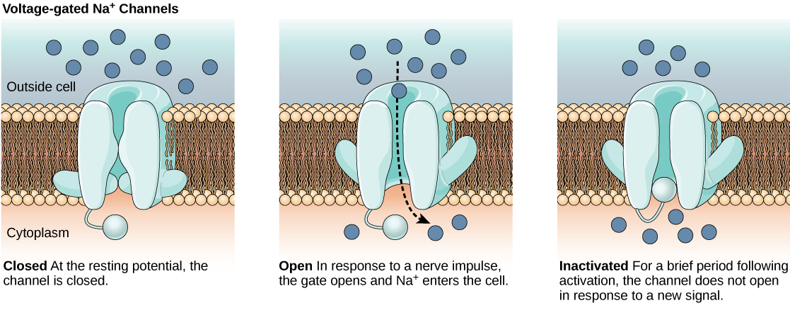
Figure 35.9図 35.9
Voltage-gated ion channels open in response to changes in membrane voltage. After activation, they become inactivated for a brief period and will no longer open in response to a signal.
電位依存性イオンチャネルは、膜電位の変化に応じて開く。活性化の後は短時間不活性化し、信号に応答して開かなくなる。
Resting Membrane Potential静止膜電位
A neuron at rest is negatively charged: the inside of a cell is approximately 70 millivolts more negative than the outside (−70 mV, note that this number varies by neuron type and by species). This voltage is called the resting membrane potential; it is caused by differences in the concentrations of ions inside and outside the cell. If the membrane were equally permeable to all ions, each type of ion would flow across the membrane and the system would reach equilibrium. Because ions cannot simply cross the membrane at will, there are different concentrations of several ions inside and outside the cell, as shown in Table 35.1. The difference in the number of positively charged potassium ions (K+) inside and outside the cell dominates the resting membrane potential (Figure 35.10). When the membrane is at rest, K+ ions accumulate inside the cell due to the activity of the Na/K pump, driving both ions against their concentration gradient. The negative resting membrane potential is created and maintained by increasing the concentration of cations outside the cell (in the extracellular fluid) relative to inside the cell (in the cytoplasm). The negative charge within the cell is created by the cell membrane being more permeable to potassium ion movement than sodium ion movement. In neurons, potassium ions are maintained at high concentrations within the cell while sodium ions are maintained at high concentrations outside of the cell. The cell possesses potassium and sodium leakage channels that allow the two cations to diffuse down their concentration gradient. However, the neurons have far more potassium leakage channels than sodium leakage channels. Therefore, potassium diffuses out of the cell at a much faster rate than sodium leaks in. Because more cations are leaving the cell than are entering, this causes the interior of the cell to be negatively charged relative to the outside of the cell. The actions of the sodium potassium pump help to maintain the resting potential, once established. Recall that sodium potassium pumps brings two K+ ions into the cell while removing three Na+ ions per ATP consumed. As more cations are expelled from the cell than taken in, the inside of the cell remains negatively charged relative to the extracellular fluid. It should be noted that chloride ions (Cl–) tend to accumulate outside of the cell because they are repelled by negatively-charged proteins within the cytoplasm.
英語のヒント
静止状態のニューロンは負に帯電している。細胞内は細胞外より約70ミリボルト負である(−70mV。この値はニューロンの種類や種によって異なる)。この電位を静止膜電位といい、細胞内外のイオン濃度の違いによって生じる。もし膜がすべてのイオンを同じように通すなら、各イオンが膜を通って流れ、系は平衡に達するだろう。イオンは自由に膜を通れるわけではないため、表35.1のように、複数のイオンの濃度が細胞内外で異なる。静止膜電位を主に決めるのは、正に帯電したカリウムイオン(K+)の細胞内外の数の違いである(図35.10)。膜が静止状態にあるとき、Na/Kポンプの働きでK+が細胞内に蓄積する。このポンプは両イオンをそれぞれの濃度勾配に逆らって運ぶ。負の静止膜電位は、細胞内(細胞質)に対する細胞外(細胞外液)の陽イオン濃度を高めることでつくられ、維持される。細胞内の負の電荷は、細胞膜がナトリウムイオンよりカリウムイオンを通しやすいことによって生じる。ニューロンでは、カリウムイオンは細胞内、ナトリウムイオンは細胞外に高濃度で保たれる。細胞にはカリウムとナトリウムの漏洩チャネルがあり、両陽イオンは濃度勾配に従って拡散できる。しかし、カリウムの漏洩チャネルはナトリウムのものよりはるかに多い。そのため、カリウムが細胞外へ拡散する速度は、ナトリウムが流入する速度よりずっと速い。入る陽イオンより出る陽イオンが多いため、細胞内は外側に対して負になる。ナトリウム・カリウムポンプは、一度成立した静止電位の維持を助ける。ATPを一分子消費するごとに、2個のK+を細胞内へ入れ、3個のNa+を外へ出すことを思い出そう。取り込まれるより排出される陽イオンが多いため、細胞内は細胞外液に対して負に保たれる。塩化物イオン(Cl–)は、細胞質内の負に帯電したタンパク質に反発されるため、細胞外に蓄積しやすい。
Ion Concentration Inside and Outside Neurons ニューロンの内外のイオン濃度 |
|||
|---|---|---|---|
Ion イオン |
Extracellular concentration (mM) 細胞外濃度(mM) |
Intracellular concentration (mM) 細胞内濃度(mM) |
Ratio outside/inside 外側/内側の比 |
Na+ |
145 |
12 |
12 |
K+ |
4 |
155 |
0.026 |
Cl− |
120 |
4 |
30 |
Organic anions (A−) 有機陰イオン(A−) |
— |
100 |
|
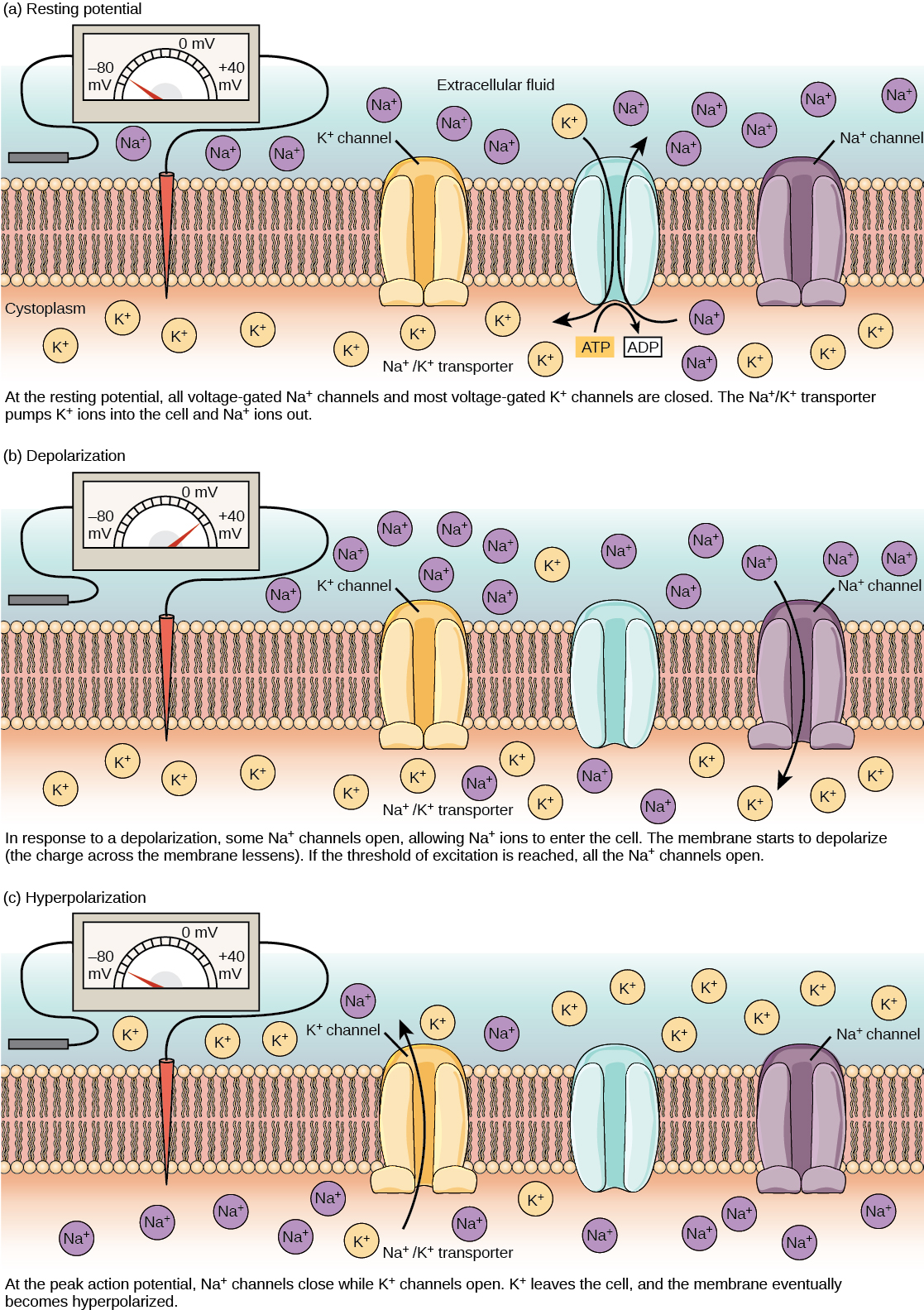


Figure 35.10図 35.10
The (a) resting membrane potential is a result of different concentrations of Na+ and K+ ions inside and outside the cell. A nerve impulse causes Na+ to enter the cell, resulting in (b) depolarization. At the peak action potential, K+ channels open and the cell becomes (c) hyperpolarized.
(a)静止膜電位は、細胞内外のNa+とK+の濃度差によって生じる。神経興奮によってNa+が細胞へ入り、(b)脱分極が起こる。活動電位の頂点でK+チャネルが開き、細胞は(c)過分極する。