第43章動物の生殖と発生
43.7 Organogenesis and Vertebrate Axis Formation43.7 器官形成と脊椎動物の体軸形成
Learning Objectives学習目標
By the end of this section, you will be able to do the following:
この節を学ぶと、次のことができるようになる。
- Describe the process of organogenesis器官形成の過程を説明する。
- Identify the anatomical axes formed in vertebrates脊椎動物で形成される解剖学的な軸を特定する。
Gastrulation leads to the formation of the three germ layers that give rise, during further development, to the different organs in the animal body. This process is called organogenesis. Organogenesis is characterized by rapid and precise movements of the cells within the embryo.
英語のヒント
原腸形成によって3つの胚葉が形成され、さらに発生が進むと、そこから動物の体のさまざまな器官が生じる。この過程を器官形成という。器官形成は、胚の内部で細胞が素早く正確に移動することを特徴とする。
Organogenesis器官形成
Organs form from the germ layers through the process of differentiation. During differentiation, the embryonic stem cells express specific sets of genes which will determine their ultimate cell type. For example, some cells in the ectoderm will express the genes specific to skin cells. As a result, these cells will differentiate into epidermal cells. The process of differentiation is regulated by cellular signaling cascades.
英語のヒント
器官は、分化という過程を通じて胚葉から形成される。分化の間、胚性幹細胞は、最終的な細胞型を決める特定の遺伝子群を発現する。例えば、外胚葉の一部の細胞は、皮膚細胞に特有の遺伝子を発現し、その結果、表皮細胞へ分化する。分化の過程は、細胞のシグナル伝達の連鎖によって調節される。
Scientists study organogenesis extensively in the lab in fruit flies (Drosophila) and the nematode Caenorhabditis elegans. Drosophila have segments along their bodies, and the patterning associated with the segment formation has allowed scientists to study which genes play important roles in organogenesis along the length of the embryo at different time points. The nematode C.elegans has roughly 1000 somatic cells and scientists have studied the fate of each of these cells during their development in the nematode life cycle. There is little variation in patterns of cell lineage between individuals, unlike in mammals where cell development from the embryo is dependent on cellular cues.
英語のヒント
科学者は実験室で、ショウジョウバエ(Drosophila)や線虫Caenorhabditis elegansを用いて器官形成を詳しく研究している。ショウジョウバエの体には体節があり、体節形成に伴うパターン形成を利用することで、胚の長軸に沿った器官形成のどの時点で、どの遺伝子が重要な役割を果たすかを調べられる。線虫C. elegansには約1,000個の体細胞があり、科学者は生活環の発生中に、それぞれの細胞がたどる運命を調べてきた。細胞系譜のパターンには、個体間でほとんど違いがない。これは、胚からの細胞の発達が細胞の合図に依存する哺乳類とは異なる。
In vertebrates, one of the primary steps during organogenesis is the formation of the neural system. The ectoderm forms epithelial cells and tissues, and neuronal tissues. During the formation of the neural system, special signaling molecules called growth factors signal some cells at the edge of the ectoderm to become epidermis cells. The remaining cells in the center form the neural plate. If the signaling by growth factors were disrupted, then the entire ectoderm would differentiate into neural tissue.
英語のヒント
脊椎動物の器官形成における主要な段階の1つは、神経系の形成である。外胚葉は上皮細胞と上皮組織、および神経組織を作る。神経系が形成されるとき、成長因子という特殊なシグナル分子が、外胚葉の縁にある一部の細胞へ、表皮細胞になるよう合図する。中央に残る細胞は神経板を形成する。成長因子によるシグナル伝達が妨げられたなら、外胚葉全体が神経組織へ分化することになる。
The neural plate undergoes a series of cell movements where it rolls up and forms a tube called the neural tube, as illustrated in Figure 43.28. In further development, the neural tube will give rise to the brain and the spinal cord.
英語のヒント
神経板では一連の細胞移動が起こり、図43.28のように巻き上がって、神経管という管を作る。その後の発生で、神経管から脳と脊髄が生じる。
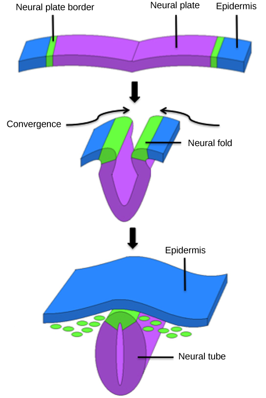

Figure 43.28図 43.28
The central region of the ectoderm forms the neural tube, which gives rise to the brain and the spinal cord.
外胚葉の中央部は神経管を形成し、そこから脳と脊髄が生じる。
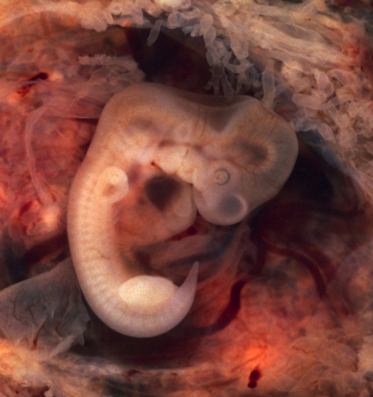
The mesoderm that lies on either side of the vertebrate neural tube will develop into the various connective tissues of the animal body. A spatial pattern of gene expression reorganizes the mesoderm into groups of cells called somites with spaces between them. The somites illustrated in Figure 43.29 will further develop into the cells that form the vertebrae and ribs, the dermis of the dorsal skin, the skeletal muscles of the back, and the skeletal muscles of the body wall and limbs. The mesoderm also forms a structure called the notochord, which is rod-shaped and forms the central axis of the animal body.
英語のヒント
脊椎動物の神経管の両側にある中胚葉は、動物の体のさまざまな結合組織へ発達する。遺伝子発現の空間的なパターンが中胚葉を再編成し、隙間を隔てて並ぶ体節という細胞群を作る。図43.29の体節はさらに発達し、椎骨と肋骨、背側の皮膚の真皮、背中の骨格筋、および体壁と四肢の骨格筋を作る細胞になる。中胚葉は脊索という構造も作る。脊索は棒状で、動物の体の中心軸を形成する。
OpenStax, Rice University; Biology 2e. (credit: modification of work by Ed Uthman) · CC BY 4.0 · Source出典

Figure 43.29図 43.29
In this five-week old human embryo, somites are segments along the length of the body. (credit: modification of work by Ed Uthman)
この5週のヒト胚では、体節が体の長さ方向に沿って並んでいる。(出典:Ed Uthmanの作品を改変)
Vertebrate Axis Formation脊椎動物の体軸形成
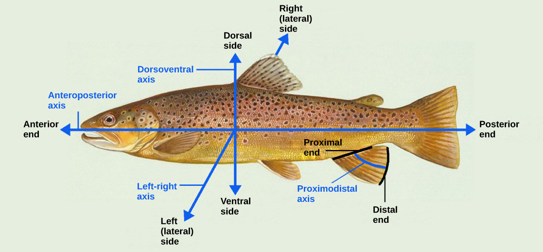
Even as the germ layers form, the ball of cells still retains its spherical shape. However, animal bodies have lateral-medial (left-right), dorsal-ventral (back-belly), and anterior-posterior (head-feet) axes, illustrated in Figure 43.30.
英語のヒント
胚葉が形成されている間も、細胞の球は球形を保っている。しかし動物の体には、図43.30のように、外側―内側(左―右)、背側―腹側(背―腹)、前―後(頭―足)の軸がある。
OpenStax, Rice University; Biology 2e. (credit: modification of work by NOAA) · CC BY 4.0 · Source出典

Figure 43.30図 43.30
Animal bodies have three axes for symmetry. (credit: modification of work by NOAA)
動物の体には、対称性に関わる3つの軸がある。(出典:NOAAの作品を改変)
How are these established? In one of the most seminal experiments ever to be carried out in developmental biology, Spemann and Mangold took dorsal cells from one embryo and transplanted them into the belly region of another embryo. They found that the transplanted embryo now had two notochords: one at the dorsal site from the original cells and another at the transplanted site. This suggested that the dorsal cells were genetically programmed to form the notochord and define the axis. Since then, researchers have identified many genes that are responsible for axis formation. Mutations in these genes leads to the loss of symmetry required for organism development.
英語のヒント
これらの軸はどのように定まるのだろうか。発生生物学で行われた、最も先駆的な実験の1つで、シュペーマンとマンゴルトは、ある胚の背側細胞を取り出し、別の胚の腹側へ移植した。移植を受けた胚には、脊索が2本できた。1本は元の細胞から背側に、もう1本は移植した場所に生じた。この結果は、背側細胞が、脊索を作って体軸を定めるように遺伝的にプログラムされていることを示唆した。それ以来、研究者は体軸形成を担う多くの遺伝子を特定している。これらの遺伝子に突然変異が起こると、生物の発生に必要な対称性が失われる。
Animal bodies have externally visible symmetry. However, the internal organs are not symmetric. For example, the heart is on the left side and the liver on the right. The formation of the central left-right axis is an important process during development. This internal asymmetry is established very early during development and involves many genes. Research is still ongoing to fully understand the developmental implications of these genes.
英語のヒント
動物の体には外見で分かる対称性があるが、内臓は対称ではない。例えば、心臓は左側、肝臓は右側にある。中央の左右軸の形成は、発生の重要な過程である。この内部の非対称性は、発生のごく初期に定まり、多くの遺伝子が関わる。これらの遺伝子が発生に及ぼす影響を十分に理解するための研究は、今も続いている。