第43章動物の生殖と発生
43.6 Fertilization and Early Embryonic Development43.6 受精と初期胚発生
Learning Objectives学習目標
By the end of this section, you will be able to do the following:
この節を学ぶと、次のことができるようになる。
- Discuss how fertilization occurs受精がどのように起こるかを論じる。
- Explain how the embryo forms from the zygote接合子から胚がどのように形成されるかを説明する。
- Discuss the role of cleavage and gastrulation in animal development動物の発生における卵割と原腸形成の役割を論じる。
The process in which an organism develops from a single-celled zygote to a multi-cellular organism is complex and well-regulated. The early stages of embryonic development are also crucial for ensuring the fitness of the organism.
英語のヒント
1細胞の接合子から多細胞の生物へ発生する過程は、複雑で厳密に調節されている。胚発生の初期段階は、その生物の適応度を確保するうえでも非常に重要である。
Fertilization受精
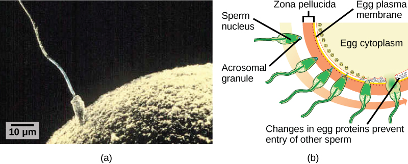
Fertilization, pictured in Figure 43.23a is the process in which gametes (an egg and sperm) fuse to form a zygote. The egg and sperm each contain one set of chromosomes. To ensure that the offspring has only one complete diploid set of chromosomes, only one sperm must fuse with one egg. In mammals, the egg is protected by a layer of extracellular matrix consisting mainly of glycoproteins called the zona pellucida. When a sperm binds to the zona pellucida, a series of biochemical events, called the acrosomal reactions, take place. In placental mammals, the acrosome contains digestive enzymes that initiate the degradation of the glycoprotein matrix protecting the egg and allowing the sperm plasma membrane to fuse with the egg plasma membrane, as illustrated in Figure 43.23b. The fusion of these two membranes creates an opening through which the sperm nucleus is transferred into the ovum. The nuclear membranes of the egg and sperm break down and the two haploid genomes condense to form a diploid genome.
英語のヒント
図43.23aに示す受精は、配偶子である卵と精子が融合して接合子を作る過程である。卵と精子は、それぞれ染色体を1組ずつもつ。子が完全な二倍体の染色体セットを1つだけもつためには、1個の卵に融合する精子は1個だけでなければならない。哺乳類の卵は、主に糖タンパク質からなる細胞外基質の層、透明帯によって守られる。精子が透明帯に結合すると、先体反応という一連の生化学的反応が起こる。有胎盤哺乳類の先体は消化酵素を含み、卵を保護する糖タンパク質の基質を分解し始める。その結果、図43.23bのように精子の細胞膜が卵の細胞膜と融合できるようになる。2つの膜が融合すると開口部ができ、精子の核がそこを通って卵へ移る。卵と精子の核膜が崩壊し、2つの半数体ゲノムが凝集して、二倍体ゲノムを形成する。
OpenStax, Rice University; Biology 2e. (credit: (b) modification of work by Mariana Ruiz Villareal; scale-bar data from Matt Russell) · CC BY 4.0 · Source出典
Figure 43.23図 43.23
(a) Fertilization is the process in which sperm and egg fuse to form a zygote. (b) Acrosomal reactions help the sperm degrade the glycoprotein matrix protecting the egg and allow the sperm to transfer its nucleus. (credit: (b) modification of work by Mariana Ruiz Villareal; scale-bar data from Matt Russell)
(a)受精は、精子と卵が融合して接合子を作る過程である。(b)先体反応は、精子が卵を保護する糖タンパク質の基質を分解し、自身の核を移すのを助ける。(出典:(b)はMariana Ruiz Villarealの作品を改変。スケールバーのデータ:Matt Russell)
To ensure that no more than one sperm fertilizes the egg, once the acrosomal reactions take place at one location of the egg membrane, the egg releases proteins in other locations to prevent other sperm from fusing with the egg. Ernest Everett Just, who was particularly skilled and knowledgeable in handling invertebrate eggs, demonstrated several mechanisms at work at different rates. The fast reaction included what Just called a "wave of negativity," in which the membrane potential of the egg cell altered quickly. Then, the slow block involved changing the membrane structure itself. If these mechanisms fail, multiple sperm can fuse with the egg, resulting in polyspermy. The resulting embryo is not genetically viable and dies within a few days.
英語のヒント
2個以上の精子が卵を受精させないよう、卵膜の1か所で先体反応が起こると、卵はほかの場所でタンパク質を放出し、ほかの精子が卵と融合するのを防ぐ。無脊椎動物の卵の扱いに特に熟練し、深い知識をもっていたアーネスト・エヴァレット・ジャストは、異なる速度で働く複数の仕組みを示した。速い反応には、ジャストが「負性の波」と呼んだものが含まれ、卵細胞の膜電位が急速に変化した。続く遅い阻止反応では、膜そのものの構造が変化した。こうした仕組みが働かなければ、複数の精子が卵と融合し、多精受精になる。生じた胚は遺伝的に生存できず、数日以内に死滅する。
Cleavage and Blastula Stage卵割と胞胚期
The development of multi-cellular organisms begins from a single-celled zygote, which undergoes rapid cell division to form the blastula. The rapid, multiple rounds of cell division are termed cleavage. Cleavage is illustrated in (Figure 43.24a). After the cleavage has produced over 100 cells, the embryo is called a blastula. The blastula is usually a spherical layer of cells (the blastoderm) surrounding a fluid-filled or yolk-filled cavity (the blastocoel). Mammals at this stage form a structure called the blastocyst, characterized by an inner cell mass that is distinct from the surrounding blastula, shown in Figure 43.24b. During cleavage, the cells divide without an increase in mass; that is, one large single-celled zygote divides into multiple smaller cells. Each cell within the blastula is called a blastomere.
英語のヒント
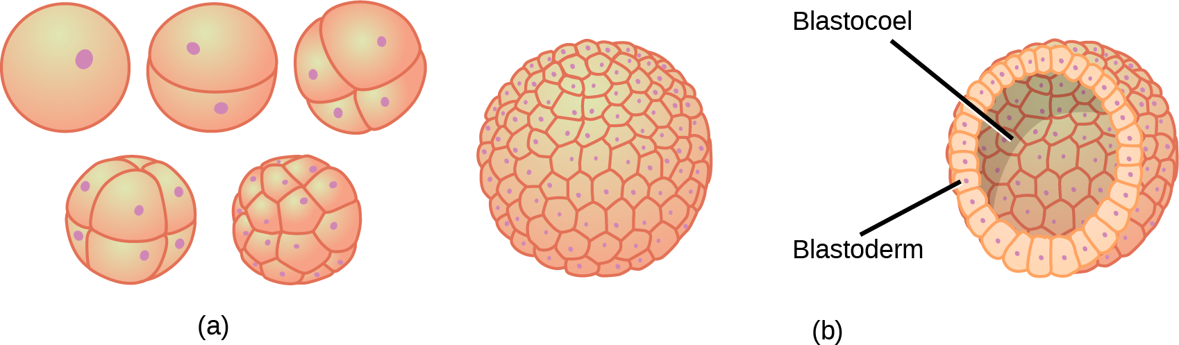
Figure 43.24図 43.24
(a) During cleavage, the zygote rapidly divides into multiple cells without increasing in size. (b) The cells rearrange themselves to form a hollow ball with a fluid-filled or yolk-filled cavity called the blastula.
(a)卵割中、接合子は大きさを増さずに急速に多数の細胞へ分裂する。(b)細胞が並び替わり、液体または卵黄で満たされた空洞をもつ中空の球、胞胚を形成する。
Cleavage can take place in two ways: holoblastic (total) cleavage or meroblastic (partial) cleavage. The type of cleavage depends on the amount of yolk in the eggs. In placental mammals (including humans) where nourishment is provided by the parent’s body, the eggs have a very small amount of yolk and undergo holoblastic cleavage. Other species, such as birds, with a lot of yolk in the egg to nourish the embryo during development, undergo meroblastic cleavage.
英語のヒント
卵割には、全割(卵全体の分裂)と部分割(卵の一部の分裂)の2つの方法がある。種類は、卵に含まれる卵黄の量によって決まる。ヒトを含む有胎盤哺乳類では、親の体から栄養が供給されるため、卵黄はごく少量で、全割が起こる。鳥類など、発生中の胚を養うために卵の中に多量の卵黄をもつ種では、部分割が起こる。
In mammals, the blastula forms the blastocyst in the next stage of development. Here the cells in the blastula arrange themselves in two layers: the inner cell mass, and an outer layer called the trophoblast. The inner cell mass is also known as the embryoblast and this mass of cells will go on to form the embryo. At this stage of development, illustrated in Figure 43.25 the inner cell mass consists of embryonic stem cells that will differentiate into the different cell types needed by the organism. The trophoblast will contribute to the placenta and nourish the embryo.
英語のヒント
哺乳類では、胞胚は次の発生段階で胚盤胞になる。細胞は内細胞塊と、栄養膜という外層の2層に並び替わる。内細胞塊は胚芽とも呼ばれ、この細胞の塊がその後、胚を形成する。図43.25に示すこの発生段階では、内細胞塊は胚性幹細胞からなり、生物に必要なさまざまな細胞型へ分化する。栄養膜は胎盤の一部となり、胚に栄養を供給する。
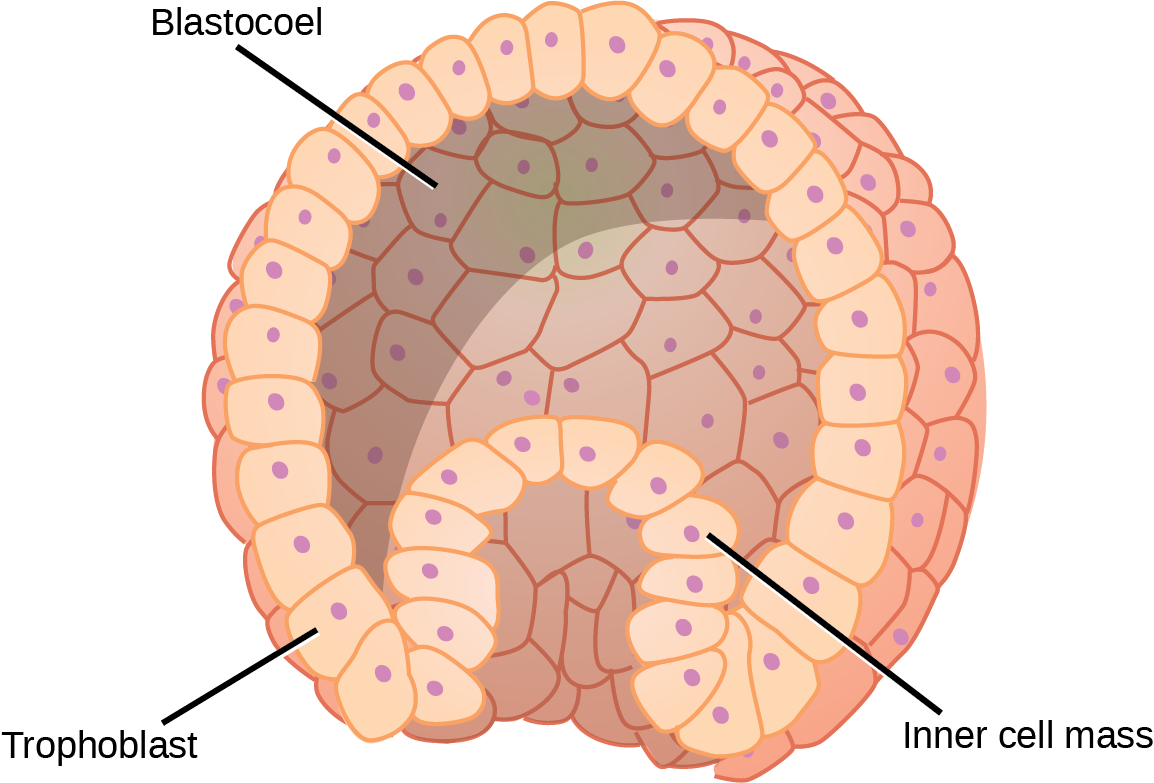


Figure 43.25図 43.25
The rearrangement of the cells in the mammalian blastula to two layers—the inner cell mass and the trophoblast—results in the formation of the blastocyst.
哺乳類の胞胚の細胞が、内細胞塊と栄養膜の2層へ並び替わると、胚盤胞が形成される。