第43章動物の生殖と発生
43.3 Human Reproductive Anatomy and Gametogenesis43.3 ヒトの生殖器の構造と配偶子形成
Learning Objectives学習目標
By the end of this section, you will be able to do the following:
この節を学ぶと、次のことができるようになる。
- Describe human male and female reproductive anatomiesヒトの男性と女性の生殖器の構造を説明する。
- Discuss the human sexual responseヒトの性反応を論じる。
- Describe spermatogenesis and oogenesis and discuss their differences and similarities精子形成と卵形成を説明し、相違点と共通点を論じる。
As animals became more complex, specific organs and organ systems developed to support specific functions for the organism. The reproductive structures that evolved in land animals allow males and females to mate, fertilize internally, and support the growth and development of offspring.
英語のヒント
動物が複雑になるにつれて、生物の特定の機能を支えるために、特定の器官や器官系が発達した。陸上動物で進化した生殖構造によって、雄と雌は交尾し、体内で受精させ、子の成長と発達を支えられるようになった。
Human Reproductive Anatomyヒトの生殖器の構造
The reproductive tissues of male and female humans develop similarly in utero until, in some cases, a low level of the hormone testosterone is released from gonads. Testosterone causes the undeveloped tissues to differentiate into organs, such as the scrotum and penis. When testosterone is absent, the tissues continue to develop into structures such as the ovaries and labia. These cells are considered bipotential: one group of cells has the potential to develop into either type of reproductive structure. Primitive gonads become testes or ovaries. Tissues that produce a penis in males produce a clitoris in females. The tissue that will become the scrotum in a male becomes the labia in a female; that is, they are homologous structures.
英語のヒント
ヒトの男性と女性の生殖組織は、子宮内では同様に発達するが、場合によっては生殖腺から少量のホルモン、テストステロンが放出される。テストステロンは、未発達の組織を陰嚢や陰茎などの器官へ分化させる。テストステロンがないと、組織は卵巣や陰唇などの構造へ発達し続ける。これらの細胞は、2方向への分化能力をもつと考えられる。1つの細胞群が、どちらの種類の生殖構造にも発達できるのである。原始生殖腺は精巣または卵巣になる。男性で陰茎を作る組織は、女性では陰核を作る。男性で陰嚢となる組織は女性では陰唇となる。つまり、これらは相同な構造である。
Male Reproductive Anatomy男性の生殖器の構造
In the male reproductive system, the scrotum houses the testicles or testes (singular: testis), including providing passage for blood vessels, nerves, and muscles related to testicular function. The testes are a pair of male reproductive organs that produce sperm and some reproductive hormones. Each testis is approximately 2.5 by 3.8 cm (1.5 by 1 in.) in size and divided into wedge-shaped lobules by connective tissue called septa. Coiled in each wedge are seminiferous tubules that produce sperm.
英語のヒント
男性の生殖系では、陰嚢が精巣を収め、精巣の機能に関係する血管、神経、筋肉の通り道も提供する。精巣は英語でtesticlesまたはtestes(単数形はtestis)といい、精子と一部の生殖ホルモンを作る一対の男性生殖器官である。各精巣の大きさは約2.5 × 3.8 cm(1.5 × 1インチ)で、中隔という結合組織によってくさび形の小葉に区切られている。それぞれの小葉には、精子を作る精細管が巻いて収まっている。
Sperm are immobile at body temperature; therefore, the scrotum and penis are external to the body, as illustrated in Figure 43.8 so that a proper temperature is maintained for motility. In land mammals, the pair of testes must be suspended outside the body at about 2° C lower than body temperature to produce viable sperm. Infertility can occur in land mammals when the testes do not descend through the abdominal cavity during fetal development.
英語のヒント
精子は体温では運動できない。そのため、図43.8のように陰嚢と陰茎は体外にあり、運動性に適した温度が維持される。陸上哺乳類では、生存可能な精子を作るために、一対の精巣が体外に下がり、体温より約2°C低く保たれる必要がある。胎児の発育中に精巣が腹腔を通って下降しないと、陸上哺乳類では不妊が起こることがある。
Sperm mature in seminiferous tubules that are coiled inside the testes, as illustrated in Figure 43.8. The walls of the seminiferous tubules are made up of the developing sperm cells, with the least developed sperm at the periphery of the tubule and the fully developed sperm in the lumen. The sperm cells are mixed with “nursemaid” cells called Sertoli cells which protect the germ cells and promote their development. Other cells mixed in the wall of the tubules are the interstitial cells of Leydig. These cells produce high levels of testosterone once the male reaches adolescence.
英語のヒント
図43.8に示すように、精子は精巣内で巻いている精細管の中で成熟する。精細管の壁は発達中の精子細胞からなり、最も未発達な精子は管の周辺部に、十分に発達した精子は内腔に位置する。精子細胞の間には、セルトリ細胞という「世話役」の細胞があり、生殖細胞を保護し、その発達を促す。精細管の壁に混在するほかの細胞として、ライディッヒ間細胞がある。これらの細胞は、男性が思春期に達すると大量のテストステロンを作る。
When the sperm have developed flagella and are nearly mature, they leave the testicles and enter the epididymis, shown in Figure 43.8. This structure resembles a comma and lies along the top and posterior portion of the testes; it is the site of sperm maturation. The sperm leave the epididymis and enter the vas deferens (or ductus deferens), which carries the sperm, behind the bladder, and forms the ejaculatory duct with the duct from the seminal vesicles. During a vasectomy, a section of the vas deferens is removed, preventing sperm from being passed out of the body during ejaculation and preventing fertilization.
英語のヒント
精子は鞭毛を発達させ、ほぼ成熟すると、精巣を出て、図43.8の精巣上体へ入る。この構造はコンマに似ていて、精巣の上部と後部に沿って位置し、精子が成熟する場所である。精子は精巣上体を出ると精管に入る。精管は膀胱の後方を通って精子を運び、精嚢からの管と合流して射精管を作る。精管切除術では精管の一部を除去し、射精時に精子が体外へ出ることを防いで受精を防ぐ。
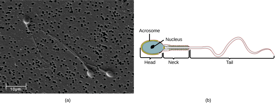
Semen is a mixture of sperm and spermatic duct secretions (about 10 percent of the total) and fluids from accessory glands that contribute most of the semen’s volume. Sperm are haploid cells, consisting of a flagellum as a tail, a neck that contains the cell’s energy-producing mitochondria, and a head that contains the genetic material. Figure 43.9 shows a micrograph of human sperm as well as a diagram of the parts of the sperm. An acrosome is found at the top of the head of the sperm. This structure contains lysosomal enzymes that can digest the protective coverings that surround the egg to help the sperm penetrate and fertilize the egg. An ejaculate will contain from two to five milliliters of fluid with from 50–120 million sperm per milliliter.
英語のヒント
精液は、精子と精路の分泌物(全体の約10%)、および精液の容積の大部分を占める付属腺の分泌液が混ざったものである。精子は半数体の細胞で、尾となる鞭毛、エネルギーを作るミトコンドリアを含む頸部、遺伝物質を含む頭部からなる。図43.9に、ヒトの精子の顕微鏡写真と各部の模式図を示す。精子の頭部の先端には、先体がある。この構造はリソソーム酵素を含み、卵を取り囲む保護層を消化することで、精子が卵へ侵入して受精するのを助ける。1回の射精で出る液体は2~5ミリリットルで、1ミリリットル当たり5,000万~1億2,000万個の精子を含む。
OpenStax, Rice University; Biology 2e. (credit b: modification of work by Mariana Ruiz Villareal; scale-bar data from Matt Russell) · CC BY 4.0 · Source出典

Figure 43.9図 43.9
Human sperm, visualized using scanning electron microscopy, have a flagellum, neck, and head. (credit b: modification of work by Mariana Ruiz Villareal; scale-bar data from Matt Russell)
走査型電子顕微鏡で見たヒトの精子には、鞭毛、頸部、頭部がある。(bの出典:Mariana Ruiz Villarealの作品を改変。スケールバーのデータ:Matt Russell)
The bulk of the semen comes from the accessory glands associated with the male reproductive system. These are the seminal vesicles, the prostate gland, and the bulbourethral gland, all of which are illustrated in Figure 43.8. The seminal vesicles are a pair of glands that lie along the posterior border of the urinary bladder. The glands make a solution that is thick, yellowish, and alkaline. As sperm are only motile in an alkaline environment, a basic pH is important to reverse the acidity of the vaginal environment. The solution also contains mucus, fructose (a sperm mitochondrial nutrient), a coagulating enzyme, ascorbic acid, and local-acting hormones called prostaglandins. The seminal vesicle glands account for 60 percent of the bulk of semen.
英語のヒント
精液の大部分は、男性の生殖系に付随する付属腺から分泌される。精嚢、前立腺、尿道球腺がそれに当たり、すべて図43.8に示されている。精嚢は膀胱の後縁に沿って位置する一対の腺で、粘りがあり、黄色がかったアルカリ性の液を作る。精子はアルカリ性の環境でしか運動できないため、膣内の酸性環境を変えるには、塩基性のpHが重要になる。この液は、粘液、フルクトース(精子のミトコンドリアの栄養源)、凝固酵素、アスコルビン酸、およびプロスタグランジンという局所で作用するホルモンも含む。精嚢の分泌物は、精液の総量の60%を占める。
The penis, illustrated in Figure 43.8, is an organ that drains urine from the renal bladder and functions as a copulatory organ during intercourse. The penis contains three tubes of erectile tissue running through the length of the organ. These consist of a pair of tubes on the dorsal side, called the corpus cavernosum, and a single tube of tissue on the ventral side, called the corpus spongiosum. This tissue will become engorged with blood, becoming erect and hard, in preparation for intercourse. The organ is inserted into the vagina culminating with an ejaculation. During intercourse, the smooth muscle sphincters at the opening to the renal bladder close and prevent urine from entering the penis. An orgasm is a two-stage process: first, glands and accessory organs connected to the testes contract, then semen (containing sperm) is expelled through the urethra during ejaculation. After intercourse, the blood drains from the erectile tissue and the penis becomes flaccid.
英語のヒント
図43.8の陰茎は、膀胱から尿を排出し、性交時には交接器として働く器官である。陰茎には、長さ方向に走る3本の勃起組織がある。背側の一対は陰茎海綿体、腹側の1本は尿道海綿体という。性交に備え、これらの組織に血液が充満して、勃起し硬くなる。陰茎は膣内に挿入され、最終的に射精が起こる。性交中は膀胱の開口部にある平滑筋の括約筋が閉じ、尿が陰茎へ入るのを防ぐ。オルガスムは2段階の過程である。まず、精巣につながる腺と付属器官が収縮し、次に射精によって精子を含む精液が尿道から排出される。性交後は勃起組織から血液が抜け、陰茎は弛緩する。
The walnut-shaped prostate gland surrounds the urethra, the connection to the urinary bladder. It has a series of short ducts that directly connect to the urethra. The gland is a mixture of smooth muscle and glandular tissue. The muscle provides much of the force needed for ejaculation to occur. The glandular tissue makes a thin, milky fluid that contains citrate (a nutrient), enzymes, and prostate specific antigen (PSA). PSA is a proteolytic enzyme that helps to liquefy the ejaculate several minutes after release from the male. Prostate gland secretions account for about 30 percent of the bulk of semen.
英語のヒント
クルミ形の前立腺は、膀胱につながる尿道を取り囲む。前立腺には、尿道へ直接つながる多数の短い導管がある。この腺は平滑筋と腺組織からなり、筋肉が射精に必要な力の多くを生み出す。腺組織は、クエン酸(栄養物質)、酵素、前立腺特異抗原(PSA)を含む、薄い乳白色の液を作る。PSAはタンパク質分解酵素で、男性から放出されて数分後に、射出された精液を液化するのを助ける。前立腺の分泌物は精液の総量の約30%を占める。
The bulbourethral gland, or Cowper’s gland, releases its secretion prior to the release of the bulk of the semen. It neutralizes any acid residue in the urethra left over from urine. This usually accounts for a couple of drops of fluid in the total ejaculate and may contain a few sperm. Withdrawal of the penis from the vagina before ejaculation to prevent pregnancy may not work if sperm are present in the bulbourethral gland secretions. The location and functions of the male reproductive organs are summarized in Table 43.1.
英語のヒント
尿道球腺(カウパー腺)は、精液の大部分が放出されるより前に分泌液を出す。この液は、尿に由来して尿道に残った酸性物質を中和する。通常、射精液全体のうち数滴を占め、少数の精子を含むことがある。尿道球腺の分泌液に精子が含まれる場合、妊娠を防ぐために射精前に陰茎を膣から抜く方法は、うまくいかない可能性がある。男性の生殖器官の位置と機能を表43.1にまとめる。
Male Reproductive Anatomy 男性の生殖器の構造 |
||
|---|---|---|
Organ 器官 |
Location 位置 |
Function 機能 |
Scrotum 陰嚢 |
External 体外 |
Carry and support testes 精巣を収め、支える |
Penis 陰茎 |
External 体外 |
Deliver urine, copulating organ 尿を排出する、交接器となる |
Testes 精巣 |
Internal 体内 |
Produce sperm and male hormones 精子と男性ホルモンを作る |
Seminal Vesicles 精嚢 |
Internal 体内 |
Contribute to semen production 精液の生成に寄与する |
Prostate Gland 前立腺 |
Internal 体内 |
Contribute to semen production 精液の生成に寄与する |
Bulbourethral Glands 尿道球腺 |
Internal 体内 |
Clean urethra at ejaculation 射精時に尿道を洗浄する |