第43章動物の生殖と発生
Female Reproductive Anatomy女性の生殖器の構造
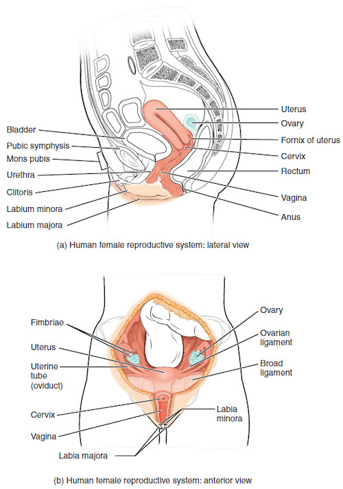
A number of reproductive structures are exterior to the female’s body. These include the breasts and the vulva, which consists of the mons pubis, clitoris, labia majora, labia minora, and the vestibular glands, all illustrated in Figure 43.10. The location and functions of the female reproductive organs are summarized in Table 43.2. The vulva is an area associated with the vestibule which includes the structures found in the inguinal (groin) area. The mons pubis is a round, fatty area that overlies the pubic symphysis. The clitoris is a structure with erectile tissue that contains a large number of sensory nerves and serves as a source of stimulation during intercourse. The labia majora are a pair of elongated folds of tissue that run posterior from the mons pubis and enclose the other components of the vulva. The labia majora derive from the same tissue that produces the scrotum in a male. The labia minora are thin folds of tissue centrally located within the labia majora. These labia protect the openings to the vagina and urethra. The mons pubis and the anterior portion of the labia majora become covered with hair during adolescence; the labia minora is hairless. The greater vestibular glands are found at the sides of the vaginal opening and provide lubrication during intercourse.
英語のヒント
女性の体の外側には、乳房や外陰部など、いくつかの生殖構造がある。外陰部は恥丘、陰核、大陰唇、小陰唇、前庭腺からなり、すべて図43.10に示す。女性の生殖器官の位置と機能は、表43.2にまとめる。外陰部は膣前庭と関連する領域で、鼠径部にある構造を含む。恥丘は、恥骨結合を覆う、丸く脂肪の多い部分である。陰核は勃起組織をもち、多数の感覚神経を含み、性交時の刺激の源となる。大陰唇は、恥丘から後方へ伸び、外陰部のほかの構成要素を囲む、細長い一対の組織のひだである。男性で陰嚢を作る組織と同じ組織に由来する。小陰唇は、大陰唇の内側の中央に位置する薄い組織のひだである。これらの陰唇は、膣口と尿道口を保護する。恥丘と大陰唇の前部は思春期に毛で覆われるが、小陰唇には毛がない。大前庭腺は膣口の両側にあり、性交時に潤滑液を供給する。
OpenStax, Rice University; Biology 2e. (credit a: modification of work by Gray's Anatomy; credit b: modification of work by CDC) · CC BY 4.0 · Source出典

Figure 43.10図 43.10
The reproductive structures of the human female are shown. (credit a: modification of work by Gray's Anatomy; credit b: modification of work by CDC)
ヒトの女性の生殖構造。(aの出典:Gray’s Anatomyの作品を改変。bの出典:CDCの作品を改変)
Female Reproductive Anatomy 女性の生殖器の構造 |
||
|---|---|---|
Organ 器官 |
Location 位置 |
Function 機能 |
Clitoris 陰核 |
External 体外 |
Sensory organ 感覚器官 |
Mons pubis 恥丘 |
External 体外 |
Fatty area overlying pubic bone 恥骨を覆う脂肪の多い部分 |
Labia majora 大陰唇 |
External 体外 |
Covers labia minora 小陰唇を覆う |
Labia minora 小陰唇 |
External 体外 |
Covers vestibule 膣前庭を覆う |
Greater vestibular glands 大前庭腺 |
External 体外 |
Secrete mucus; lubricate vagina 粘液を分泌し、膣を潤滑にする |
Breasts 乳房 |
External 体外 |
Produce and deliver milk 乳を作り、送り出す |
Ovaries 卵巣 |
Internal 体内 |
Carry and develop eggs 卵を収め、発達させる |
Oviducts (Fallopian tubes) 卵管 |
Internal 体内 |
Transport egg to uterus 卵を子宮へ運ぶ |
Uterus 子宮 |
Internal 体内 |
Support developing embryo 発育中の胚を支える |
Vagina 膣 |
Internal 体内 |
Common tube for intercourse, birth canal, passing menstrual flow 性交、産道、月経血の通路に共通して使われる管 |
The breasts consist of mammary glands and fat. The size of the breast is determined by the amount of fat deposited behind the gland. Each gland consists of 15 to 25 lobes that have ducts that empty at the nipple and that supply the nursing child with nutrient- and antibody-rich milk to aid development and protect the child.
英語のヒント
乳房は乳腺と脂肪からなる。乳房の大きさは、腺の後ろに蓄積する脂肪の量で決まる。各乳腺は15~25の葉からなり、それぞれに乳頭へ開く導管がある。これらを通じて、授乳中の子に栄養と抗体に富む乳を与え、発達を助け、子を保護する。
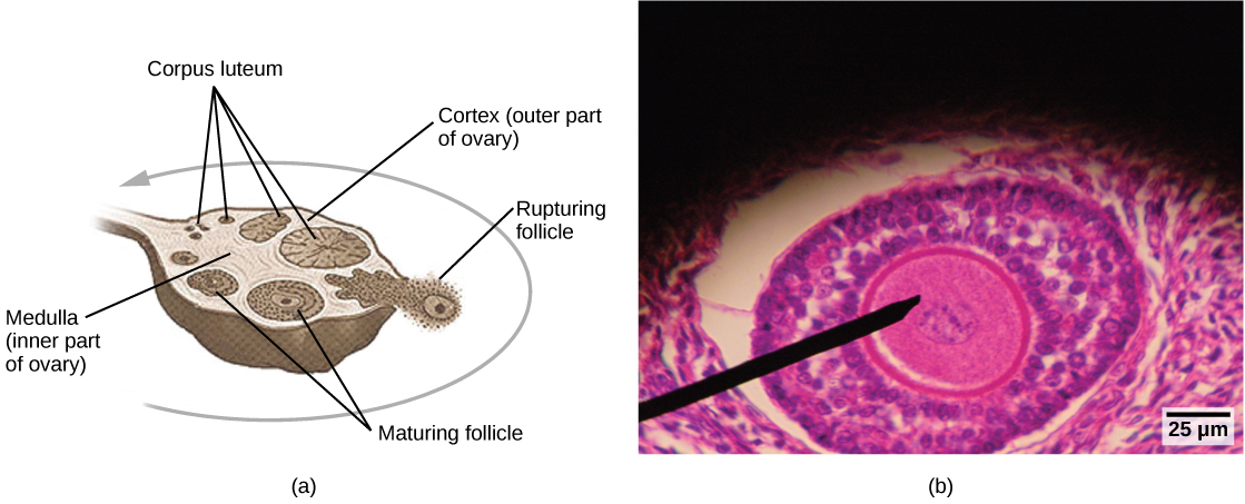
Internal female reproductive structures include ovaries, oviducts, the uterus, and the vagina, shown in Figure 43.10. The pair of ovaries is held in place in the abdominal cavity by a system of ligaments. Ovaries consist of a medulla and cortex: the medulla contains nerves and blood vessels to supply the cortex with nutrients and remove waste. The outer layers of cells of the cortex are the functional parts of the ovaries. The cortex is made up of follicular cells that surround eggs that develop during fetal development in utero. During the menstrual period, a batch of follicular cells develops and prepares the eggs for release. At ovulation, one follicle ruptures and one egg is released, as illustrated in Figure 43.11a.
英語のヒント
OpenStax, Rice University; Biology 2e. (credit a: modification of work by NIH; scale-bar data from Matt Russell) · CC BY 4.0 · Source出典

Figure 43.11図 43.11
Oocytes develop in (a) follicles, located in the ovary. At the beginning of the menstrual cycle, the follicle matures. At ovulation, the follicle ruptures, releasing the egg. The follicle becomes a corpus luteum, which eventually degenerates. The (b) follicle in this light micrograph has an oocyte at its center. (credit a: modification of work by NIH; scale-bar data from Matt Russell)
卵母細胞は、卵巣内の(a)卵胞で発達する。月経周期の初めに卵胞が成熟する。排卵時には卵胞が破裂し、卵を放出する。卵胞は黄体となり、最終的に退化する。(b)この光学顕微鏡写真の卵胞には、中央に卵母細胞がある。(aの出典:NIHの作品を改変。スケールバーのデータ:Matt Russell)
The oviducts, or fallopian tubes, extend from the uterus in the lower abdominal cavity to the ovaries, but they are not in contact with the ovaries. The lateral ends of the oviducts flare out into a trumpet-like structure and have a fringe of finger-like projections called fimbriae, illustrated in Figure 43.10b. When an egg is released at ovulation, the fimbrae help the nonmotile egg enter into the tube and passage to the uterus. The walls of the oviducts are ciliated and are made up mostly of smooth muscle. The cilia beat toward the middle, and the smooth muscle contracts in the same direction, moving the egg toward the uterus. Fertilization usually takes place within the oviducts and the developing embryo is moved toward the uterus for development. It usually takes the egg or embryo a week to travel through the oviduct. Sterilization in females is called a tubal ligation; it is analogous to a vasectomy in males in that the oviducts are severed and sealed.
英語のヒント
卵管は、下腹部の腹腔にある子宮から卵巣へ向かって伸びるが、卵巣とは接していない。卵管の外側の端はラッパ状に広がり、図43.10bに示す卵管采という指状の突起で縁取られる。排卵によって卵が放出されると、卵管采は、自力で動けない卵が卵管へ入り、子宮へ向かうのを助ける。卵管の壁には繊毛があり、主に平滑筋からなる。繊毛は中央に向かって動き、平滑筋も同じ向きに収縮して、卵を子宮へ送る。受精は通常、卵管内で起こり、発生中の胚は、さらに発育するために子宮へ運ばれる。卵または胚が卵管を通過するには、通常1週間かかる。女性の不妊手術は卵管結紮と呼ばれる。卵管を切断して閉じる点で、男性の精管切除術に相当する。
The uterus is a structure about the size of a woman’s fist. This is lined with an endometrium rich in blood vessels and mucus glands. The uterus supports the developing embryo and fetus during gestation. The thickest portion of the wall of the uterus is made of smooth muscle. Contractions of the smooth muscle in the uterus aid in passing the baby through the vagina during labor. A portion of the lining of the uterus sloughs off during each menstrual period, and then builds up again in preparation for an implantation. Part of the uterus, called the cervix, protrudes into the top of the vagina. The cervix functions as the birth canal.
英語のヒント
子宮は、女性の握りこぶしほどの大きさの構造である。内側は、血管と粘液腺に富む子宮内膜に覆われる。子宮は、妊娠中に発育する胚と胎児を支える。子宮壁の最も厚い部分は平滑筋からなり、その収縮が分娩時に赤ちゃんを膣から送り出すのを助ける。月経のたびに子宮内膜の一部が剥がれ落ち、着床に備えて再び厚くなる。子宮の一部である子宮頸部は、膣の上部へ突き出し、産道として機能する。
The vagina is a muscular tube that serves several purposes. It allows menstrual flow to leave the body. It is the receptacle for the penis during intercourse and the vessel for the delivery of offspring. It is lined by stratified squamous epithelial cells to protect the underlying tissue.
英語のヒント
膣は、いくつかの役割をもつ筋肉性の管である。月経血を体外へ出し、性交時には陰茎を受け入れ、出産時には子の通り道となる。内側は重層扁平上皮細胞に覆われ、その下の組織を保護する。