第43章動物の生殖と発生
Sexual Response during Intercourse性交中の性反応
The sexual response in humans is both psychological and physiological. People with testes-based or ovary-based reproductive systems experience sexual arousal through psychological and physical stimulation. There are four phases of the sexual response. During phase one, called excitement, vasodilation leads to vasocongestion in erectile tissues. The nipples, clitoris, labia, and penis engorge with blood and become enlarged. Vaginal secretions are released to lubricate the vagina to facilitate intercourse. During the second phase, called the plateau, stimulation continues, the outer third of the vaginal wall enlarges with blood, and breathing and heart rate increase.
英語のヒント
ヒトの性反応には心理的側面と生理的側面の両方がある。精巣を中心とする生殖系をもつ人も、卵巣を中心とする生殖系をもつ人も、心理的・身体的な刺激によって性的興奮を経験する。性反応には4つの段階がある。第1段階の興奮期では、血管拡張によって勃起組織に血液がうっ滞する。乳頭、陰核、陰唇、陰茎に血液が充満し、大きくなる。膣を潤滑にして性交を容易にする分泌液が放出される。第2段階の高原期では刺激が続き、膣壁の外側3分の1が血液で膨らみ、呼吸数と心拍数が増加する。
During phase three, or orgasm, rhythmic, involuntary contractions of muscles occur. In the male, the reproductive accessory glands and tubules constrict placing semen in the urethra, then the urethra contracts expelling the semen through the penis. In the female, the uterus and vaginal muscles contract in waves that may last slightly less than a second each. During phase four, or resolution, the processes described in the first three phases reverse themselves and return to their normal state. Males experience a refractory period in which they cannot maintain an erection or ejaculate for a period of time ranging from minutes to hours.
英語のヒント
第3段階のオルガスム期では、筋肉が律動的に、不随意に収縮する。男性では、生殖系の付属腺や管が収縮して精液を尿道へ送り、続いて尿道が収縮し、陰茎から精液を排出する。女性では、子宮と膣の筋肉が波状に収縮し、各収縮は1秒弱続くことがある。第4段階の消退期では、最初の3段階で起こった過程が逆転し、通常の状態へ戻る。男性には不応期があり、数分から数時間の間、勃起を維持したり射精したりできない。
Gametogenesis (Spermatogenesis and Oogenesis)配偶子形成(精子形成と卵形成)
Gametogenesis, the production of sperm and eggs, takes place through the process of meiosis. During meiosis, two cell divisions separate the paired chromosomes in the nucleus and then separate the chromatids that were made during an earlier stage of the cell’s life cycle. Meiosis produces haploid cells with half of each pair of chromosomes normally found in diploid cells. The production of sperm is called spermatogenesis and the production of eggs is called oogenesis.
英語のヒント
精子と卵を作る配偶子形成は、減数分裂によって進む。減数分裂では、2回の細胞分裂によって、まず核内の対をなす染色体が分かれ、次に細胞周期の前の段階で作られた染色分体が分かれる。減数分裂は、通常の二倍体細胞にある染色体の各対から1本ずつをもつ、半数体細胞を作る。精子を作る過程を精子形成、卵を作る過程を卵形成という。
Spermatogenesis精子形成
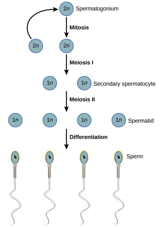
Figure 43.12図 43.12
During spermatogenesis, four sperm result from each primary spermatocyte.
精子形成では、1個の一次精母細胞から4個の精子が生じる。
Spermatogenesis, illustrated in Figure 43.12, occurs in the wall of the seminiferous tubules (Figure 43.8), with stem cells at the periphery of the tube and the spermatozoa at the lumen of the tube. Immediately under the capsule of the tubule are diploid, undifferentiated cells. These stem cells, called spermatogonia (singular: spermatagonium), go through mitosis with one offspring going on to differentiate into a sperm cell and the other giving rise to the next generation of sperm.
英語のヒント
Meiosis starts with a cell called a primary spermatocyte. At the end of the first meiotic division, a haploid cell is produced called a secondary spermatocyte. This cell is haploid and must go through another meiotic cell division. The cell produced at the end of meiosis is called a spermatid and when it reaches the lumen of the tubule and grows a flagellum, it is called a sperm cell. Four sperm result from each primary spermatocyte that goes through meiosis.
英語のヒント
減数分裂は、一次精母細胞という細胞から始まる。最初の減数分裂が終わると、二次精母細胞という半数体の細胞ができる。この細胞は半数体であり、さらにもう1回の減数分裂を行わなければならない。減数分裂の最後にできる細胞は精子細胞と呼ばれる。精細管の内腔へ達して鞭毛を伸ばすと、精子と呼ばれるようになる。減数分裂を行う1個の一次精母細胞から、4個の精子が生じる。
Stem cells are deposited during gestation and are present at birth through the beginning of adolescence, but in an inactive state. During adolescence, gonadotropic hormones from the anterior pituitary cause the activation of these cells and the production of viable sperm. This continues into old age.
英語のヒント
幹細胞は妊娠中に形成され、出生時から思春期の初めまで存在するが、その間は不活性な状態にある。思春期になると、下垂体前葉の性腺刺激ホルモンが、これらの細胞を活性化し、生存可能な精子の生成を引き起こす。この過程は老年期まで続く。
Oogenesis卵形成
Oogenesis, illustrated in Figure 43.13, occurs in the outermost layers of the ovaries. As with sperm production, oogenesis starts with a germ cell, called an oogonium (plural: oogonia), but this cell undergoes mitosis to increase in number, eventually resulting in up to about one to two million cells in the embryo.
英語のヒント
図43.13に示す卵形成は、卵巣の最外層で起こる。精子の形成と同様に、卵形成も卵原細胞という生殖細胞から始まる。ただし、この細胞は有糸分裂によって数を増やし、最終的に胚の中で約100万~200万個に達する。
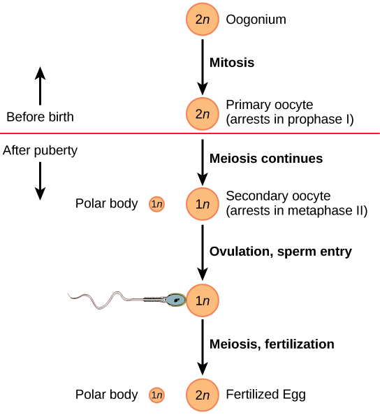


Figure 43.13図 43.13
The process of oogenesis occurs in the ovary’s outermost layer.
卵形成は卵巣の最外層で起こる。
The cell starting meiosis is called a primary oocyte, as shown in Figure 43.13. This cell will start the first meiotic division and be arrested in its progress in the first prophase stage. At the time of birth, all future eggs are in the prophase stage. At adolescence, anterior pituitary hormones cause the development of a number of follicles in an ovary. This results in the primary oocyte finishing the first meiotic division. The cell divides unequally, with most of the cellular material and organelles going to one cell, called a secondary oocyte, and only one set of chromosomes and a small amount of cytoplasm going to the other cell. This second cell is called a polar body and usually dies. A secondary meiotic arrest occurs, this time at the metaphase II stage. At ovulation, this secondary oocyte will be released and travel toward the uterus through the oviduct. If the secondary oocyte is fertilized, the cell continues through the meiosis II, producing a second polar body and a fertilized egg containing all 46 chromosomes of a human being, half of them coming from the sperm.
英語のヒント
図43.13に示すように、減数分裂を始める細胞は一次卵母細胞と呼ばれる。この細胞は最初の減数分裂を始め、その前期で進行が停止する。出生時には、将来卵となる細胞はすべて前期にある。思春期には、下垂体前葉のホルモンが卵巣内のいくつかの卵胞を発達させる。その結果、一次卵母細胞が最初の減数分裂を完了する。細胞は不均等に分かれ、大部分の細胞物質と細胞小器官は二次卵母細胞という一方の細胞へ入り、もう一方には染色体1組と少量の細胞質だけが入る。後者は極体と呼ばれ、通常は死滅する。減数分裂は再び停止するが、今度は減数第2分裂中期である。排卵時、この二次卵母細胞は放出され、卵管を通って子宮へ向かう。受精すると減数第2分裂が進み、第2の極体と、ヒトの染色体46本すべてをもつ受精卵が生じる。その半分は精子に由来する。
Egg production begins before birth, is arrested during meiosis until puberty, and then individual cells continue through at each menstrual cycle. One egg is produced from each meiotic process, with the extra chromosomes and chromatids going into polar bodies that degenerate and are reabsorbed by the body.
英語のヒント
卵の生成は出生前に始まり、減数分裂の途中で思春期まで停止した後、月経周期ごとに個々の細胞で進む。各減数分裂の過程から1個の卵が生じ、余分な染色体と染色分体は極体へ入る。極体は退化し、体に再吸収される。