第42章免疫系
Primary Centers of the Immune System免疫系の主要な拠点
Although the immune system is characterized by circulating cells throughout the body, the regulation, maturation, and intercommunication of immune factors occur at specific sites. The blood circulates immune cells, proteins, and other factors through the body. Approximately 0.1 percent of all cells in the blood are leukocytes, which encompass monocytes (the precursor of macrophages) and lymphocytes. The majority of cells in the blood are erythrocytes (red blood cells). Lymph is a watery fluid that bathes tissues and organs with protective white blood cells and does not contain erythrocytes. Cells of the immune system can travel between the distinct lymphatic and blood circulatory systems, which are separated by interstitial space, by a process called extravasation (passing through to surrounding tissue).
英語のヒント
免疫系の特徴は細胞が体中を循環することであるが、免疫因子の調節、成熟、相互の情報伝達は、特定の場所で行われる。血液は免疫細胞、タンパク質、その他の因子を全身に循環させる。血液中の全細胞の約0.1%が白血球で、単球(マクロファージの前駆細胞)やリンパ球が含まれる。血液中の細胞の大部分は赤血球である。リンパは水のような液体で、防御を担う白血球とともに組織や器官を浸すが、赤血球を含まない。リンパ系と血液循環系は別々で、その間を間質の空間が隔てている。免疫系の細胞は、血管外遊出(周囲の組織へ通り抜けること)という過程で、両者の間を移動できる。
The cells of the immune system originate from hematopoietic stem cells in the bone marrow. Cytokines stimulate these stem cells to differentiate into immune cells. B cell maturation occurs in the bone marrow, whereas naïve T cells transit from the bone marrow to the thymus for maturation. In the thymus, immature T cells that express TCRs complementary to self-antigens are destroyed. This process helps prevent autoimmune responses.
英語のヒント
免疫系の細胞は、骨髄の造血幹細胞から生じる。サイトカインが幹細胞を刺激し、免疫細胞へ分化させる。B細胞は骨髄で成熟する一方、ナイーブT細胞は成熟するために骨髄から胸腺へ移動する。胸腺では、自己抗原に相補的なTCRを発現する未熟なT細胞が破壊される。この過程は、自己免疫応答を防ぐのに役立つ。
On maturation, T and B lymphocytes circulate to various destinations. Lymph nodes scattered throughout the body, as illustrated in Figure 42.20, house large populations of T and B cells, dendritic cells, and macrophages. Lymph gathers antigens as it drains from tissues. These antigens then are filtered through lymph nodes before the lymph is returned to circulation. APCs in the lymph nodes capture and process antigens and inform nearby lymphocytes about potential pathogens.
英語のヒント
Tリンパ球とBリンパ球は成熟すると、さまざまな場所へ循環する。図42.20のように全身に散在するリンパ節には、多数のT細胞、B細胞、樹状細胞、マクロファージが存在する。リンパは組織から流れ出す際に抗原を集める。循環へ戻る前に、これらの抗原はリンパ節で濾し取られる。リンパ節内のAPCは抗原を捕らえて処理し、近くのリンパ球へ、病原体となり得るものの情報を伝える。
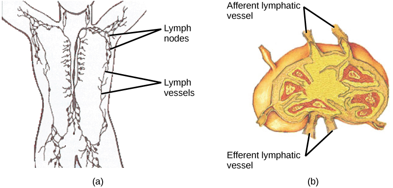
OpenStax, Rice University; Biology 2e. (credit: modification of work by NIH, NCI) · CC BY 4.0 · Source出典
Figure 42.20図 42.20
(a) Lymphatic vessels carry a clear fluid called lymph throughout the body. The liquid enters (b) lymph nodes through afferent vessels. Lymph nodes are filled with lymphocytes that purge infecting cells. The lymph then exits through efferent vessels. (credit: modification of work by NIH, NCI)
(a)リンパ管は、リンパという透明な液を体中へ運ぶ。この液は輸入管を通って(b)リンパ節に入る。リンパ節は感染細胞を取り除くリンパ球で満たされている。その後、リンパは輸出管から出る。(出典:NIH、NCIの作品を改変)
The spleen houses B and T cells, macrophages, dendritic cells, and NK cells. The spleen, shown in Figure 42.21, is the site where APCs that have trapped foreign particles in the blood can communicate with lymphocytes. Antibodies are synthesized and secreted by activated plasma cells in the spleen, and the spleen filters foreign substances and antibody-complexed pathogens from the blood. Functionally, the spleen is to the blood as lymph nodes are to the lymph.
英語のヒント
脾臓にはB細胞とT細胞、マクロファージ、樹状細胞、NK細胞が存在する。図42.21の脾臓は、血液中の異物粒子を捕らえたAPCが、リンパ球と情報をやり取りできる場所である。脾臓内の活性化した形質細胞が抗体を合成・分泌する。また、脾臓は血液から異物や抗体と複合体を作った病原体を濾し取る。機能の面では、リンパ節がリンパに対して果たす役割を、脾臓が血液に対して果たしている。
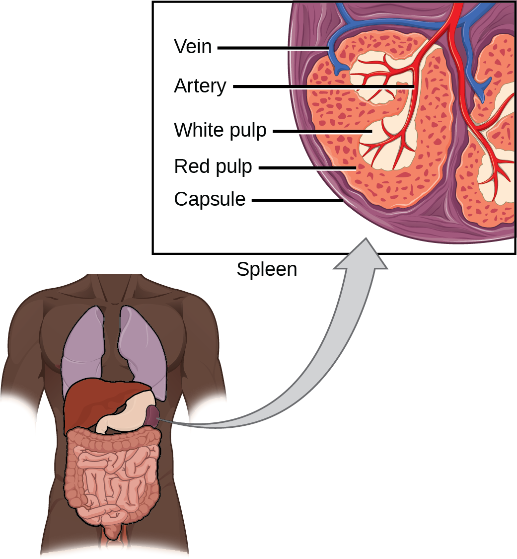


Figure 42.21図 42.21
The spleen is similar to a lymph node but is much larger and filters blood instead of lymph. Blood enters the spleen through arteries and exits through veins. The spleen contains two types of tissue: red pulp and white pulp. Red pulp consists of cavities that store blood. Within the red pulp, damaged red blood cells are removed and replaced by new ones. White pulp is rich in lymphocytes that remove antigen-coated bacteria from the blood. (credit: modification of work by NCI)
脾臓はリンパ節に似ているが、はるかに大きく、リンパの代わりに血液を濾過する。血液は動脈から脾臓に入り、静脈から出る。脾臓には赤脾髄と白脾髄の2種類の組織がある。赤脾髄は血液を蓄える空間からなる。ここでは、損傷した赤血球が除かれ、新しいものに置き換わる。白脾髄はリンパ球に富み、抗原で覆われた細菌を血液から除く。(出典:NCIの作品を改変)
Section Summary節のまとめ
The adaptive immune response is a slower-acting, longer-lasting, and more specific response than the innate response. However, the adaptive response requires information from the innate immune system to function. APCs display antigens via MHC molecules to complementary naïve T cells. In response, the T cells differentiate and proliferate, becoming TH cells or CTLs. TH cells stimulate B cells that have engulfed and presented pathogen-derived antigens. B cells differentiate into plasma cells that secrete antibodies, whereas CTLs induce apoptosis in intracellularly infected or cancerous cells. Memory cells persist after a primary exposure to a pathogen. If reexposure occurs, memory cells differentiate into effector cells without input from the innate immune system. The mucosal immune system is largely independent from the systemic immune system but functions in a parallel fashion to protect the extensive mucosal surfaces of the body.
獲得免疫応答は、自然免疫応答より作用が遅いが、長く持続し、特異性が高い。ただし、機能するには自然免疫系からの情報が必要である。APCはMHC分子を介して、相補的なナイーブT細胞へ抗原を提示する。T細胞はそれに応答して分化・増殖し、TH細胞またはCTLとなる。TH細胞は、病原体由来の抗原を取り込んで提示したB細胞を刺激する。B細胞は抗体を分泌する形質細胞に分化し、CTLは細胞内感染した細胞やがん細胞にアポトーシスを誘導する。病原体への最初の曝露の後も、記憶細胞は存続する。再びさらされると、自然免疫系からの入力なしにエフェクター細胞へ分化する。粘膜免疫系は全身免疫系から大きく独立しているが、並行した仕組みで働き、体の広範な粘膜表面を保護する。
Visual Connection Questions図を使った問題
Exercises 7–9練習問題 7–9
Exercise 7問題 7
Figure 42.11 Which of the following statements about T cells is false?
図42.11 T細胞について、次の記述のうち誤っているものはどれか。
- Helper T cells release cytokines while cytotoxic T cells kill the infected cell.ヘルパーT細胞はサイトカインを放出し、細胞傷害性T細胞は感染細胞を殺す。
- Helper T cells are CD4+, while cytotoxic T cells are CD8+.ヘルパーT細胞はCD4+で、細胞傷害性T細胞はCD8+である。
- MHC II is a receptor found on most body cells, while MHC I is a receptor found on immune cells only.MHC IIは体の大部分の細胞にある受容体で、MHC Iは免疫細胞だけにある受容体である。
- The T cell receptor is found on both CD4+ and CD8+ T cells.T細胞受容体は、CD4+とCD8+ の両方のT細胞に存在する。
Solution解き方
Exercise 8問題 8
Figure 42.14 Based on what you know about MHC receptors, why do you think an organ transplanted from an incompatible donor to a recipient will be rejected?
図42.14 MHC受容体について知っていることに基づいて考えよう。不適合な提供者から受容者へ移植された器官は、なぜ拒絶されると考えられるか。
Solution解き方
Figure 42.14 MHC receptors differ from person to person. Thus, MHC receptors on an incompatible donor are considered “non-self” and are rejected by the immune system.
図42.14 MHC受容体は人によって異なる。そのため、不適合な提供者のMHC受容体は「非自己」と見なされ、免疫系によって拒絶される。
Exercise 9問題 9
Figure 42.16 The Rh antigen is found on Rh-positive red blood cells. During pregnancy, Rh-negative people can usually carry an Rh-positive fetus to term without difficulty. However, a second Rh-positive fetus may trigger an immune attack that causes hemolytic disease of the newborn. Why do you think hemolytic disease is only a problem during the second or subsequent pregnancies?
図42.16 Rh抗原はRh陽性の赤血球に存在する。Rh陰性の人が妊娠した場合、通常はRh陽性の胎児を問題なく満期まで妊娠し続けることができる。しかし、2回目のRh陽性胎児の妊娠では、新生児溶血性疾患を起こす免疫攻撃が引き起こされる場合がある。なぜ溶血性疾患が問題になるのは、2回目以降の妊娠だけなのだと考えられるか。
Solution解き方
Figure 42.16 If the blood of the mother and fetus mixes, memory cells that recognize the Rh antigen can form late in the first pregnancy. During subsequent pregnancies, these memory cells launch an immune attack on the fetal blood cells. Injection of anti-Rh antibody during the first pregnancy prevents the immune response from occurring.
図42.16 母親と胎児の血液が混ざると、最初の妊娠の後期に、Rh抗原を認識する記憶細胞ができることがある。その後の妊娠では、これらの記憶細胞が胎児の血球への免疫攻撃を始める。最初の妊娠中に抗Rh抗体を注射することで、この免疫応答が起こるのを防げる。