第42章免疫系
Natural Killer Cellsナチュラルキラー細胞
Lymphocytes are leukocytes that are histologically identifiable by their large, darkly staining nuclei; they are small cells with very little cytoplasm, as shown in Figure 42.6. Infected cells are identified and destroyed by natural killer (NK) cells, lymphocytes that can kill cells infected with viruses or tumor cells (abnormal cells that uncontrollably divide and invade other tissue). T cells and B cells of the adaptive immune system also are classified as lymphocytes. T cells are lymphocytes that mature in the thymus gland, and B cells are lymphocytes that mature in the bone marrow. NK cells identify intracellular infections, especially from viruses, by the altered expression of major histocompatibility complex (MHC) I molecules on the surface of infected cells. MHC I molecules are proteins on the surfaces of all nucleated cells, thus they are scarce on red blood cells and platelets which are non-nucleated. The function of MHC I molecules is to display fragments of proteins from the infectious agents within the cell to T cells; healthy cells will be ignored, while “non-self” or foreign proteins will be attacked by the immune system. MHC II molecules are found mainly on cells containing antigens (“non-self proteins”) and on lymphocytes. MHC II molecules interact with helper T cells to trigger the appropriate immune response, which may include the inflammatory response.
英語のヒント
リンパ球は、大きく濃く染まる核によって組織学的に識別される白血球である。図42.6のように、細胞質がごく少ない小さな細胞である。感染細胞はナチュラルキラー(NK)細胞によって識別され、破壊される。NK細胞は、ウイルス感染細胞や腫瘍細胞(制御を失って分裂し、他の組織へ侵入する異常細胞)を殺せるリンパ球である。獲得免疫系のT細胞とB細胞もリンパ球に分類される。T細胞は胸腺で成熟するリンパ球であり、B細胞は骨髄で成熟するリンパ球である。NK細胞は、感染細胞表面の主要組織適合遺伝子複合体(MHC)I分子の発現変化により、特にウイルスによる細胞内感染を識別する。MHC I分子は、すべての有核細胞の表面にあるタンパク質である。そのため、核のない赤血球や血小板では乏しい。MHC I分子は、細胞内の感染因子に由来するタンパク質の断片をT細胞に提示する。健康な細胞は無視されるが、「非自己」または外来のタンパク質は免疫系に攻撃される。MHC II分子は主に、抗原(「非自己のタンパク質」)を含む細胞とリンパ球に見られる。ヘルパーT細胞と相互作用し、炎症応答などの適切な免疫応答を引き起こす。
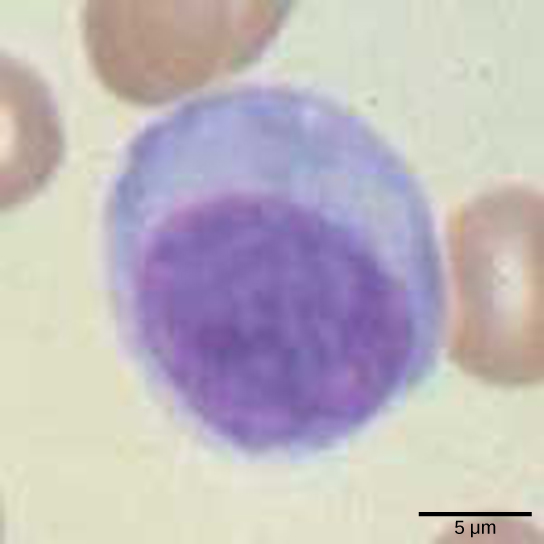
Figure 42.6図 42.6
Lymphocytes, such as NK cells, are characterized by their large nuclei that actively absorb Wright stain and therefore appear dark colored under a microscope.
NK細胞などのリンパ球は、大きな核をもつことが特徴である。核はライト染色をよく取り込むため、顕微鏡では濃い色に見える。
An infected cell (or a tumor cell) is usually incapable of synthesizing and displaying MHC I molecules appropriately. The metabolic resources of cells infected by some viruses produce proteins that interfere with MHC I processing and/or trafficking to the cell surface. The reduced MHC I on host cells varies from virus to virus and results from active inhibitors being produced by the viruses. This process can deplete host MHC I molecules on the cell surface, which NK cells detect as “unhealthy” or “abnormal” while searching for cellular MHC I molecules. Similarly, the dramatically altered gene expression of tumor cells leads to expression of extremely deformed or absent MHC I molecules that also signal “unhealthy” or “abnormal.”
英語のヒント
感染細胞(または腫瘍細胞)は、通常、MHC I分子を適切に合成・提示できない。一部のウイルスに感染した細胞では、細胞の代謝資源によって、MHC Iの加工や細胞表面への輸送を妨げるタンパク質が作られる。宿主細胞上のMHC Iの減少の程度はウイルスによって異なり、ウイルスが作る能動的な阻害因子に由来する。この過程によって宿主の細胞表面のMHC I分子が減少すると、細胞のMHC I分子を探しているNK細胞は、それを「不健康」または「異常」と検出する。同様に、腫瘍細胞で大きく変化した遺伝子発現は、著しく変形したMHC I分子の発現、またはその欠如をもたらし、これも「不健康」や「異常」の合図となる。
NK cells are always active; an interaction with normal, intact MHC I molecules on a healthy cell disables the killing sequence, and the NK cell moves on. After the NK cell detects an infected or tumor cell, its cytoplasm secretes granules comprised of perforin, a destructive protein that creates a pore in the target cell. Granzymes are released along with the perforin in the immunological synapse. A granzyme is a protease that digests cellular proteins and induces the target cell to undergo programmed cell death, or apoptosis. Phagocytic cells then digest the cell debris left behind. NK cells are constantly patrolling the body and are an effective mechanism for controlling potential infections and preventing cancer progression.
英語のヒント
NK細胞は常に活動している。健康な細胞の正常で損なわれていないMHC I分子と相互作用すると、細胞を殺す一連の反応が止まり、NK細胞は先へ進む。NK細胞が感染細胞や腫瘍細胞を見つけると、その細胞質からパーフォリンを含む顆粒が分泌される。パーフォリンは、標的細胞に孔を作る破壊作用のあるタンパク質である。免疫シナプスでは、パーフォリンとともにグランザイムが放出される。グランザイムはプロテアーゼで、細胞のタンパク質を分解し、標的細胞にプログラムされた細胞死、すなわちアポトーシスを誘導する。その後、食細胞が残された細胞の残骸を消化する。NK細胞は絶えず体内を巡回しており、起こり得る感染を抑え、がんの進行を防ぐための有効な仕組みとなっている。
Complement補体
An array of approximately 20 types of soluble proteins, called a complement system, functions to destroy extracellular pathogens. Cells of the liver and macrophages synthesize complement proteins continuously; these proteins are abundant in the blood serum and are capable of responding immediately to infecting microorganisms. The complement system is so named because it is complementary to the antibody response of the adaptive immune system. Complement proteins bind to the surfaces of microorganisms and are particularly attracted to pathogens that are already bound by antibodies. Binding of complement proteins occurs in a specific and highly regulated sequence, with each successive protein being activated by cleavage and/or structural changes induced upon binding of the preceding protein(s). After the first few complement proteins bind, a cascade of sequential binding events follows in which the pathogen rapidly becomes coated in complement proteins.
英語のヒント
補体系と呼ばれる約20種類の可溶性タンパク質の集まりは、細胞外の病原体を破壊する働きをする。肝臓の細胞とマクロファージは、補体タンパク質を絶えず合成している。これらは血清中に豊富にあり、感染してきた微生物に直ちに応答できる。獲得免疫系の抗体応答を補うため、補体系という名が付いている。補体タンパク質は微生物の表面に結合し、特に、すでに抗体が結合した病原体に引き寄せられる。その結合は、厳密に調節された特定の順序で起こる。先行するタンパク質の結合によって誘導される切断や構造変化を通じ、後続のタンパク質が順に活性化される。最初の数個の補体タンパク質が結合すると、次々に結合が起こる連鎖反応が続き、病原体は急速に補体タンパク質で覆われる。
Complement proteins perform several functions. The proteins serve as a marker to indicate the presence of a pathogen to phagocytic cells, such as macrophages and B cells, and enhance engulfment; this process is called opsonization. Certain complement proteins can combine to form attack complexes that open pores in microbial cell membranes. These structures destroy pathogens by causing their contents to leak, as illustrated in Figure 42.7.
英語のヒント
補体タンパク質にはいくつかの機能がある。マクロファージやB細胞などの食細胞に、病原体の存在を知らせる目印として働き、取り込みを促す。この過程をオプソニン化という。特定の補体タンパク質は結合して攻撃複合体を作り、微生物の細胞膜に孔を開ける。この構造は、図42.7のように内容物を漏出させて病原体を破壊する。
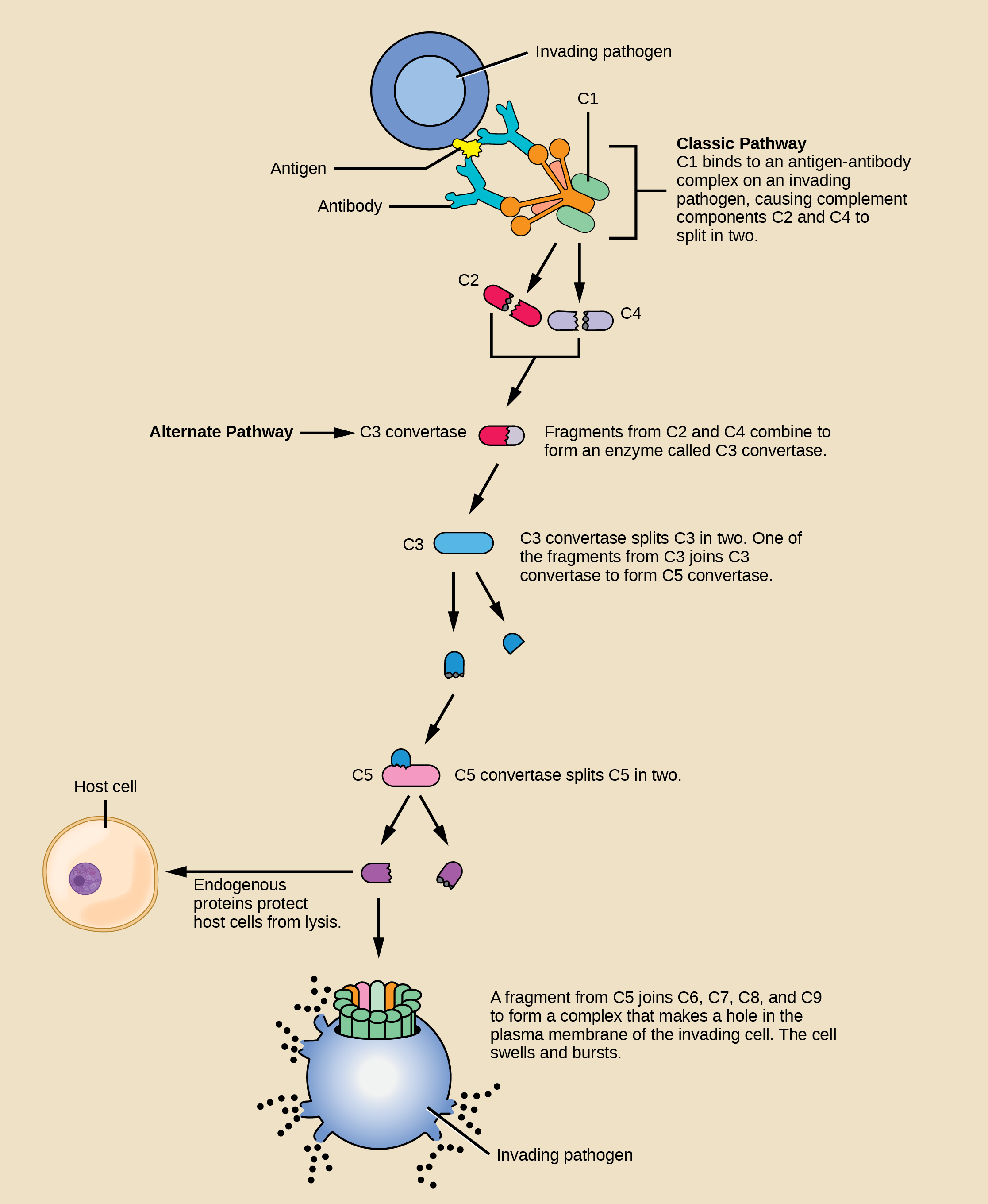


Figure 42.7図 42.7
The classic pathway for the complement cascade involves the attachment of several initial complement proteins to an antibody-bound pathogen followed by rapid activation and binding of many more complement proteins and the creation of destructive pores in the microbial cell envelope and cell wall. The alternate pathway does not involve antibody activation. Rather, C3 convertase spontaneously breaks down C3. Endogenous regulatory proteins prevent the complement complex from binding to host cells. Pathogens lacking these regulatory proteins are lysed. (credit: modification of work by NIH)
補体カスケードの古典経路では、抗体が結合した病原体に最初のいくつかの補体タンパク質が付着する。続いて、さらに多くの補体タンパク質が迅速に活性化・結合し、微生物の細胞外被や細胞壁に破壊的な孔を作る。代替経路には抗体による活性化は関わらず、C3転換酵素が自発的にC3を分解する。内在性の調節タンパク質は、補体複合体が宿主細胞に結合するのを防ぐ。これらの調節タンパク質をもたない病原体は溶解される。(出典:NIHの作品を改変)