第42章免疫系
Cytotoxic T Lymphocytes細胞傷害性Tリンパ球
CTLs, a subclass of T cells, function to clear infections directly. The cell-mediated part of the adaptive immune system consists of CTLs that attack and destroy infected cells. CTLs are particularly important in protecting against viral infections; this is because viruses replicate within cells where they are shielded from extracellular contact with circulating antibodies. When APCs phagocytize pathogens and present MHC I-embedded antigens to naïve CD8+ T cells that express complementary TCRs, the CD8+ T cells become activated to proliferate according to clonal selection. These resulting CTLs then identify non-APCs displaying the same MHC I-embedded antigens (for example, viral proteins)—for example, the CTLs identify infected host cells.
英語のヒント
T細胞の一群であるCTLは、感染を直接除去する働きをする。獲得免疫系の細胞性の部分は、感染細胞を攻撃して破壊するCTLからなる。CTLはウイルス感染に対する防御で特に重要である。ウイルスは細胞内で複製し、循環中の抗体が細胞外から接触することを免れているためである。APCが病原体を食作用で取り込み、MHC Iに組み込んだ抗原を、相補的なTCRを発現するナイーブCD8+T細胞に提示すると、CD8+T細胞は活性化され、クローン選択に従って増殖する。こうして生じたCTLは次に、同じMHC Iに組み込まれた抗原(例えばウイルスタンパク質)を提示する、APC以外の細胞を識別する。つまり、感染した宿主細胞などを識別する。
Intracellularly, infected cells typically die after the infecting pathogen replicates to a sufficient concentration and lyses the cell, as many viruses do. CTLs attempt to identify and destroy infected cells before the pathogen can replicate and escape, thereby halting the progression of intracellular infections. CTLs also support NK lymphocytes to destroy early cancers. Cytokines secreted by the TH1 response that stimulates macrophages also stimulate CTLs and enhance their ability to identify and destroy infected cells and tumors.
英語のヒント
細胞内感染では、多くのウイルスに見られるように、病原体が十分な濃度まで増殖して細胞を溶解し、その後に感染細胞が死ぬのが一般的である。CTLは、病原体が増殖して脱出する前に感染細胞を識別・破壊し、細胞内感染の進行を止めようとする。CTLはまた、NKリンパ球による初期のがんの破壊を助ける。マクロファージを刺激するTH1応答によって分泌されるサイトカインは、CTLも刺激し、感染細胞や腫瘍を識別・破壊する能力を高める。
CTLs sense MHC I-embedded antigens by directly interacting with infected cells via their TCRs. Binding of TCRs with antigens activates CTLs to release perforin and granzyme, degradative enzymes that will induce apoptosis of the infected cell. Recall that this is a similar destruction mechanism to that used by NK cells. In this process, the CTL does not become infected and is not harmed by the secretion of perforin and granzymes. In fact, the functions of NK cells and CTLs are complementary and maximize the removal of infected cells, as illustrated in Figure 42.14. If the NK cell cannot identify the “missing self” pattern of down-regulated MHC I molecules, then the CTL can identify it by the complex of MHC I with foreign antigens, which signals “altered self.” Similarly, if the CTL cannot detect antigen-embedded MHC I because the receptors are depleted from the cell surface, NK cells will destroy the cell instead. CTLs also emit cytokines, such as interferons, that alter surface protein expression in other infected cells, such that the infected cells can be easily identified and destroyed. Moreover, these interferons can also prevent virally infected cells from releasing virus particles.
英語のヒント
CTLはTCRを介して感染細胞と直接相互作用し、MHC Iに組み込まれた抗原を感知する。TCRと抗原が結合すると、CTLは活性化され、感染細胞にアポトーシスを誘導する分解酵素であるパーフォリンとグランザイムを放出する。これはNK細胞が使うものと似た破壊機構であることを思い出そう。この過程でCTL自身が感染することはなく、パーフォリンやグランザイムの分泌で害を受けることもない。実際、NK細胞とCTLの機能は相補的であり、図42.14のように感染細胞の除去を最大限に高める。NK細胞が、MHC I分子の減少による「自己の欠如」というパターンを識別できなくても、CTLは「変化した自己」を示す、MHC Iと外来抗原の複合体によって識別できる。同様に、細胞表面の受容体が減少してCTLが抗原を組み込んだMHC Iを検出できない場合には、代わりにNK細胞がその細胞を破壊する。CTLはインターフェロンなどのサイトカインも放出する。これらは他の感染細胞の表面タンパク質の発現を変え、感染細胞を容易に識別・破壊できるようにする。さらに、インターフェロンは、ウイルス感染細胞からのウイルス粒子の放出を防ぐこともできる。
Plasma cells and CTLs are collectively called effector cells: they represent differentiated versions of their naïve counterparts, and they are involved in bringing about the immune defense of killing pathogens and infected host cells.
英語のヒント
形質細胞とCTLは、まとめてエフェクター細胞と呼ばれる。それぞれに対応するナイーブ細胞が分化したものであり、病原体や感染した宿主細胞を殺す免疫防御を実行する。
Mucosal Surfaces and Immune Tolerance粘膜表面と免疫寛容
The innate and adaptive immune responses discussed thus far comprise the systemic immune system (affecting the whole body), which is distinct from the mucosal immune system. Mucosal immunity is formed by mucosa-associated lymphoid tissue, which functions independently of the systemic immune system, and which has its own innate and adaptive components. Mucosa-associated lymphoid tissue (MALT), illustrated in Figure 42.15, is a collection of lymphatic tissue that combines with epithelial tissue lining the mucosa throughout the body. This tissue functions as the immune barrier and response in areas of the body with direct contact to the external environment. The systemic and mucosal immune systems use many of the same cell types. Foreign particles that make their way to MALT are taken up by absorptive epithelial cells called M cells and delivered to APCs located directly below the mucosal tissue. M cells function in the transport described, and are located in the Peyer’s patch, a lymphoid nodule. APCs of the mucosal immune system are primarily dendritic cells, with B cells and macrophages having minor roles. Processed antigens displayed on APCs are detected by T cells in the MALT and at various mucosal induction sites, such as the tonsils, adenoids, appendix, or the mesenteric lymph nodes of the intestine. Activated T cells then migrate through the lymphatic system and into the circulatory system to mucosal sites of infection.
英語のヒント
ここまで扱った自然免疫応答と獲得免疫応答は、全身免疫系(体全体に作用する)を構成し、粘膜免疫系とは区別される。粘膜免疫は粘膜関連リンパ組織によって担われる。この組織は全身免疫系から独立して機能し、独自の自然免疫と獲得免疫の構成要素をもつ。図42.15に示す粘膜関連リンパ組織(MALT)は、体中の粘膜を覆う上皮組織と組み合わさった、リンパ組織の集まりである。外部環境に直接触れる体の領域で、免疫の障壁と応答を担う。全身免疫系と粘膜免疫系は、多くの同じ種類の細胞を使う。MALTに到達した異物粒子は、M細胞という吸収性上皮細胞に取り込まれ、粘膜組織のすぐ下にあるAPCへ届けられる。M細胞はこの輸送を行い、リンパ小節であるパイエル板に存在する。粘膜免疫系のAPCは主に樹状細胞であり、B細胞やマクロファージの役割は小さい。APCが提示する処理済みの抗原は、MALTや、扁桃、アデノイド、虫垂、腸の腸間膜リンパ節などの粘膜免疫の誘導部位で、T細胞に検出される。活性化したT細胞は次にリンパ系から循環系へ移り、粘膜の感染部位へ向かう。
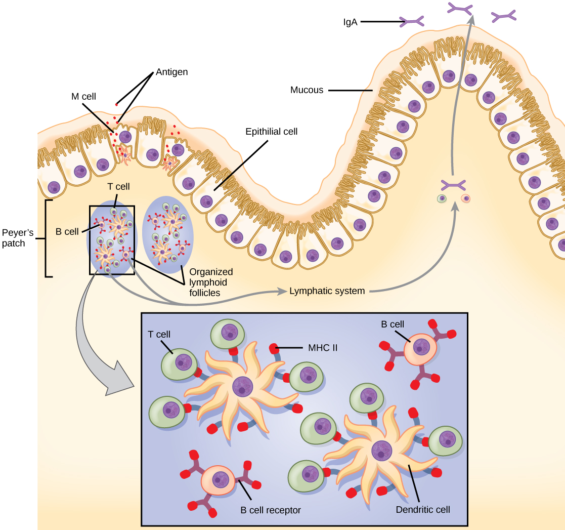

Figure 42.15図 42.15
The topology and function of intestinal MALT is shown. Pathogens are taken up by M cells in the intestinal epithelium and excreted into a pocket formed by the inner surface of the cell. The pocket contains antigen-presenting cells such as dendritic cells, which engulf the antigens, then present them with MHC II molecules on the cell surface. The dendritic cells migrate to an underlying tissue called a Peyer’s patch. Antigen-presenting cells, T cells, and B cells aggregate within the Peyer’s patch, forming organized lymphoid follicles. There, some T cells and B cells are activated. Other antigen-loaded dendritic cells migrate through the lymphatic system where they activate B cells, T cells, and plasma cells in the lymph nodes. The activated cells then return to MALT tissue effector sites. IgA and other antibodies are secreted into the intestinal lumen.
腸のMALTの配置と機能。腸上皮のM細胞が病原体を取り込み、細胞の内側表面が作るポケットへ排出する。ポケットには樹状細胞などの抗原提示細胞があり、抗原を取り込んだ後、MHC II分子とともに細胞表面に提示する。樹状細胞は、その下のパイエル板という組織へ移動する。抗原提示細胞、T細胞、B細胞はパイエル板内に集まり、組織化されたリンパ濾胞を作る。そこで一部のT細胞とB細胞が活性化する。抗原を取り込んだ別の樹状細胞はリンパ系を通って移動し、リンパ節でB細胞、T細胞、形質細胞を活性化する。活性化した細胞は、その後MALT組織の作用部位へ戻る。IgAなどの抗体が腸管腔へ分泌される。
MALT is a crucial component of a functional immune system because mucosal surfaces, such as the nasal passages, are the first tissues onto which inhaled or ingested pathogens are deposited. The mucosal tissue includes the mouth, pharynx, and esophagus, and the gastrointestinal, respiratory, and urogenital tracts.
英語のヒント
鼻腔などの粘膜表面は、吸い込んだり摂取したりした病原体が最初に付着する組織である。このため、MALTは免疫系が機能するうえで不可欠な構成要素である。粘膜組織には、口、咽頭、食道、そして消化管、気道、泌尿生殖路が含まれる。
The immune system has to be regulated to prevent wasteful, unnecessary responses to harmless substances, and more importantly so that it does not attack “self.” The acquired ability to prevent an unnecessary or harmful immune response to a detected foreign substance known not to cause disease is described as immune tolerance. Immune tolerance is crucial for maintaining mucosal homeostasis given the tremendous number of foreign substances (such as food proteins) that APCs of the oral cavity, pharynx, and gastrointestinal mucosa encounter. Immune tolerance is brought about by specialized APCs in the liver, lymph nodes, small intestine, and lung that present harmless antigens to an exceptionally diverse population of regulatory T (Treg) cells, specialized lymphocytes that suppress local inflammation and inhibit the secretion of stimulatory immune factors. The combined result of Treg cells is to prevent immunologic activation and inflammation in undesired tissue compartments and to allow the immune system to focus on pathogens instead. In addition to promoting immune tolerance of harmless antigens, other subsets of Treg cells are involved in the prevention of the autoimmune response, which is an inappropriate immune response to host cells or self-antigens. Another Treg class suppresses immune responses to harmful pathogens after the infection has cleared to minimize host cell damage induced by inflammation and cell lysis.
英語のヒント
免疫系は、無害な物質への無駄で不要な応答を防ぎ、さらに重要なこととして「自己」を攻撃しないよう、調節されなければならない。病気を起こさないと分かっている外来物質を検出しても、不要または有害な免疫応答を起こさないようにする、獲得された能力を免疫寛容という。口腔、咽頭、消化管粘膜のAPCは、食物タンパク質など膨大な数の外来物質に出会う。そのため、免疫寛容は粘膜の恒常性の維持に不可欠である。肝臓、リンパ節、小腸、肺の特殊なAPCが、非常に多様な制御性T(Treg)細胞に無害な抗原を提示し、免疫寛容をもたらす。制御性T細胞は、局所の炎症と、刺激性の免疫因子の分泌を抑える特殊なリンパ球である。Treg細胞の作用を合わせると、望ましくない組織区画での免疫活性化と炎症が防がれ、免疫系は病原体への対応に集中できる。無害な抗原への免疫寛容を促すものに加え、Treg細胞の別の集団は、宿主細胞や自己抗原への不適切な免疫応答である自己免疫応答を防ぐ。さらに別のTreg細胞の群は、感染が除去された後、有害な病原体への免疫応答を抑え、炎症や細胞溶解による宿主細胞の損傷を最小限にする。