第37章内分泌系
37.5 Endocrine Glands37.5 内分泌腺
Learning Objectives学習目標
By the end of this section, you will be able to do the following:
この節を学ぶと、次のことができるようになる。
- Describe the role of different glands in the endocrine system内分泌系における各種の腺の役割を述べる。
- Explain how the different glands work together to maintain homeostasis恒常性を維持するために、各種の腺がどのように協働するか説明する。
Both the endocrine and nervous systems use chemical signals to communicate and regulate the body's physiology. The endocrine system releases hormones that act on target cells to regulate development, growth, energy metabolism, reproduction, and many behaviors. The nervous system releases neurotransmitters or neurohormones that regulate neurons, muscle cells, and endocrine cells. Because the neurons can regulate the release of hormones, the nervous and endocrine systems work in a coordinated manner to regulate the body's physiology.
内分泌系と神経系は、どちらも化学的な信号で情報を伝え、体の生理機能を調節する。内分泌系はホルモンを放出し、標的細胞に作用させて、発達、成長、エネルギー代謝、生殖、多くの行動を調節する。神経系は、ニューロン、筋細胞、内分泌細胞を調節する神経伝達物質や神経ホルモンを放出する。ニューロンはホルモン放出を調節できるため、神経系と内分泌系は協調して体の生理機能を調節する。
Hypothalamic-Pituitary Axis視床下部・下垂体系
The hypothalamus in vertebrates integrates the endocrine and nervous systems. The hypothalamus is an endocrine organ located in the diencephalon of the brain. It receives input from the body and other brain areas and initiates endocrine responses to environmental changes. The hypothalamus acts as an endocrine organ, synthesizing hormones and transporting them along axons to the posterior pituitary gland. It synthesizes and secretes regulatory hormones that control the endocrine cells in the anterior pituitary gland. The hypothalamus contains autonomic centers that control endocrine cells in the adrenal medulla via neuronal control.
英語のヒント
脊椎動物の視床下部は、内分泌系と神経系を統合する。脳の間脳にある内分泌器官であり、体や脳の他の領域から入力を受け、環境の変化に対する内分泌反応を開始する。視床下部は内分泌器官としてホルモンを合成し、軸索に沿って下垂体後葉へ輸送する。また、下垂体前葉の内分泌細胞を制御する調節ホルモンを合成・分泌する。視床下部には、神経を介して副腎髄質の内分泌細胞を制御する自律神経の中枢もある。
The pituitary gland, sometimes called the hypophysis or “master gland” is located at the base of the brain in the sella turcica, a groove of the sphenoid bone of the skull, illustrated in Figure 37.15. It is attached to the hypothalamus via a stalk called the pituitary stalk (or infundibulum). The anterior portion of the pituitary gland is regulated by releasing or release-inhibiting hormones produced by the hypothalamus, and the posterior pituitary receives signals via neurosecretory cells to release hormones produced by the hypothalamus. The pituitary has two distinct regions—the anterior pituitary and the posterior pituitary—which between them secrete nine different peptide or protein hormones. The posterior lobe of the pituitary gland contains axons of the hypothalamic neurons.
英語のヒント
下垂体はhypophysis、あるいは「内分泌の司令塔」とも呼ばれ、図37.15のように脳底部にある。位置するのは、頭蓋骨の蝶形骨のくぼみであるトルコ鞍である。下垂体茎(漏斗)という柄によって、視床下部につながる。下垂体前葉は視床下部の放出ホルモンや放出抑制ホルモンによって調節される。後葉は神経分泌細胞を介して信号を受け、視床下部でつくられたホルモンを放出する。前葉と後葉という異なる二つの領域を合わせて、九種類のペプチドまたはタンパク質ホルモンを分泌する。後葉には視床下部ニューロンの軸索が含まれる。
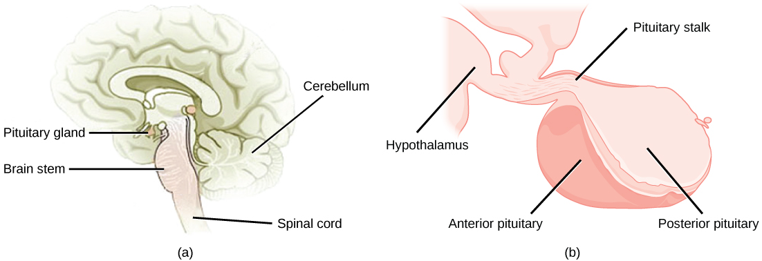
OpenStax, Rice University; Biology 2e. (credit a: modification of work by NCI; credit b: modification of work by Gray’s Anatomy) · CC BY 4.0 · Source出典

Figure 37.15図 37.15
The pituitary gland is located at (a) the base of the brain and (b) connected to the hypothalamus by the pituitary stalk. (credit a: modification of work by NCI; credit b: modification of work by Gray’s Anatomy)
下垂体は(a)脳底部にあり、(b)下垂体茎によって視床下部とつながる。(出典a:NCIの作品を改変、b:Gray’s Anatomyの作品を改変)
Anterior Pituitary下垂体前葉
The anterior pituitary gland, or adenohypophysis, is surrounded by a capillary network that extends from the hypothalamus, down along the infundibulum, and to the anterior pituitary. This capillary network is a part of the hypophyseal portal system that carries substances from the hypothalamus to the anterior pituitary and hormones from the anterior pituitary into the circulatory system. A portal system carries blood from one capillary network to another; therefore, the hypophyseal portal system allows hormones produced by the hypothalamus to be carried directly to the anterior pituitary without first entering the circulatory system.
英語のヒント
下垂体前葉、すなわち腺性下垂体は、視床下部から漏斗に沿って下行し、前葉へ続く毛細血管網に囲まれている。この網は下垂体門脈系の一部であり、視床下部から前葉へ物質を運び、前葉から循環系へホルモンを運ぶ。門脈系は、一つの毛細血管網から別の毛細血管網へ血液を送る。したがって下垂体門脈系では、視床下部のホルモンは最初に全身の循環系へ入ることなく、前葉へ直接運ばれる。
The anterior pituitary produces seven hormones: growth hormone (GH), prolactin (PRL), thyroid-stimulating hormone (TSH), melanin-stimulating hormone (MSH), adrenocorticotropic hormone (ACTH), follicle-stimulating hormone (FSH), and luteinizing hormone (LH). Anterior pituitary hormones are sometimes referred to as tropic hormones, because they control the functioning of other organs. While these hormones are produced by the anterior pituitary, their production is controlled by regulatory hormones produced by the hypothalamus. These regulatory hormones can be releasing hormones or inhibiting hormones, causing more or less of the anterior pituitary hormones to be secreted. These travel from the hypothalamus through the hypophyseal portal system to the anterior pituitary where they exert their effect. Negative feedback then regulates how much of these regulatory hormones are released and how much anterior pituitary hormone is secreted.
英語のヒント
下垂体前葉は七つのホルモンをつくる。成長ホルモン(GH)、プロラクチン(PRL)、甲状腺刺激ホルモン(TSH)、メラニン刺激ホルモン(MSH)、副腎皮質刺激ホルモン(ACTH)、卵胞刺激ホルモン(FSH)、黄体形成ホルモン(LH)である。他の器官の働きを制御するため、刺激ホルモンとも呼ばれる。これらをつくるのは下垂体前葉だが、その産生を制御するのは視床下部の調節ホルモンである。調節ホルモンには放出ホルモンと抑制ホルモンがあり、前葉から分泌されるホルモン量を増減させる。視床下部から下垂体門脈系を通って前葉へ運ばれ、そこで作用する。続いて負のフィードバックにより、これらの調節ホルモンがどれだけ放出され、下垂体前葉のホルモンがどれだけ分泌されるかが調節される。
Posterior Pituitary下垂体後葉
The posterior pituitary is significantly different in structure from the anterior pituitary. It is a part of the brain, extending down from the hypothalamus, and contains mostly nerve fibers and neuroglial cells, which support axons that extend from the hypothalamus to the posterior pituitary. The posterior pituitary and the infundibulum together are referred to as the neurohypophysis.
下垂体後葉の構造は、前葉と大きく異なる。視床下部から下方に伸びる脳の一部であり、主に神経線維とグリア細胞からなる。グリア細胞は、視床下部から後葉まで伸びる軸索を支える。後葉と漏斗を合わせて神経性下垂体と呼ぶ。
The hormones antidiuretic hormone (ADH), also known as vasopressin, and oxytocin are produced by neurons in the hypothalamus and transported within these axons along the infundibulum to the posterior pituitary. They are released into the circulatory system via neural signaling from the hypothalamus. These hormones are considered to be posterior pituitary hormones, even though they are produced by the hypothalamus, because that is where they are released into the circulatory system. The posterior pituitary itself does not produce hormones, but instead stores hormones produced by the hypothalamus and releases them into the bloodstream.
英語のヒント
バソプレシンとも呼ばれる抗利尿ホルモン(ADH)とオキシトシンは、視床下部のニューロンでつくられ、その軸索内を漏斗に沿って後葉へ運ばれる。視床下部からの神経信号によって、循環系へ放出される。これらが視床下部でつくられるにもかかわらず下垂体後葉ホルモンとされるのは、後葉から循環系へ放出されるからである。後葉自体はホルモンをつくらず、視床下部でつくられたホルモンを貯蔵し、血中へ放出する。