第37章内分泌系
Regulation of Metabolic Rates by Thyroid Hormones甲状腺ホルモンによる代謝率の調節
The basal metabolic rate, which is the amount of calories required by the body at rest, is determined by two hormones produced by the thyroid gland: thyroxine, also known as tetraiodothyronine or T4, and triiodothyronine, also known as T3. These hormones affect nearly every cell in the body except for the adult brain, uterus, testes, blood cells, and spleen. They are transported across the plasma membrane of target cells and bind to receptors on the mitochondria resulting in increased ATP production. In the nucleus, T3 and T4 activate genes involved in energy production and glucose oxidation. This results in increased rates of metabolism and body heat production, which is known as the hormone’s calorigenic effect.
英語のヒント
基礎代謝率は、安静時に体が必要とするカロリー量であり、甲状腺のつくる二つのホルモンによって決まる。チロキシン(テトラヨードチロニン、T4)と、トリヨードチロニン(T3)である。成人の脳、子宮、精巣、血球、脾臓を除き、ほぼすべての体細胞に作用する。標的細胞の細胞膜を越えて運ばれ、ミトコンドリアの受容体に結合してATP産生を増やす。核内では、T3 とT4 がエネルギー産生とグルコース酸化に関わる遺伝子を活性化する。その結果、代謝率と体熱の産生が増加する。これをホルモンの熱産生作用という。
T3 and T4 release from the thyroid gland is stimulated by thyroid-stimulating hormone (TSH), which is produced by the anterior pituitary. TSH binding at the receptors of the follicle of the thyroid triggers the production of T3 and T4 from a glycoprotein called thyroglobulin. Thyroglobulin is present in the follicles of the thyroid, and is converted into thyroid hormones with the addition of iodine. Iodine is formed from iodide ions that are actively transported into the thyroid follicle from the bloodstream. A peroxidase enzyme then attaches the iodine to the tyrosine amino acid found in thyroglobulin. T3 has three iodine ions attached, while T4 has four iodine ions attached. T3 and T4 are then released into the bloodstream, with T4 being released in much greater amounts than T3. As T3 is more active than T4 and is responsible for most of the effects of thyroid hormones, tissues of the body convert T4 to T3 by the removal of an iodine ion. Most of the released T3 and T4 becomes attached to transport proteins in the bloodstream and is unable to cross the plasma membrane of cells. These protein-bound molecules are only released when blood levels of the unattached hormone begin to decline. In this way, a week’s worth of reserve hormone is maintained in the blood. Increased T3 and T4 levels in the blood inhibit the release of TSH, which results in lower T3 and T4 release from the thyroid.
英語のヒント
甲状腺からのT3 とT4 の放出は、下垂体前葉のつくる甲状腺刺激ホルモン(TSH)によって促される。TSHが甲状腺濾胞の受容体に結合すると、サイログロブリンという糖タンパク質からT3とT4がつくられる。サイログロブリンは甲状腺濾胞内にあり、ヨウ素が加わると甲状腺ホルモンに変わる。血液から甲状腺濾胞へ能動輸送されたヨウ化物イオンから、ヨウ素がつくられる。次にペルオキシダーゼという酵素が、サイログロブリン中のアミノ酸チロシンにヨウ素を結合させる。T3には三つ、T4には四つのヨウ素イオンが結合している。T3とT4は血中へ放出され、T4の放出量はT3よりはるかに多い。T3はT4より活性が高く、甲状腺ホルモンの作用の大部分を担うため、体の組織はT4からヨウ素イオンを一つ除き、T3へ変える。放出されたT3 とT4 の大部分は、血中の輸送タンパク質に結合し、細胞膜を通れない。このタンパク質に結合した分子が遊離するのは、結合していないホルモンの血中濃度が下がり始めたときだけである。こうして血中には一週間分の予備のホルモンが保たれる。血中のT3 とT4 が増えると、TSH放出が抑えられ、その結果、甲状腺からのT3 とT4 の放出が減る。
The follicular cells of the thyroid require iodides (anions of iodine) in order to synthesize T3 and T4. Iodides obtained from the diet are actively transported into follicle cells resulting in a concentration that is approximately 30 times higher than in blood. The typical diet in North America provides more iodine than required due to the addition of iodide to table salt. Inadequate iodine intake, which occurs in many developing countries, results in an inability to synthesize T3 and T4 hormones. The thyroid gland enlarges in a condition called goiter, which is caused by overproduction of TSH without the formation of thyroid hormone. Thyroglobulin is contained in a fluid called colloid, and TSH stimulation results in higher levels of colloid accumulation in the thyroid. In the absence of iodine, this is not converted to thyroid hormone, and colloid begins to accumulate more and more in the thyroid gland, leading to goiter.
英語のヒント
甲状腺の濾胞細胞がT3 とT4を合成するには、ヨウ化物イオン(ヨウ素の陰イオン)が必要である。食事から得たヨウ化物イオンは、濾胞細胞へ能動輸送され、血中のおよそ30倍の濃度になる。北米の一般的な食事では、食塩へのヨウ化物添加により、必要量を超えるヨウ素が供給される。多くの発展途上国ではヨウ素摂取が不足し、T3 とT4 を合成できなくなる。甲状腺ホルモンが形成されないままTSHが過剰につくられると、甲状腺が大きくなる甲状腺腫が起こる。サイログロブリンはコロイドという液中に含まれ、TSHの刺激は甲状腺内へのコロイドの蓄積を増やす。ヨウ素がなければ、これは甲状腺ホルモンへ変わらず、コロイドが甲状腺にますます蓄積して甲状腺腫に至る。
Disorders can arise from both the underproduction and overproduction of thyroid hormones. Hypothyroidism, underproduction of the thyroid hormones, can cause a low metabolic rate leading to weight gain, sensitivity to cold, and reduced mental activity, among other symptoms. In children, hypothyroidism can cause cretinism, which can lead to intellectual disabilities and growth defects. Hyperthyroidism, the overproduction of thyroid hormones, can lead to an increased metabolic rate and its effects: weight loss, excess heat production, sweating, and an increased heart rate. Graves’ disease is one example of a hyperthyroid condition.
英語のヒント
甲状腺ホルモンの産生不足と過剰産生は、どちらも障害を起こしうる。産生不足である甲状腺機能低下症では、代謝率が低下し、体重増加、寒さへの敏感さ、精神活動の低下などが生じる。小児ではクレチン症を起こし、知的障害や成長障害につながることがある。過剰産生である甲状腺機能亢進症は代謝率を高め、体重減少、過剰な熱産生、発汗、心拍数増加を生じうる。バセドウ病は甲状腺機能亢進症の一例である。
Hormonal Control of Blood Calcium Levels血中カルシウム濃度のホルモン調節
Regulation of blood calcium concentrations is important for generation of muscle contractions and nerve impulses, which are electrically stimulated. If calcium levels get too high, membrane permeability to sodium decreases and membranes become less responsive. If calcium levels get too low, membrane permeability to sodium increases and convulsions or muscle spasms can result.
英語のヒント
血中カルシウム濃度の調節は、電気的刺激で起こる筋収縮と神経インパルスの発生に重要である。カルシウム濃度が高すぎると、膜のナトリウム透過性が下がり、膜が反応しにくくなる。低すぎるとナトリウム透過性が上がり、けいれんや筋攣縮が起こる場合がある。
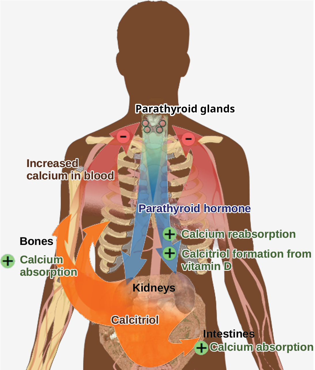
Blood calcium levels are regulated by parathyroid hormone (PTH), which is produced by the parathyroid glands, as illustrated in Figure 37.12. PTH is released in response to low blood Ca2+ levels. PTH increases Ca2+ levels by targeting the skeleton, the kidneys, and the intestine. In the skeleton, PTH stimulates osteoclasts, which causes bone to be reabsorbed, releasing Ca2+ from bone into the blood. PTH also inhibits osteoblasts, reducing Ca2+ deposition in bone. In the intestines, PTH increases dietary Ca2+ absorption, and in the kidneys, PTH stimulates reabsorption of the Ca2+. While PTH acts directly on the kidneys to increase Ca2+ reabsorption, its effects on the intestine are indirect. PTH triggers the formation of calcitriol, an active form of vitamin D, which acts on the intestines to increase absorption of dietary calcium. PTH release is inhibited by rising blood calcium levels.
英語のヒント
図37.12のように、血中カルシウム濃度を調節するのは、副甲状腺がつくる副甲状腺ホルモン(PTH)である。PTHは血中Ca2+の低下に応じて放出される。骨格、腎臓、腸を標的とし、Ca2+ 濃度を高める。骨格では破骨細胞を刺激し、骨を再吸収させて、Ca2+ を骨から血中へ放出させる。骨芽細胞も抑制し、骨へのCa2+ 沈着を減らす。腸では食事由来のCa2+の吸収を増やし、腎臓ではCa2+の再吸収を促す。PTHは腎臓には直接作用してCa2+再吸収を増やすが、腸への作用は間接的である。活性型ビタミンDであるカルシトリオールの形成を促し、それが腸に作用して食事由来のカルシウムの吸収を増やす。血中カルシウムが上昇すると、PTH放出は抑制される。
OpenStax, Rice University; Biology 2e. (credit: modification of work by Mikael Häggström) · CC BY 4.0 · Source出典

Figure 37.12図 37.12
Parathyroid hormone (PTH) is released in response to low blood calcium levels. It increases blood calcium levels by targeting the skeleton, the kidneys, and the intestine. (credit: modification of work by Mikael Häggström)
副甲状腺ホルモン(PTH)は、血中カルシウム濃度の低下に応じて放出される。骨格、腎臓、腸を標的として血中濃度を上げる。(出典:Mikael Häggströmの作品を改変)
Hyperparathyroidism results from an overproduction of parathyroid hormone. This results in excessive calcium being removed from bones and introduced into blood circulation, producing structural weakness of the bones, which can lead to deformation and fractures, plus nervous system impairment due to high blood calcium levels. Hypoparathyroidism, the underproduction of PTH, results in extremely low levels of blood calcium, which causes impaired muscle function and may result in tetany (severe sustained muscle contraction).
英語のヒント
副甲状腺機能亢進症は、副甲状腺ホルモンの過剰産生によって起こる。骨から過剰なカルシウムが取り除かれて血液循環へ入り、骨の構造が弱くなるため、変形や骨折につながりうる。血中カルシウムの増加による神経系の障害も生じる。PTHの産生不足である副甲状腺機能低下症では、血中カルシウムが著しく低下する。筋機能が損なわれ、テタニー(強く持続する筋収縮)を起こすことがある。
The hormone calcitonin, which is produced by the parafollicular or C cells of the thyroid, has the opposite effect on blood calcium levels as does PTH. Calcitonin decreases blood calcium levels by inhibiting osteoclasts, stimulating osteoblasts, and stimulating calcium excretion by the kidneys. This results in calcium being added to the bones to promote structural integrity. Calcitonin is most important in children (when it stimulates bone growth), during pregnancy (when it reduces bone loss), and during prolonged starvation (because it reduces bone mass loss). In healthy nonpregnant, unstarved adults, the role of calcitonin is unclear.
英語のヒント
甲状腺の傍濾胞細胞、すなわちC細胞がつくるカルシトニンは、血中カルシウムにPTHと逆の作用を及ぼす。破骨細胞を抑え、骨芽細胞と腎臓のカルシウム排泄を促して、血中カルシウムを下げる。その結果、骨にカルシウムが加わり、構造の強さが保たれる。カルシトニンが最も重要なのは、骨の成長を促す小児期、骨量の減少を抑える妊娠中、同じく骨量減少を抑える長期の飢餓時である。妊娠も飢餓もない健康な成人では、その役割は明らかでない。