第37章内分泌系
Thyroid Gland甲状腺
The thyroid gland is located in the neck, just below the larynx and in front of the trachea, as shown in Figure 37.16. It is a butterfly-shaped gland with two lobes that are connected by the isthmus. It has a dark red color due to its extensive vascular system. When the thyroid swells due to dysfunction, it can be felt under the skin of the neck.
英語のヒント
甲状腺は図37.16のように、首の喉頭のすぐ下、気管の前にある。峡部によって二つの葉がつながる、蝶の形をした腺である。血管が豊富なため暗赤色をしている。機能障害で腫大すると、首の皮膚の下に触れてわかる。
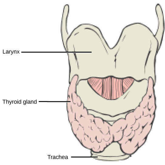
Figure 37.16図 37.16
This illustration shows the location of the thyroid gland.
甲状腺の位置を示す図。
The thyroid gland is made up of many spherical thyroid follicles, which are lined with a simple cuboidal epithelium. These follicles contain a viscous fluid, called colloid, which stores the glycoprotein thyroglobulin, the precursor to the thyroid hormones. The follicles produce hormones that can be stored in the colloid or released into the surrounding capillary network for transport to the rest of the body via the circulatory system.
英語のヒント
甲状腺は、単層立方上皮で内張りされた、多数の球状の甲状腺濾胞からなる。濾胞にはコロイドという粘性のある液があり、甲状腺ホルモンの前駆体である糖タンパク質サイログロブリンを蓄える。濾胞がつくるホルモンは、コロイドに貯蔵されるか、周囲の毛細血管網へ放出され、循環系を通して全身に運ばれる。
Thyroid follicle cells synthesize the hormone thyroxine, which is also known as T4 because it contains four atoms of iodine, and triiodothyronine, also known as T3 because it contains three atoms of iodine. Follicle cells are stimulated to release stored T3 and T4 by thyroid stimulating hormone (TSH), which is produced by the anterior pituitary. These thyroid hormones increase the rates of mitochondrial ATP production.
甲状腺の濾胞細胞は、ヨウ素原子を四つ含むためT4とも呼ばれるチロキシンと、三つ含むためT3とも呼ばれるトリヨードチロニンを合成する。下垂体前葉がつくる甲状腺刺激ホルモン(TSH)は、濾胞細胞を刺激して貯蔵されたT3 とT4 を放出させる。これらの甲状腺ホルモンはミトコンドリアのATP産生速度を高める。
A third hormone, calcitonin, is produced by parafollicular cells of the thyroid. Calcitonin release is not controlled by TSH, but instead is released when calcium ion concentrations in the blood rise. Calcitonin functions to help regulate calcium concentrations in body fluids. It acts in the bones to inhibit osteoclast activity and in the kidneys to stimulate excretion of calcium. The combination of these two events lowers body fluid levels of calcium.
第三のホルモンであるカルシトニンは、甲状腺の傍濾胞細胞がつくる。放出を制御するのはTSHではなく、血中カルシウムイオン濃度の上昇である。カルシトニンは、体液のカルシウム濃度調節を助ける。骨では破骨細胞の活動を抑え、腎臓ではカルシウム排泄を促す。この二つが合わさって、体液のカルシウム濃度が下がる。
Parathyroid Glands副甲状腺
Most people have four parathyroid glands; however, the number can vary from two to six. These glands are located on the posterior surface of the thyroid gland, as shown in Figure 37.17. Normally, there is a superior gland and an inferior gland associated with each of the thyroid’s two lobes. Each parathyroid gland is covered by connective tissue and contains many secretory cells that are associated with a capillary network.
多くの人には四つの副甲状腺があるが、数は二〜六個の範囲で変わる。図37.17のように、甲状腺の後面に位置する。通常は甲状腺の二つの葉それぞれに、上と下の副甲状腺が一つずつある。各副甲状腺は結合組織に包まれ、毛細血管網に接する多数の分泌細胞を含む。
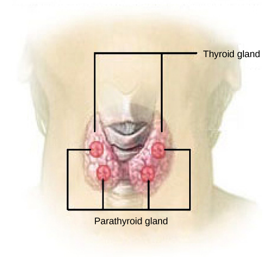
Figure 37.17図 37.17
The parathyroid glands are located on the posterior of the thyroid gland. (credit: modification of work by NCI)
副甲状腺は甲状腺の後面にある。(出典:NCIの作品を改変)
The parathyroid glands produce parathyroid hormone (PTH). PTH increases blood calcium concentrations when calcium ion levels fall below normal. PTH (1) enhances reabsorption of Ca2+ by the kidneys, (2) stimulates osteoclast activity and inhibits osteoblast activity, and (3) it stimulates synthesis and secretion of calcitriol by the kidneys, which enhances Ca2+ absorption by the digestive system. PTH is produced by chief cells of the parathyroid. PTH and calcitonin work in opposition to one another to maintain homeostatic Ca2+ levels in body fluids. Another type of cells, oxyphil cells, exist in the parathyroid but their function is not known.
英語のヒント
副甲状腺は副甲状腺ホルモン(PTH)をつくる。カルシウムイオンが正常値より低下すると、PTHは血中カルシウム濃度を上げる。(1)腎臓でのCa2+再吸収を増やし、(2)破骨細胞の活動を促すとともに骨芽細胞の活動を抑え、(3)腎臓にカルシトリオールを合成・分泌させ、消化器系でのCa2+吸収を増やす。PTHをつくるのは副甲状腺の主細胞である。PTHとカルシトニンは互いに逆向きに作用し、体液のCa2+を恒常的な水準に保つ。副甲状腺には好酸性細胞という別の細胞もあるが、その機能はわかっていない。
Adrenal Glands副腎
The adrenal glands are associated with the kidneys; one gland is located on top of each kidney as illustrated in Figure 37.18. The adrenal glands consist of an outer adrenal cortex and an inner adrenal medulla. These regions secrete different hormones.
副腎は腎臓に付随する腺で、図37.18のように左右の腎臓の上に一つずつ位置する。外側の副腎皮質と内側の副腎髄質からなり、それぞれ異なるホルモンを分泌する。
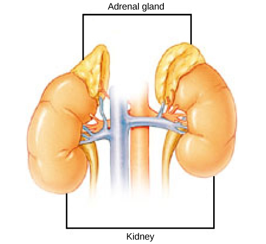


Figure 37.18図 37.18
The location of the adrenal glands on top of the kidneys is shown. (credit: modification of work by NCI)
腎臓の上に位置する副腎。(出典:NCIの作品を改変)
Adrenal Cortex副腎皮質
The adrenal cortex is made up of layers of epithelial cells and associated capillary networks. These layers form three distinct regions: an outer zona glomerulosa that produces mineralocorticoids, a middle zona fasciculata that produces glucocorticoids, and an inner zona reticularis that produces androgens.
英語のヒント
副腎皮質は、上皮細胞の層と、それに伴う毛細血管網からなる。三つの領域に分かれ、外側の球状帯は鉱質コルチコイド、中間の束状帯は糖質コルチコイド、内側の網状帯はアンドロゲンをつくる。
The main mineralocorticoid is aldosterone, which regulates the concentration of Na+ ions in urine, sweat, pancreas, and saliva. Aldosterone release from the adrenal cortex is stimulated by a decrease in blood concentrations of sodium ions, blood volume, or blood pressure, or by an increase in blood potassium levels.
主な鉱質コルチコイドはアルドステロンであり、尿、汗、膵臓、唾液のNa+濃度を調節する。血中ナトリウム濃度、血液量、血圧の低下、または血中カリウム濃度の上昇によって、副腎皮質からの放出が促される。
The three main glucocorticoids are cortisol, corticosterone, and cortisone. The glucocorticoids stimulate the synthesis of glucose and gluconeogenesis (converting a non-carbohydrate to glucose) by liver cells and they promote the release of fatty acids from adipose tissue. These hormones increase blood glucose levels to maintain levels within a normal range between meals. These hormones are secreted in response to ACTH and levels are regulated by negative feedback.
英語のヒント
主な糖質コルチコイドは、コルチゾール、コルチコステロン、コルチゾンの三つである。肝細胞でのグルコース合成と糖新生(糖質以外の物質をグルコースに変換すること)を促し、脂肪組織からの脂肪酸放出も促す。食間の血糖値を正常範囲に保つために血糖を上げる。ACTHに応じて分泌され、その濃度は負のフィードバックで調節される。
The adrenal cortex also produces small amounts of testosterone precursor, although the role of this additional hormone production is not fully understood. Testosterone is a type of androgen that promotes a suite of characteristics such as the growth and development of the testes and penis, increased skeletal and muscular growth, enlargement of the larynx, increased growth and redistribution of body hair, and increased sexual drive. Testosterone secretion is regulated by both the hypothalamus and the anterior pituitary gland. The hypothalamus sends releasing hormones that stimulate the release of gonadotropins from the anterior pituitary gland. Testosterone produced in small amounts in the adrenal cortex may work with sex hormones released from the gonads.
英語のヒント
副腎皮質は少量のテストステロン前駆体もつくるが、この付加的なホルモン産生の役割は十分には解明されていない。テストステロンはアンドロゲンの一種で、精巣と陰茎の成長・発達、骨格と筋肉の成長促進、喉頭の拡大、体毛の増加と分布変化、性欲の増大という一連の特徴を促す。分泌は視床下部と下垂体前葉の両方で調節される。視床下部は放出ホルモンを送り、前葉からの性腺刺激ホルモン放出を促す。副腎皮質で少量つくられるテストステロンは、性腺から放出される性ホルモンとともに働く可能性がある。