第4章細胞の構造
4.5 The Cytoskeleton4.5 細胞骨格
Learning Objectives学習目標
By the end of this section, you will be able to do the following:
この節を学ぶと、次のことができるようになる。
- Describe the cytoskeleton細胞骨格を説明する。
- Compare the roles of microfilaments, intermediate filaments, and microtubulesマイクロフィラメント、中間径フィラメント、微小管の役割を比較する。
- Compare and contrast cilia and flagella繊毛と鞭毛の共通点と相違点を比較する。
- Summarize the differences among the components of prokaryotic cells, animal cells, and plant cells原核細胞、動物細胞、植物細胞の構成要素の違いを要約する。
If you were to remove all the organelles from a cell, would the plasma membrane and the cytoplasm be the only components left? No. Within the cytoplasm, there would still be ions and organic molecules, plus a network of protein fibers that help maintain the cell's shape, secure some organelles in specific positions, allow cytoplasm and vesicles to move within the cell, and enable cells within multicellular organisms to move. Collectively, scientists call this network of protein fibers the cytoskeleton. There are three types of fibers within the cytoskeleton: microfilaments, intermediate filaments, and microtubules (Figure 4.22). Here, we will examine each.
英語のヒント
細胞からすべての細胞小器官を取り除いたら、残る構成要素は細胞膜と細胞質だけになるだろうか。そうではない。細胞質内にはイオンや有機分子に加え、タンパク質の繊維の網目が残る。この網目は、細胞の形を保ち、一部の細胞小器官を特定の位置に固定し、細胞質や小胞が細胞内を移動できるようにし、多細胞生物の細胞自体も移動できるようにする。このタンパク質の繊維の網目を、まとめて細胞骨格という。細胞骨格には、マイクロフィラメント、中間径フィラメント、微小管という3種類の繊維がある(図4.22)。ここでは、それぞれを見ていく。
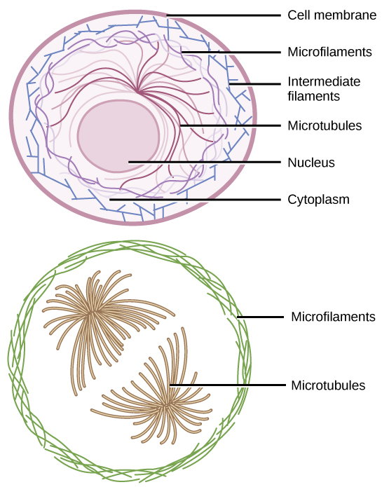
Figure 4.22図 4.22
Microfilaments thicken the cortex around the cell's inner edge. Like rubber bands, they resist tension. There are microtubules in the cell's interior where they maintain their shape by resisting compressive forces. There are intermediate filaments throughout the cell that hold organelles in place.
マイクロフィラメントは、細胞の内縁に沿った皮層を厚くする。ゴムバンドのように張力に抵抗する。細胞内部には微小管があり、圧縮力に抵抗して形を保つ。中間径フィラメントは細胞全体にあり、細胞小器官を所定の位置に保つ。
Microfilamentsマイクロフィラメント
Of the three types of protein fibers in the cytoskeleton, microfilaments are the narrowest. They function in cellular movement, have a diameter of about 7 nm, and are comprised of two globular protein intertwined strands, which we call actin (Figure 4.23). For this reason, we also call microfilaments actin filaments.
英語のヒント
細胞骨格を構成する3種類のタンパク質繊維のうち、マイクロフィラメントは最も細い。細胞運動に働き、直径は約7 nmである。アクチンと呼ばれる球状タンパク質からなる2本の鎖が、互いに絡み合ってできている(図4.23)。このため、マイクロフィラメントをアクチンフィラメントとも呼ぶ。
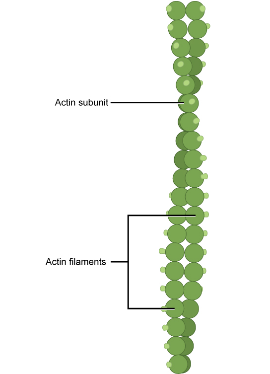
Figure 4.23図 4.23
Two intertwined actin strands comprise microfilaments.
絡み合った2本のアクチン鎖が、マイクロフィラメントを構成する。
ATP powers actin to assemble its filamentous form, which serves as a track for the movement of a motor protein we call myosin. This enables actin to engage in cellular events requiring motion, such as cell division in eukaryotic cells and cytoplasmic streaming, which is the cell cytoplasm's circular movement in plant cells. Actin and myosin are plentiful in muscle cells. When your actin and myosin filaments slide past each other, your muscles contract.
英語のヒント
ATPのエネルギーによって、アクチンはフィラメント状に組み立てられる。それは、ミオシンというモータータンパク質が動くための軌道となる。これにより、アクチンは真核細胞の細胞分裂や原形質流動など、動きを必要とする細胞内の出来事に関われる。原形質流動とは、植物細胞内で細胞質が循環する動きである。アクチンとミオシンは筋細胞に豊富にある。両者のフィラメントが互いに滑り合うと、筋肉が収縮する。
Microfilaments also provide some rigidity and shape to the cell. They can depolymerize (disassemble) and reform quickly, thus enabling a cell to change its shape and move. White blood cells (your body’s infection-fighting cells) make good use of this ability. They can move to an infection site and phagocytize the pathogen.
英語のヒント
マイクロフィラメントは細胞にある程度の剛性と形も与える。すばやく脱重合(分解)して再形成できるため、細胞は形を変えて移動できる。白血球、つまり体内で感染と闘う細胞は、この能力を有効に使う。感染部位へ移動し、病原体を貪食できる。
Intermediate Filaments中間径フィラメント
Several strands of fibrous proteins that are wound together comprise intermediate filaments (Figure 4.24). Cytoskeleton elements get their name from the fact that their diameter, 8 to 10 nm, is between those of microfilaments and microtubules.
英語のヒント
中間径フィラメントは、繊維状タンパク質の複数の鎖がより合わさってできている(図4.24)。その直径は8〜10 nmで、マイクロフィラメントと微小管の中間にあることから、この細胞骨格要素に名がつけられた。
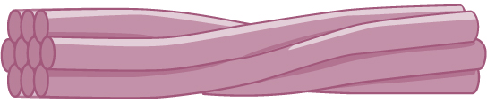
Figure 4.24図 4.24
Intermediate filaments consist of several intertwined strands of fibrous proteins.
中間径フィラメントは、繊維状タンパク質の複数の鎖が絡み合ってできている。
Intermediate filaments have no role in cell movement. Their function is purely structural. They bear tension, thus maintaining the cell's shape, and anchor the nucleus and other organelles in place. Figure 4.22 shows how intermediate filaments create a supportive scaffolding inside the cell.
英語のヒント
中間径フィラメントは、細胞運動には関与しない。その機能は専ら構造を支えることである。張力を受け止めて細胞の形を保ち、核やほかの細胞小器官を所定の位置につなぎ留める。図4.22は、中間径フィラメントが細胞内に支持用の足場を作る様子を示す。
The intermediate filaments are the most diverse group of cytoskeletal elements. Several fibrous protein types are in the intermediate filaments. You are probably most familiar with keratin, the fibrous protein that strengthens your hair, nails, and the skin's epidermis.
英語のヒント
中間径フィラメントは、細胞骨格要素の中で最も多様な集団であり、複数の種類の繊維状タンパク質を含む。最もよく知られているのは、おそらく毛髪、爪、皮膚の表皮を強くする繊維状タンパク質、ケラチンだろう。
Microtubules微小管
As their name implies, microtubules are small hollow tubes. Polymerized dimers of α-tubulin and β-tubulin, two globular proteins, comprise the microtubule's walls (Figure 4.25). With a diameter of about 25 nm, microtubules are cytoskeletons' widest components. They help the cell resist compression, provide a track along which vesicles move through the cell, and pull replicated chromosomes to opposite ends of a dividing cell. Like microfilaments, microtubules can disassemble and reform quickly.
英語のヒント
微小管は、その名が示すとおり、小さな中空の管である。二つの球状タンパク質、α-チューブリンとβ-チューブリンの二量体が重合し、微小管の壁を作っている(図4.25)。直径は約25 nmで、細胞骨格の構成要素の中で最も太い。微小管は、細胞が圧縮に抵抗するのを助け、小胞が細胞内を移動する軌道を提供し、複製された染色体を分裂中の細胞の両端へ引っ張る。マイクロフィラメントと同様に、微小管もすばやく分解・再形成できる。
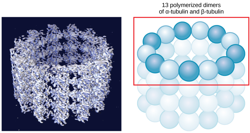


Figure 4.25図 4.25
Microtubules are hollow. Their walls consist of 13 polymerized dimers of α-tubulin and β-tubulin (right image). The left image shows the tube's molecular structure.
微小管は中空である。壁は、α-チューブリンとβ-チューブリンの重合した二量体13個からなる(右)。左の図は、管の分子構造を示す。
Microtubules are also the structural elements of flagella, cilia, and centrioles (the latter are the centrosome's two perpendicular bodies). In animal cells, the centrosome is the microtubule-organizing center. In eukaryotic cells, flagella and cilia are quite different structurally from their counterparts in prokaryotes, as we discuss below.
英語のヒント
微小管は、鞭毛、繊毛、中心小体の構造要素でもある。最後の中心小体とは、中心体を構成する互いに垂直な二つの構造である。動物細胞では、中心体が微小管形成中心となる。以下で述べるように、真核細胞の鞭毛と繊毛は、原核生物の対応する構造とは大きく異なる。