第4章細胞の構造
Electron Microscopes電子顕微鏡
In contrast to light microscopes, electron microscopes (Figure 4.2b) use a beam of electrons instead of a beam of light. Not only does this allow for higher magnification and, thus, more detail (Figure 4.3), it also provides higher resolving power. The method to prepare the specimen for viewing with an electron microscope kills the specimen. Electrons have short wavelengths (shorter than photons) that move best in a vacuum, so we cannot view living cells with an electron microscope.
英語のヒント
In a scanning electron microscope, a beam of electrons moves back and forth across a cell’s surface, creating details of cell surface characteristics. In a transmission electron microscope, the electron beam penetrates the cell and provides details of a cell’s internal structures. As you might imagine, electron microscopes are significantly more bulky and expensive than light microscopes.
英語のヒント
走査電子顕微鏡では、電子線が細胞表面を往復して走り、表面の特徴を詳しく示す。透過電子顕微鏡では、電子線が細胞を通り抜け、内部構造の細部を示す。想像できるように、電子顕微鏡は光学顕微鏡よりもかなり大きく、高価である。
OpenStax, Rice University; Biology 2e. (credit a: modification of work by CDC/Armed Forces Institute of Pathology, Charles N. Farmer, Rocky Mountain Laboratories; credit b: modification of work by NIAID, NIH; scale-bar data from Matt Russell) · CC BY 4.0 · Source出典

Figure 4.3図 4.3
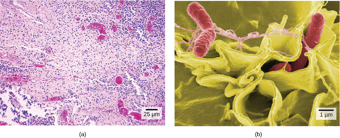
(a) These Salmonella bacteria appear as tiny purple dots when viewed with a light microscope. (b) This scanning electron microscope micrograph shows Salmonella bacteria (in red) invading human cells (yellow). Even though subfigure (b) shows a different Salmonella specimen than subfigure (a), you can still observe the comparative increase in magnification and detail. (credit a: modification of work by CDC/Armed Forces Institute of Pathology, Charles N. Farmer, Rocky Mountain Laboratories; credit b: modification of work by NIAID, NIH; scale-bar data from Matt Russell)
(a)光学顕微鏡で見ると、これらのサルモネラ菌は小さな紫色の点に見える。(b)走査電子顕微鏡写真では、サルモネラ菌(赤)がヒトの細胞(黄)に侵入している。(b)と(a)は別のサルモネラ菌標本だが、倍率と細部の見え方がどれほど増しているかを比べられる。(出典a:CDC/米軍病理学研究所、Charles N. Farmer、ロッキー・マウンテン研究所の作品を改変。出典b:NIAID、NIHの作品を改変。スケールバーのデータ:Matt Russell)
Cell Theory細胞説
The microscopes we use today are far more complex than those that Dutch shopkeeper Antony van Leeuwenhoek, used in the 1600s. Skilled in crafting lenses, van Leeuwenhoek observed the movements of single-celled organisms, which he collectively termed “animalcules.”
英語のヒント
現在の顕微鏡は、1600年代にオランダの商店主アントニ・ファン・レーウェンフックが使っていたものより、はるかに複雑である。レンズ作りに熟練していたレーウェンフックは、単細胞生物の運動を観察し、それらをまとめて「アニマルキュール(微小動物)」と名付けた。
In the 1665 publication Micrographia, experimental scientist Robert Hooke coined the term “cell” for the box-like structures he observed when viewing cork tissue through a lens. In the 1670s, van Leeuwenhoek discovered bacteria and protozoa. Later advances in lenses, microscope construction, and staining techniques enabled other scientists to see some components inside cells.
英語のヒント
実験科学者ロバート・フックは、1665年刊の『ミクログラフィア』で、レンズを通してコルク組織を観察した際に見えた箱状の構造を「cell(細胞)」と名付けた。1670年代には、レーウェンフックが細菌と原生動物を発見した。その後、レンズ、顕微鏡の製作、染色技術が進歩し、ほかの科学者たちも細胞内の一部の構成要素を見られるようになった。
By the late 1830s, botanist Matthias Schleiden and zoologist Theodor Schwann were studying tissues and proposed the unified cell theory, which states that one or more cells comprise all living things, the cell is the basic unit of life, and new cells arise from existing cells. Rudolf Virchow later made important contributions to this theory.
英語のヒント
1830年代後半までに、植物学者マティアス・シュライデンと動物学者テオドール・シュワンは組織を研究し、統一的な細胞説を提唱した。この説は、すべての生物が一つ以上の細胞からなり、細胞が生命の基本単位で、新しい細胞は既存の細胞から生じるとする。後にルドルフ・フィルヒョウも、この理論に重要な貢献をした。
Section Summaryこの節のまとめ
A cell is the smallest unit of life. Most cells are so tiny that we cannot see them with the naked eye. Therefore, scientists use microscopes to study cells. Electron microscopes provide higher magnification, higher resolution, and more detail than light microscopes. The unified cell theory states that one or more cells comprise all organisms, the cell is the basic unit of life, and new cells arise from existing cells.
細胞は生命の最小単位である。ほとんどの細胞は非常に小さく、肉眼では見えない。そのため、科学者は顕微鏡を使って細胞を研究する。電子顕微鏡は光学顕微鏡よりも倍率と分解能が高く、細部をより詳しく観察できる。統一的な細胞説では、すべての生物は1個以上の細胞からなり、細胞は生命の基本単位であり、新しい細胞は既存の細胞から生じるとされる。