第4章細胞の構造
Peroxisomesペルオキシソーム
Peroxisomes are small, round organelles enclosed by single membranes. They carry out oxidation reactions that break down fatty acids and amino acids. They also detoxify many poisons that may enter the body. (Many of these oxidation reactions release hydrogen peroxide, H2O2, which would be damaging to cells; however, when these reactions are confined to peroxisomes, enzymes safely break down the H2O2 into oxygen and water.) For example, peroxisomes in liver cells detoxify alcohol. Glyoxysomes, which are specialized peroxisomes in plants, are responsible for converting stored fats into sugars. Plant cells contain many different types of peroxisomes that play a role in metabolism, pathogene defense, and stress response, to mention a few.
英語のヒント
ペルオキシソームは、1枚の膜で囲まれた小さな球状の細胞小器官である。脂肪酸やアミノ酸を分解する酸化反応を行い、体内に入りうる多くの毒物を解毒する。これらの酸化反応の多くは、細胞に損傷を与えうる過酸化水素H2O2を放出する。しかし、反応がペルオキシソーム内に限定されていれば、酵素がH2O2を酸素と水に安全に分解する。例えば、肝細胞のペルオキシソームはアルコールを解毒する。植物の特殊なペルオキシソームであるグリオキシソームは、蓄えられた脂肪を糖に変える役割を担う。植物細胞には多様なペルオキシソームがあり、代謝、病原体に対する防御、ストレス応答などの役割を果たす。
Vesicles and Vacuoles小胞と液胞
Vesicles and vacuoles are membrane-bound sacs that function in storage and transport. Other than the fact that vacuoles are somewhat larger than vesicles, there is a very subtle distinction between them. Vesicle membranes can fuse with either the plasma membrane or other membrane systems within the cell. Additionally, some agents such as enzymes within plant vacuoles break down macromolecules. The vacuole's membrane does not fuse with the membranes of other cellular components.
英語のヒント
小胞と液胞は、貯蔵と輸送に働く、膜で囲まれた袋である。液胞のほうが小胞よりやや大きいことを除けば、両者の違いはごくわずかである。小胞の膜は、細胞膜とも、細胞内のほかの膜系とも融合できる。また、植物の液胞内にある酵素などの物質は、高分子を分解する。液胞の膜は、ほかの細胞構成要素の膜とは融合しない。
Animal Cells versus Plant Cells動物細胞と植物細胞の比較
At this point, you know that each eukaryotic cell has a plasma membrane, cytoplasm, a nucleus, ribosomes, mitochondria, peroxisomes, and in some, vacuoles, but there are some striking differences between animal and plant cells. While both animal and plant cells have microtubule organizing centers (MTOCs), animal cells also have centrioles associated with the MTOC: a complex we call the centrosome. Animal cells each have a centrosome and lysosomes; whereas, most plant cells do not. Plant cells have a cell wall, chloroplasts and other specialized plastids, and a large central vacuole; whereas, animal cells do not.
英語のヒント
ここまでで、真核細胞には細胞膜、細胞質、核、リボソーム、ミトコンドリア、ペルオキシソームがあり、一部には液胞もあることが分かった。しかし、動物細胞と植物細胞には、いくつか際立った違いがある。動物細胞にも植物細胞にも微小管形成中心(MTOC)があるが、動物細胞にはさらに、MTOCに付随する中心小体がある。この複合体を中心体という。動物細胞はそれぞれ中心体とリソソームをもつが、大部分の植物細胞にはない。植物細胞には細胞壁、葉緑体とそのほかの特殊化した色素体、大きな中央液胞があるが、動物細胞にはない。
The Centrosome中心体
The centrosome is a microtubule-organizing center found near the nuclei of animal cells. It contains a pair of centrioles, two structures that lie perpendicular to each other (Figure 4.15). Each centriole is a cylinder of nine triplets of microtubules.
英語のヒント
中心体は、動物細胞の核の近くにある微小管形成中心である。互いに直角に配置された二つの構造、1対の中心小体を含む(図4.15)。それぞれの中心小体は、3本組の微小管が9組集まった円筒である。
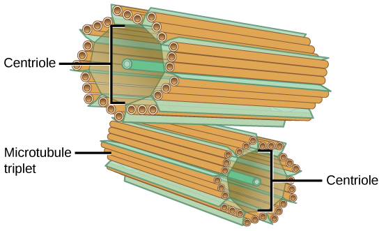
Figure 4.15図 4.15
The centrosome consists of two centrioles that lie at right angles to each other. Each centriole is a cylinder comprised of nine triplets of microtubules. Nontubulin proteins (indicated by the green lines) hold the microtubule triplets together.
中心体は、互いに直角に配置された二つの中心小体からなる。中心小体はそれぞれ、3本組の微小管が9組集まった円筒である。チューブリン以外のタンパク質(緑色の線で示す)が、3本組の微小管どうしをつなぎ留めている。
The centrosome (the organelle where all microtubules originate) replicates itself before a cell divides, and the centrioles appear to have some role in pulling the duplicated chromosomes to opposite ends of the dividing cell. However, the centriole's exact function in cell division isn’t clear, because cells that have had the centrosome removed can still divide, and plant cells, which lack centrosomes, are capable of cell division.
英語のヒント
中心体(すべての微小管が生じる細胞小器官)は、細胞が分裂する前に自身を複製する。中心小体は、複製された染色体を分裂中の細胞の両端へ引っ張る際に、何らかの役割を果たすようである。しかし、細胞分裂における中心小体の正確な機能は明らかでない。中心体を除去しても細胞は分裂でき、中心体をもたない植物細胞も細胞分裂が可能だからである。
Lysosomesリソソーム
Animal cells have another set of organelles that most plant cells do not: lysosomes. The lysosomes are the cell’s “garbage disposal.” In plant cells, the digestive processes take place in vacuoles. Enzymes within the lysosomes aid in breaking down proteins, polysaccharides, lipids, nucleic acids, and even worn-out organelles. These enzymes are active at a much lower pH than the cytoplasm's. Therefore, the pH within lysosomes is more acidic than the cytoplasm's pH. Many reactions that take place in the cytoplasm could not occur at a low pH, so again, the advantage of compartmentalizing the eukaryotic cell into organelles is apparent.
英語のヒント
動物細胞には、大部分の植物細胞にはない別の細胞小器官、リソソームもある。リソソームは細胞の「ごみ処理装置」である。植物細胞では、消化の過程は液胞で行われる。リソソーム内の酵素は、タンパク質、多糖、脂質、核酸、さらには使い古された細胞小器官の分解も助ける。これらの酵素は、細胞質よりもはるかに低いpHで活性を示す。そのため、リソソーム内のpHは細胞質よりも酸性である。細胞質で起こる多くの反応は、低いpHでは起こりえない。この点からも、真核細胞を細胞小器官に区画化する利点が分かる。
The Cell Wall細胞壁
If you examine Figure 4.8, the plant cell diagram, you will see a structure external to the plasma membrane. This is the cell wall, a rigid covering that protects the cell, provides structural support, and gives shape to the cell. Fungal and some protistan cells also have cell walls. While the prokaryotic cell walls' chief component is peptidoglycan, the major organic molecule in the plant (and some protists') cell wall is cellulose (Figure 4.16), a polysaccharide comprised of glucose units. Have you ever noticed that when you bite into a raw vegetable, like celery, it crunches? That’s because you are tearing the celery cells' rigid cell walls with your teeth.
英語のヒント
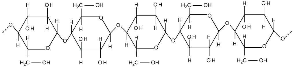


Figure 4.16図 4.16
Cellulose is a long chain of β-glucose molecules connected by a 1-4 linkage. The dashed lines at each end of the figure indicate a series of many more glucose units. The size of the page makes it impossible to portray an entire cellulose molecule.
セルロースは、β-グルコース分子が1-4結合でつながった長い鎖である。図の両端の破線は、さらに多数のグルコース単位が連続していることを示す。ページの大きさの都合上、セルロース分子の全体を描くことはできない。