第4章細胞の構造
4.1 Studying Cells4.1 細胞を調べる
Learning Objectives学習目標
By the end of this section, you will be able to do the following:
この節を学ぶと、次のことができるようになる。
- Describe the role of cells in organisms生物における細胞の役割を説明する。
- Compare and contrast light microscopy and electron microscopy光学顕微鏡法と電子顕微鏡法の共通点と相違点を比較する。
- Summarize cell theory細胞説を要約する。
A cell is the smallest unit of a living thing. Whether comprised of one cell (like bacteria) or many cells (like a human), we call it an organism. Thus, cells are the basic building blocks of all organisms.
英語のヒント
細胞は、生物の最小単位である。細菌のように一つの細胞からできていても、ヒトのように多くの細胞からできていても、それを生物と呼ぶ。したがって、細胞はすべての生物の基本的な構成単位である。
Several cells of one kind that interconnect with each other and perform a shared function form tissues. These tissues combine to form an organ (your stomach, heart, or brain), and several organs comprise an organ system (such as the digestive system, circulatory system, or nervous system). Several systems that function together form an organism (like a human being). Here, we will examine the structure and function of cells.
英語のヒント
同じ種類の細胞がいくつかつながり合い、共通の機能を果たして組織を作る。組織が組み合わさると、胃、心臓、脳などの器官となる。複数の器官は、消化器系、循環器系、神経系などの器官系を構成する。複数の系がともに働くことで、ヒトのような一つの生物が成り立つ。ここでは、細胞の構造と機能を調べる。
There are many types of cells, which scientists group into one of two broad categories: prokaryotic and eukaryotic. For example, we classify both animal and plant cells as eukaryotic cells; whereas, we classify bacterial cells as prokaryotic. Before discussing the criteria for determining whether a cell is prokaryotic or eukaryotic, we will first examine how biologists study cells.
英語のヒント
細胞には多くの種類があり、大きく原核細胞と真核細胞の二つに分類される。例えば、動物細胞と植物細胞はどちらも真核細胞であり、細菌の細胞は原核細胞である。細胞が原核か真核かを決める基準を論じる前に、まず生物学者がどのように細胞を研究するかを見ていこう。
Microscopy顕微鏡観察
Cells vary in size. With few exceptions, we cannot see individual cells with the naked eye, so scientists use microscopes (micro- = “small”; -scope = “to look at”) to study them. A microscope is an instrument that magnifies an object. We photograph most cells with a microscope, so we can call these images micrographs.
英語のヒント
細胞の大きさはさまざまである。少数の例外を除くと、個々の細胞を肉眼では見られない。そのため、科学者は顕微鏡で細胞を調べる(microscopeのmicro-は「小さい」、-scopeは「見る」の意味)。顕微鏡は物体を拡大する装置である。細胞の写真の多くは顕微鏡で撮影するので、顕微鏡写真と呼ぶことができる。
The optics of a microscope’s lenses change the image orientation that the user sees. A specimen that is right-side up and facing right on the microscope slide will appear upside-down and facing left when one views through a microscope, and vice versa. Similarly, if one moves the slide left while looking through the microscope, it will appear to move right, and if one moves it down, it will seem to move up. This occurs because microscopes use two sets of lenses to magnify the image. Because of the manner by which light travels through the lenses, this two lens system produces an inverted image (binocular, or dissecting microscopes, work in a similar manner, but include an additional magnification system that makes the final image appear to be upright).
英語のヒント
顕微鏡のレンズの光学的な性質によって、観察者に見える像の向きは変わる。スライドガラス上で上下が正しく、右を向いた標本は、顕微鏡を通して見ると上下が逆になり、左を向いて見える。逆の場合も同じである。同様に、顕微鏡をのぞきながらスライドを左へ動かすと像は右へ動き、下へ動かすと上へ動いて見える。これは顕微鏡が二組のレンズで像を拡大するためである。光がレンズを通る仕方によって、この二レンズ系は倒立像を作る。双眼顕微鏡や解剖顕微鏡も同じように働くが、さらに追加の拡大系があり、最終的な像は正立して見える。
Light Microscopes光学顕微鏡
To give you a sense of cell size, a typical human red blood cell is about eight millionths of a meter or eight micrometers (abbreviated as eight μm) in diameter. A pin head is about two thousandths of a meter (two mm) in diameter. That means about 250 red blood cells could fit on a pinhead.
英語のヒント
細胞の大きさを実感するために、典型的なヒトの赤血球を考えよう。直径は約100万分の8メートル、すなわち8マイクロメートル(8 μmと略記)である。ピンの頭の直径は約1000分の2メートル、すなわち2 mmである。したがって、ピンの頭には赤血球がおよそ250個並ぶことになる。
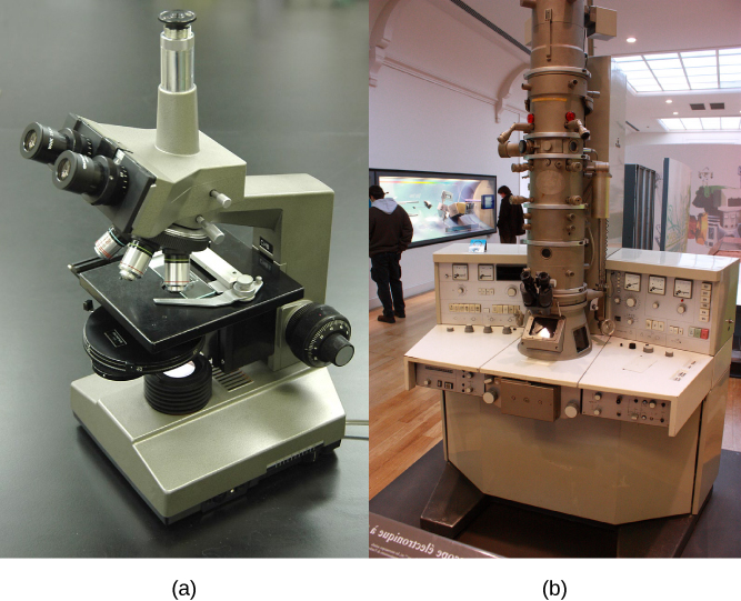
Most student microscopes are light microscopes (Figure 4.2a). Visible light passes and bends through the lens system to enable the user to see the specimen. Light microscopes are advantageous for viewing living organisms, but since individual cells are generally transparent, their components are not distinguishable unless they are colored with special stains. Staining, however, usually kills the cells.
英語のヒント
学生が使う顕微鏡の多くは光学顕微鏡である(図4.2a)。可視光がレンズ系を通過し屈折することで、標本が見えるようになる。光学顕微鏡は生きた生物を見るのに適している。しかし個々の細胞は一般に透明なので、特殊な染色剤で色を付けなければ構成部分を見分けられない。ただし、染色によって通常は細胞が死んでしまう。
Light microscopes that undergraduates commonly use in the laboratory magnify up to approximately 400 times. Two parameters that are important in microscopy are magnification and resolving power. Magnification is the process of enlarging an object in appearance. Resolving power is the microscope's ability to distinguish two adjacent structures as separate: the higher the resolution, the better the image's clarity and detail. When one uses oil immersion lenses to study small objects, magnification usually increases to 1,000 times. In order to gain a better understanding of cellular structure and function, scientists typically use electron microscopes.
英語のヒント
大学生が実験室で普通に使う光学顕微鏡は、約400倍まで拡大する。顕微鏡観察で重要な二つの指標は、倍率と分解能である。倍率は、物体を見かけ上どれだけ大きくするかを表す。分解能は、隣り合う二つの構造を別々のものとして識別する顕微鏡の能力である。分解能が高いほど、像は鮮明になり細部がよく分かる。小さな対象を油浸レンズで調べる場合、倍率は通常1000倍まで上がる。細胞の構造と機能をさらに詳しく理解するため、科学者は一般に電子顕微鏡を使う。
OpenStax, Rice University; Biology 2e. (credit a: modification of work by "GcG"/Wikimedia Commons; credit b: modification of work by Evan Bench) · CC BY 4.0 · Source出典

Figure 4.2図 4.2
(a) Most light microscopes in a college biology lab can magnify cells up to approximately 400 times and have a resolution of about 200 nanometers. (b) Electron microscopes provide a much higher magnification, 100,000x, and have a resolution of 50 picometers. (credit a: modification of work by "GcG"/Wikimedia Commons; credit b: modification of work by Evan Bench)
(a)大学の生物学実験室にある光学顕微鏡の多くは、細胞を約400倍まで拡大でき、分解能は約200ナノメートルである。(b)電子顕微鏡ははるかに高い10万倍の倍率と、50ピコメートルの分解能を持つ。(出典a:「GcG」/ウィキメディア・コモンズの作品を改変。出典b:Evan Benchの作品を改変)