第4章細胞の構造
The Nucleus核
Typically, the nucleus is the most prominent organelle in a cell (Figure 4.8). The nucleus (plural = nuclei) houses the cell’s DNA and directs the synthesis of ribosomes and proteins. Let’s look at it in more detail (Figure 4.11).
英語のヒント
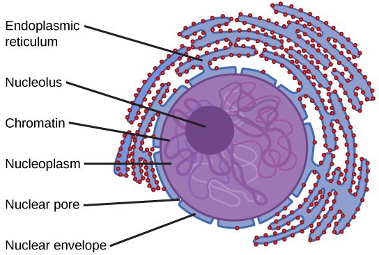
Figure 4.11図 4.11
The nucleus stores chromatin (DNA plus proteins) in a gel-like substance called the nucleoplasm. The nucleolus is a condensed chromatin region where ribosome synthesis occurs. We call the nucleus' boundary the nuclear envelope. It consists of two phospholipid bilayers: an outer and an inner membrane. The nuclear membrane is continuous with the endoplasmic reticulum. Nuclear pores allow substances to enter and exit the nucleus.
核は、核質と呼ばれるゲル状の物質の中にクロマチン(DNAとタンパク質)を保持する。核小体は、リボソームの合成が起こる、クロマチンが凝縮した領域である。核の境界を核膜という。核膜は、外膜と内膜という二つのリン脂質二重層からなる。核膜は小胞体とつながっている。核膜孔を通って、物質が核に出入りする。
The Nuclear Envelope核膜
The nuclear envelope is a double-membrane structure that constitutes the nucleus' outermost portion (Figure 4.11). Both the nuclear envelope's inner and outer membranes are phospholipid bilayers.
英語のヒント
核膜は、核の最も外側の部分を構成する二重の膜構造である(図4.11)。核膜の内膜と外膜は、どちらもリン脂質二重層である。
The nuclear envelope is punctuated with pores that control the passage of ions, molecules, and RNA between the nucleoplasm and cytoplasm. The nucleoplasm is the semi-solid fluid inside the nucleus, where we find the chromatin and the nucleolus.
英語のヒント
核膜には多数の孔があり、核質と細胞質の間を通過するイオン、分子、RNAを調節している。核質は核内の半固体状の液体で、その中にクロマチンと核小体が存在する。
Chromatin and Chromosomesクロマチンと染色体
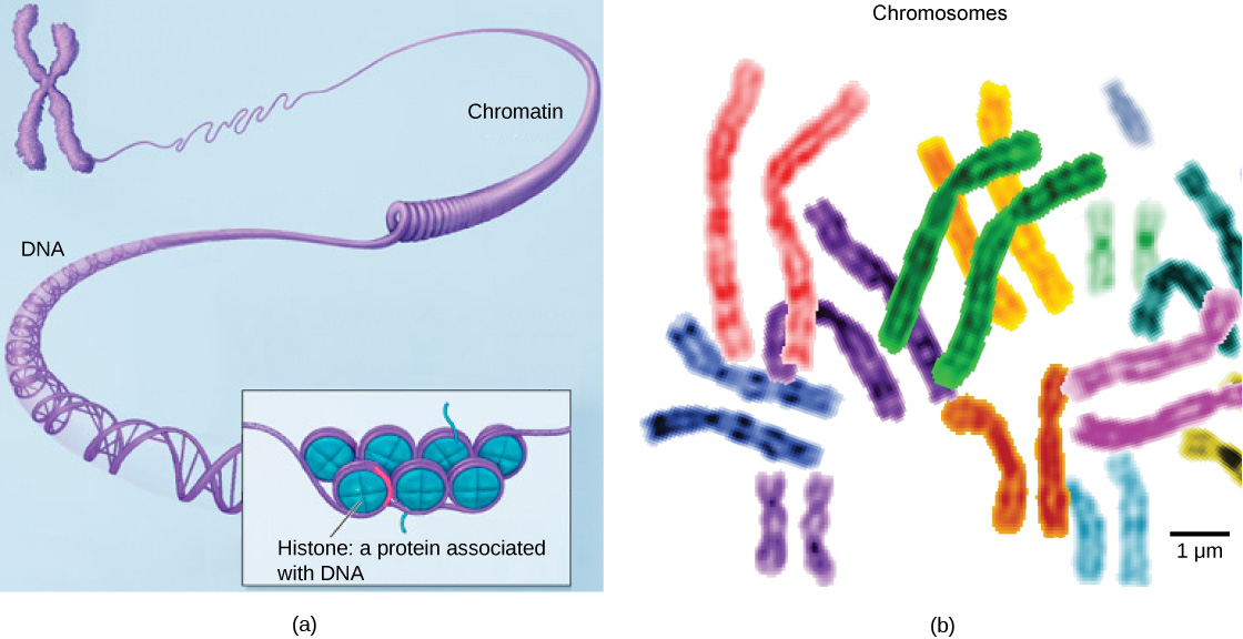
To understand chromatin, it is helpful to first explore chromosomes, structures within the nucleus that are made up of DNA, the hereditary material. You may remember that in prokaryotes, DNA is organized into a single circular chromosome. In eukaryotes, chromosomes are linear structures. Every eukaryotic species has a specific number of chromosomes in the nucleus of each cell. For example, in humans, the chromosome number is 46, while in fruit flies, it is eight. Chromosomes are only visible and distinguishable from one another when the cell is getting ready to divide. When the cell is in the growth and maintenance phases of its life cycle, proteins attach to chromosomes, and they resemble an unwound, jumbled bunch of threads. We call these unwound protein-chromosome complexes chromatin (Figure 4.12). Chromatin describes the material that makes up the chromosomes both when condensed and decondensed.
英語のヒント
クロマチンを理解するには、まず染色体を調べるとよい。染色体は核内にある構造で、遺伝物質であるDNAからできている。原核生物では、DNAが1本の環状染色体としてまとまっていることを思い出そう。真核生物では、染色体は線状である。真核生物では、種ごとに各細胞の核内の染色体数が決まっている。例えば、ヒトでは46本、ショウジョウバエでは8本である。染色体が見えるようになり、互いを区別できるのは、細胞が分裂の準備をしているときだけである。細胞が生活環の成長・維持の段階にあるときは、タンパク質が染色体に結合し、染色体はほどけて絡み合った糸の束のように見える。こうした、ほどけたタンパク質と染色体の複合体をクロマチンという(図4.12)。クロマチンとは、凝縮した状態でもほどけた状態でも、染色体を構成している物質を指す。
OpenStax, Rice University; Biology 2e. (credit b: modification of work by NIH; scale-bar data from Matt Russell) · CC BY 4.0 · Source出典
Figure 4.12図 4.12
(a) This image shows various levels of chromatin's organization (DNA and protein). (b) This image shows paired chromosomes. (credit b: modification of work by NIH; scale-bar data from Matt Russell)
(a)クロマチン(DNAとタンパク質)のさまざまな構成段階を示す。(b)対になった染色体を示す。(bのクレジット:NIHの作品を改変。スケールバーのデータ:Matt Russell)
The Nucleolus核小体
We already know that the nucleus directs the synthesis of ribosomes, but how does it do this? Some chromosomes have sections of DNA that encode ribosomal RNA. A darkly staining area within the nucleus called the nucleolus (plural = nucleoli) aggregates the ribosomal RNA with associated proteins to assemble the ribosomal subunits that are then transported out through the pores in the nuclear envelope to the cytoplasm.
英語のヒント
核がリボソーム合成を指揮することはすでに分かったが、どのように行うのだろうか。一部の染色体には、リボソームRNAをコードするDNAの領域がある。核小体(単数形nucleolus、複数形nucleoli)と呼ばれる核内の濃く染まる領域は、リボソームRNAを関連するタンパク質と集め、リボソームのサブユニットを組み立てる。そのサブユニットは、核膜の孔を通って細胞質へ運ばれる。
Ribosomesリボソーム
Ribosomes are the cellular structures responsible for protein synthesis. When we view them through an electron microscope, ribosomes appear either as clusters (polyribosomes) or single, tiny dots that float freely in the cytoplasm. They may be attached to the plasma membrane's cytoplasmic side or the endoplasmic reticulum's cytoplasmic side and the nuclear envelope's outer membrane (Figure 4.8). Electron microscopy shows us that ribosomes, which are large protein and RNA complexes, consist of two subunits, large and small (Figure 4.13). Ribosomes receive their “orders” for protein synthesis from the nucleus where the DNA transcribes into messenger RNA (mRNA). The mRNA travels to the ribosomes, which translate the code provided by the sequence of the nitrogenous bases in the mRNA into a specific order of amino acids in a protein. Amino acids are the building blocks of proteins.
英語のヒント
リボソームは、タンパク質合成を担う細胞構造である。電子顕微鏡で見ると、リボソームは集団(ポリリボソーム)として、あるいは細胞質中を自由に浮かぶ、小さな点として見える。細胞膜の細胞質側や、小胞体の細胞質側、核膜の外膜に付着していることもある(図4.8)。電子顕微鏡観察によって、リボソームはタンパク質とRNAの大きな複合体で、大と小の二つのサブユニットからなることが分かる(図4.13)。リボソームはタンパク質合成の「指令」を核から受け取る。核ではDNAがメッセンジャーRNA(mRNA)へと転写される。mRNAはリボソームに移動する。リボソームは、mRNA内の窒素を含む塩基の配列が与える暗号を、タンパク質中の特定のアミノ酸配列へと翻訳する。アミノ酸はタンパク質の構成要素である。
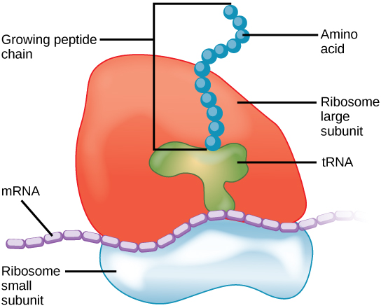


Figure 4.13図 4.13
A large subunit (top) and a small subunit (bottom) comprise ribosomes. During protein synthesis, ribosomes assemble amino acids into proteins.
大サブユニット(上)と小サブユニット(下)がリボソームを構成する。タンパク質合成の間、リボソームはアミノ酸を組み立ててタンパク質を作る。
Because protein synthesis is an essential function of all cells (including enzymes, hormones, antibodies, pigments, structural components, and surface receptors), there are ribosomes in practically every cell. Ribosomes are particularly abundant in cells that synthesize large amounts of protein. For example, the pancreas is responsible for creating several digestive enzymes and the cells that produce these enzymes contain many ribosomes. Thus, we see another example of form following function.
英語のヒント
タンパク質合成はすべての細胞に欠かせない機能であり、酵素、ホルモン、抗体、色素、構造の構成要素、表面の受容体などを作る。そのため、事実上すべての細胞にリボソームが存在する。特に、大量のタンパク質を合成する細胞にはリボソームが豊富にある。例えば、膵臓はいくつかの消化酵素を作る役割を担い、これらの酵素を作る細胞は多数のリボソームを含む。ここにも、形態が機能に従う例が見られる。
Mitochondriaミトコンドリア
Scientists often call mitochondria (singular = mitochondrion) “powerhouses” or “energy factories” of both plant and animal cells because they are responsible for making adenosine triphosphate (ATP), the cell’s main energy-carrying molecule. ATP represents the cell's short-term stored energy. Cellular respiration is the process of making ATP using the chemical energy in glucose and other nutrients. In mitochondria, this process uses oxygen and produces carbon dioxide as a waste product. In fact, the carbon dioxide that you exhale with every breath comes from the cellular reactions that produce carbon dioxide as a byproduct.
英語のヒント
ミトコンドリア(複数形mitochondria、単数形mitochondrion)は、細胞の主なエネルギー運搬分子であるアデノシン三リン酸(ATP)を作る役割を担うため、植物細胞でも動物細胞でも、しばしば「発電所」や「エネルギー工場」と呼ばれる。ATPは、細胞内に短期間蓄えられたエネルギーを表す。細胞呼吸は、グルコースなどの栄養素がもつ化学エネルギーを利用してATPを作る過程である。ミトコンドリアでは、この過程で酸素が使われ、老廃物として二酸化炭素が生じる。実際、呼吸のたびに吐き出す二酸化炭素は、副産物として二酸化炭素を生じる細胞内の反応に由来する。
In keeping with our theme of form following function, it is important to point out that muscle cells have a very high concentration of mitochondria that produce ATP. Your muscle cells need considerable energy to keep your body moving. When your cells don’t get enough oxygen, they do not make much ATP. Instead, producing lactic acid accompanies the small amount of ATP they make in the absence of oxygen.
英語のヒント
形態が機能に従うというテーマに沿って、筋細胞にはATPを作るミトコンドリアが非常に高い密度で存在することを指摘しておこう。筋細胞は、体を動かし続けるためにかなりのエネルギーを必要とする。細胞に十分な酸素が供給されないと、ATPはあまり作られない。酸素がないときに細胞が作る少量のATPには、乳酸の生成が伴う。
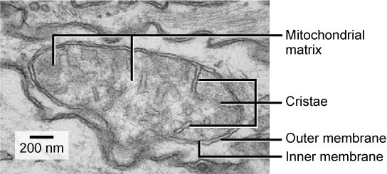
Mitochondria are oval-shaped, double membrane organelles (Figure 4.14) that have their own ribosomes and DNA. Each membrane is a phospholipid bilayer embedded with proteins. The inner layer has folds called cristae. We call the area surrounded by the folds the mitochondrial matrix. The cristae and the matrix have different roles in cellular respiration.
英語のヒント
ミトコンドリアは、独自のリボソームとDNAをもち、二重の膜に囲まれた楕円形の細胞小器官である(図4.14)。それぞれの膜は、タンパク質が埋め込まれたリン脂質二重層である。内側の膜には、クリステと呼ばれるひだがある。ひだに囲まれた領域をミトコンドリアマトリックスという。クリステとマトリックスは、細胞呼吸で異なる役割を果たす。
OpenStax, Rice University; Biology 2e. (credit: modification of work by Matthew Britton; scale-bar data from Matt Russell) · CC BY 4.0 · Source出典

Figure 4.14図 4.14
This electron micrograph shows a mitochondrion through an electron microscope. This organelle has an outer membrane and an inner membrane. The inner membrane contains folds, called cristae, which increase its surface area. We call the space between the two membranes the intermembrane space, and the space inside the inner membrane the mitochondrial matrix. ATP synthesis takes place on the inner membrane. (credit: modification of work by Matthew Britton; scale-bar data from Matt Russell)
電子顕微鏡で見たミトコンドリア。この細胞小器官には外膜と内膜がある。内膜にはクリステと呼ばれるひだがあり、表面積を増やしている。二つの膜の間を膜間腔、内膜の内側をミトコンドリアマトリックスという。ATP合成は内膜で行われる。(クレジット:Matthew Brittonの作品を改変。スケールバーのデータ:Matt Russell)