第4章細胞の構造
The Golgi Apparatusゴルジ体
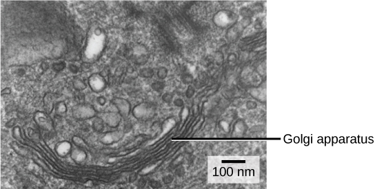
We have already mentioned that vesicles can bud from the ER and transport their contents elsewhere, but where do the vesicles go? Before reaching their final destination, the lipids or proteins within the transport vesicles still need sorting, packaging, and tagging so that they end up in the right place. Sorting, tagging, packaging, and distributing lipids and proteins takes place in the Golgi apparatus (also called the Golgi body), a series of flattened membranous sacs (Figure 4.20).
英語のヒント
小胞が小胞体から出芽し、内容物を別の場所へ運べることは、すでに述べた。では、小胞はどこへ行くのだろうか。輸送小胞内の脂質やタンパク質は、最終目的地に到着する前に、正しい場所へ行き着くよう、さらに仕分け、包装、目印づけを受ける必要がある。脂質とタンパク質の仕分け、目印づけ、包装、配送は、扁平な膜の袋が連なったゴルジ装置、別名ゴルジ体で行われる(図4.20)。
OpenStax, Rice University; Biology 2e. (credit: modification of work by Louisa Howard) · CC BY 4.0 · Source出典
Figure 4.20図 4.20
The Golgi apparatus in this white blood cell is visible as a stack of semicircular, flattened rings in the lower portion of the image. You can see several vesicles near the Golgi apparatus. (credit: modification of work by Louisa Howard)
この白血球のゴルジ体は、画像下部に、半円形で扁平な輪が積み重なったように見える。ゴルジ体の近くには、いくつかの小胞が見える。(クレジット:Louisa Howardの作品を改変)
The side of the Golgi apparatus that is closer to the ER is called the cis face. The opposite side is the trans face. The transport vesicles that formed from the ER travel to the cis face, fuse with it, and empty their contents into the Golgi apparatus' lumen. As the proteins and lipids travel through the Golgi, they undergo further modifications that allow them to be sorted. The most frequent modification is adding short sugar molecule chains. These newly modified proteins and lipids then tag with phosphate groups or other small molecules in order to travel to their proper destinations.
英語のヒント
ゴルジ体のうち、小胞体に近い側をシス面、反対側をトランス面という。小胞体から形成された輸送小胞はシス面へ移動して融合し、内容物をゴルジ体の内腔へ放出する。タンパク質や脂質は、ゴルジ体を通る間に、仕分けを可能にするさらなる修飾を受ける。最もよくある修飾は、短い糖分子の鎖を付け加えることである。新たに修飾されたタンパク質や脂質には、適切な目的地へ行くために、リン酸基などの小分子で目印がつけられる。
Finally, the modified and tagged proteins are packaged into secretory vesicles that bud from the Golgi's trans face. While some of these vesicles deposit their contents into other cell parts where they will be used, other secretory vesicles fuse with the plasma membrane and release their contents outside the cell.
英語のヒント
最後に、修飾され目印のついたタンパク質は、ゴルジ体のトランス面から出芽する分泌小胞に詰め込まれる。小胞の一部は、内容物をその使用場所である細胞内のほかの部分へ届ける。一方、別の分泌小胞は細胞膜と融合し、内容物を細胞外へ放出する。
In another example of form following function, cells that engage in a great deal of secretory activity (such as salivary gland cells that secrete digestive enzymes or immune system cells that secrete antibodies) have an abundance of Golgi.
英語のヒント
消化酵素を分泌する唾液腺細胞や、抗体を分泌する免疫系の細胞のように、盛んに分泌を行う細胞にはゴルジ体が豊富にある。これも、形態が機能に従う例である。
In plant cells, the Golgi apparatus has the additional role of synthesizing polysaccharides, some of which are incorporated into the cell wall and some of which other cell parts use.
英語のヒント
植物細胞では、ゴルジ体にはさらに多糖を合成する役割がある。合成された多糖の一部は細胞壁に組み込まれ、別の一部は細胞のほかの部分で使われる。
Lysosomesリソソーム
In addition to their role as the digestive component and organelle-recycling facility of animal cells, lysosomes are part of the endomembrane system. Lysosomes also use their hydrolytic enzymes to destroy pathogens (disease-causing organisms) that might enter the cell. A good example of this occurs in macrophages, a group of white blood cells which are part of your body’s immune system. In a process that scientists call phagocytosis or endocytosis, a section of the macrophage's plasma membrane invaginates (folds in) and engulfs a pathogen. The invaginated section, with the pathogen inside, then pinches itself off from the plasma membrane and becomes a vesicle. The vesicle fuses with a lysosome. The lysosome’s hydrolytic enzymes then destroy the pathogen (Figure 4.21).
英語のヒント
リソソームは、動物細胞の消化装置、細胞小器官の再利用施設として働くほか、内膜系の一部でもある。また、加水分解酵素を使い、細胞に侵入しうる病原体(病気を起こす生物)を破壊する。その良い例は、体の免疫系の一部を担う白血球の一群、マクロファージで見られる。食作用またはエンドサイトーシスと呼ばれる過程では、マクロファージの細胞膜の一部が陥入、つまり内側に折れ込み、病原体を包み込む。次に、病原体を内側に含んだ陥入部分が細胞膜からくびれ切れ、小胞になる。小胞はリソソームと融合し、リソソームの加水分解酵素が病原体を破壊する(図4.21)。
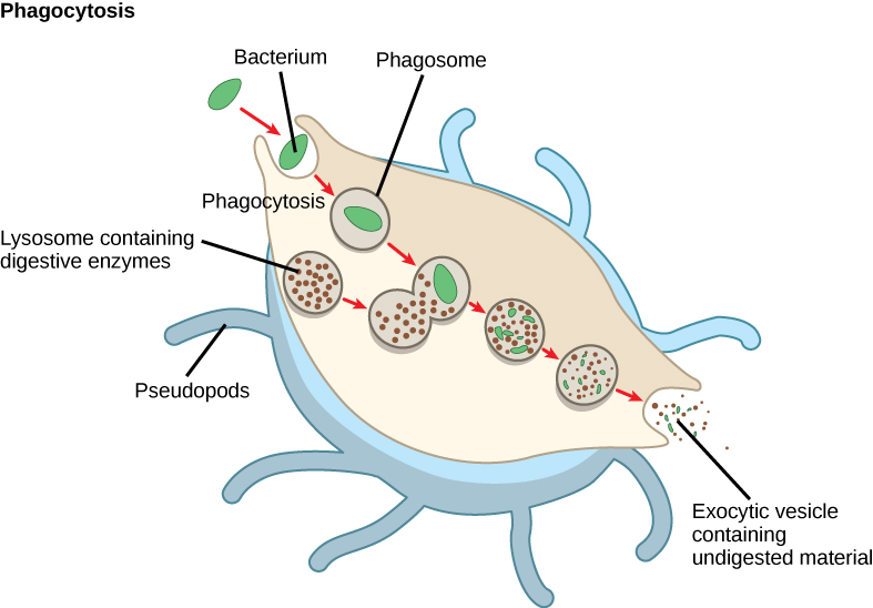


Figure 4.21図 4.21
A macrophage has engulfed (phagocytized) a potentially pathogenic bacterium and then fuses with lysosomes within the cell to destroy the pathogen. Other organelles are present in the cell but for simplicity we do not show them.
マクロファージが、病原性をもちうる細菌を包み込んだ(食作用を行った)後、細胞内のリソソームと融合させて病原体を破壊する。細胞内にはほかの細胞小器官もあるが、簡略化のため示していない。