第4章細胞の構造
4.2 Prokaryotic Cells4.2 原核細胞
Learning Objectives学習目標
By the end of this section, you will be able to do the following:
この節を学ぶと、次のことができるようになる。
- Name examples of prokaryotic and eukaryotic organisms原核生物と真核生物の例を挙げる。
- Compare and contrast prokaryotic and eukaryotic cells原核細胞と真核細胞の共通点と相違点を比較する。
- Describe the relative sizes of different cellsさまざまな細胞の相対的な大きさを説明する。
- Explain why cells must be small細胞が小さくなければならない理由を説明する。
Cells fall into one of two broad categories: prokaryotic and eukaryotic. We classify only the predominantly single-celled organisms Bacteria and Archaea as prokaryotes (pro- = “before”; -kary- = “nucleus”). Animal cells, plants, fungi, and protists are all eukaryotes (eu- = “true”).
英語のヒント
細胞は、原核細胞と真核細胞という二つの大きな分類のどちらかに属する。原核生物に分類されるのは、主に単細胞である細菌と古細菌だけである(pro-は「前」、-kary-は「核」を意味する)。動物の細胞、植物、菌類、原生生物はすべて真核生物である(eu-は「真の」を意味する)。
Components of Prokaryotic Cells原核細胞の構成要素
All cells share four common components: 1) a plasma membrane, an outer covering that separates the cell’s interior from its surrounding environment; 2) cytoplasm, consisting of a jelly-like cytosol within the cell in which there are other cellular components; 3) DNA, the cell's genetic material; and 4) ribosomes, which synthesize proteins. However, prokaryotes differ from eukaryotic cells in several ways.
英語のヒント
すべての細胞には、次の四つの構成要素が共通している。1)細胞膜。細胞内部を周囲の環境から隔てる外側の覆いである。2)細胞質。細胞内のゼリー状の細胞質基質からなり、その中にほかの細胞構成要素が存在する。3)DNA。細胞の遺伝物質である。4)リボソーム。タンパク質を合成する。しかし、原核生物の細胞と真核細胞にはいくつかの違いがある。
A prokaryote is a simple, mostly single-celled (unicellular) organism that lacks a nucleus, or any other membrane-bound organelle. We will shortly come to see that this is significantly different in eukaryotes. Prokaryotic DNA is in the cell's central part: the nucleoid (Figure 4.5).
英語のヒント
原核生物は、核も、そのほかの膜で囲まれた細胞小器官ももたない、単純で大部分が単細胞の生物である。まもなく見るように、この点は真核生物と大きく異なる。原核生物のDNAは、細胞の中央部にある核様体に存在する(図4.5)。
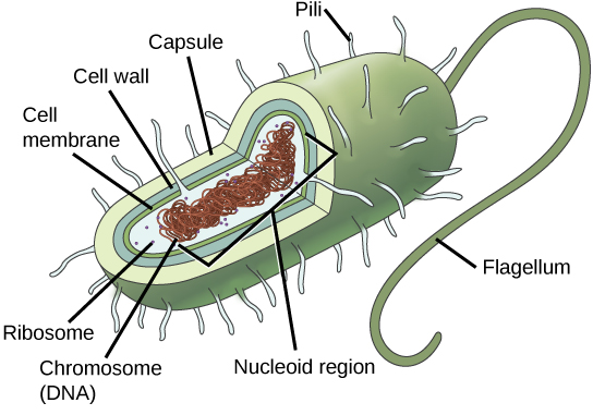
Figure 4.5図 4.5
This figure shows the generalized structure of a prokaryotic cell. All prokaryotes have chromosomal DNA localized in a nucleoid, ribosomes, a cell membrane, and a cell wall. The other structures shown are present in some, but not all, bacteria.
原核細胞の一般的な構造。すべての原核生物は、核様体に局在する染色体DNA、リボソーム、細胞膜、細胞壁をもつ。図に示したそのほかの構造は、一部の細菌には存在するが、すべての細菌にあるわけではない。
Most bacteria have a peptidoglycan cell wall and many have a polysaccharide capsule (Figure 4.5). The cell wall acts as an extra layer of protection, helps the cell maintain its shape, and prevents dehydration. The capsule enables the cell to attach to surfaces in its environment. Some prokaryotes have flagella, pili, or fimbriae. Flagella are used for locomotion. Pili exchange genetic material during conjugation, the process by which one bacterium transfers genetic material to another through direct contact. Bacteria use fimbriae to attach to a host cell.
英語のヒント
ほとんどの細菌はペプチドグリカンからなる細胞壁をもち、多くは多糖からなる莢膜ももつ(図4.5)。細胞壁は保護の層をさらに加え、細胞の形を保つのを助け、脱水を防ぐ。莢膜があると、細胞は周囲の環境にある表面に付着できる。一部の原核生物は、鞭毛、性線毛、または付着線毛をもつ。鞭毛は移動に使われる。性線毛は接合の際に遺伝物質をやり取りする。接合とは、直接接触することで、ある細菌が別の細菌に遺伝物質を移す過程である。細菌は付着線毛を使って宿主の細胞に付着する。
Cell Size細胞の大きさ
At 0.1 to 5.0 μm in diameter, prokaryotic cells are significantly smaller than eukaryotic cells, which have diameters ranging from 10 to 100 μm (Figure 4.6). The prokaryotes' small size allows ions and organic molecules that enter them to quickly diffuse to other parts of the cell. Similarly, any wastes produced within a prokaryotic cell can quickly diffuse. This is not the case in eukaryotic cells, which have developed different structural adaptations to enhance intracellular transport.
英語のヒント
原核細胞の直径は0.1〜5.0 μmであり、直径10〜100 μmの真核細胞よりもはるかに小さい(図4.6)。原核生物は小さいので、細胞内に入ったイオンや有機分子は、細胞のほかの部分へすばやく拡散できる。原核細胞の内部で生じた老廃物も、同じようにすばやく拡散できる。真核細胞ではそうはいかないため、細胞内の輸送を促進するさまざまな構造上の適応が発達している。
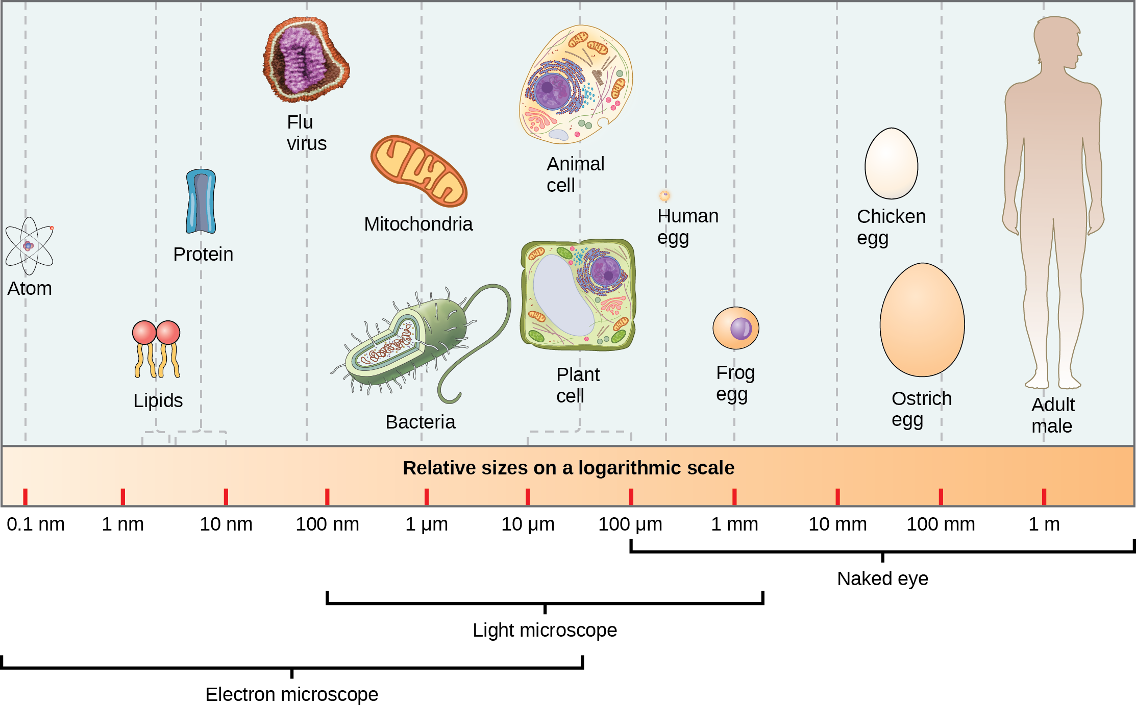


Figure 4.6図 4.6
This figure shows relative sizes of microbes on a logarithmic scale (recall that each unit of increase in a logarithmic scale represents a 10-fold increase in the quantity measured).
微生物の相対的な大きさを対数目盛で示している。対数目盛では目盛が一つ増えるごとに、測定している量が10倍になることを思い出そう。
Small size, in general, is necessary for all cells, whether prokaryotic or eukaryotic. Let’s examine why that is so. First, we’ll consider the area and volume of a typical cell. Not all cells are spherical in shape, but most tend to approximate a sphere. You may remember from your high school geometry course that the formula for the surface area of a sphere is 4πr2, while the formula for its volume is 4πr3/3. Thus, as the radius of a cell increases, its surface area increases as the square of its radius, but its volume increases as the cube of its radius (much more rapidly). Therefore, as a cell increases in size, its surface area-to-volume ratio decreases. This same principle would apply if the cell had a cube shape (Figure 4.7). If the cell grows too large, the plasma membrane will not have sufficient surface area to support the rate of diffusion required for the increased volume. In other words, as a cell grows, it becomes less efficient. One way to become more efficient is to divide. Other ways are to increase surface area by foldings of the cell membrane, become flat or thin and elongated, or develop organelles that perform specific tasks. These adaptations lead to developing more sophisticated cells, which we call eukaryotic cells.
英語のヒント
一般に、原核細胞でも真核細胞でも、細胞には小さいことが必要である。その理由を考えよう。まず、典型的な細胞の表面積と体積を考える。すべての細胞が球形とは限らないが、多くはおおむね球に近い形をしている。高校の幾何で学んだように、球の表面積の公式は4πr2、体積の公式は4πr3/3である。したがって、細胞の半径が大きくなると、表面積は半径の2乗に比例して増えるが、体積は半径の3乗に比例し、はるかに速く増える。そのため、細胞が大きくなるにつれて、体積に対する表面積の比は小さくなる。この原理は、細胞が立方体の形でも当てはまる(図4.7)。細胞が大きくなりすぎると、増えた体積に必要な拡散速度を支えるだけの表面積が細胞膜になくなる。言い換えれば、細胞は成長すると効率が低下する。効率を高める一つの方法は分裂することである。ほかには、細胞膜を折り畳んで表面積を増やす、平たくなる、細長くなる、特定の仕事をする細胞小器官を発達させる、といった方法がある。これらの適応によって、真核細胞と呼ばれる、より高度な細胞が発達する。