第38章筋骨格系
38.4 Muscle Contraction and Locomotion38.4 筋の収縮と移動
Learning Objectives学習目標
By the end of this section, you will be able to do the following:
この節を終えると、次のことができるようになる。
- Classify the different types of muscle tissueさまざまな種類の筋組織を分類する。
- Explain the role of muscles in locomotion移動における筋の役割を説明する。
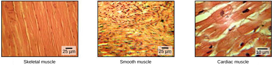
Muscle cells are specialized for contraction. Muscles allow for motions such as walking, and they also facilitate bodily processes such as respiration and digestion. The body contains three types of muscle tissue: skeletal muscle, cardiac muscle, and smooth muscle (Figure 38.33).
筋細胞は、収縮に特化している。筋は歩行などの運動を可能にし、呼吸や消化などの体内の過程も助ける。体には、骨格筋、心筋、平滑筋という3種類の筋組織がある(図38.33)。
OpenStax, Rice University; Biology 2e. (credit: modification of work by NCI, NIH; scale-bar data from Matt Russell) · CC BY 4.0 · Source出典
Figure 38.33図 38.33
The body contains three types of muscle tissue: skeletal muscle, smooth muscle, and cardiac muscle, visualized here using light microscopy. Skeletal muscle cells are long, cylindrical cells with multiple nuclei. Smooth muscle cells are short, tapered at each end, and have only one plump nucleus in each. Cardiac muscle cells are branched and striated, but short. The cytoplasm may branch, and they have one nucleus in the center of the cell. (credit: modification of work by NCI, NIH; scale-bar data from Matt Russell)
体には、骨格筋、平滑筋、心筋の3種類の筋組織があり、ここでは光学顕微鏡で示している。骨格筋細胞は、複数の核をもつ長い円柱状の細胞である。平滑筋細胞は短く、両端が先細りになっており、それぞれに1個のふっくらした核がある。心筋細胞は短く、枝分かれして横紋をもつ。細胞質は枝分かれすることがあり、細胞の中央に1個の核がある。(出典:NCI、NIHの作品を改変;スケールバーのデータ:Matt Russell)
Skeletal muscle tissue forms skeletal muscles, which attach to bones or skin and control locomotion and any movement that can be consciously controlled. Because it can be controlled by thought, skeletal muscle is also called voluntary muscle. Skeletal muscles are long and cylindrical in appearance; when viewed under a microscope, skeletal muscle tissue has a striped or striated appearance. The striations are caused by the regular arrangement of contractile proteins (actin and myosin). Actin is a globular contractile protein that interacts with myosin for muscle contraction. Skeletal muscle also has multiple nuclei present in a single cell.
英語のヒント
骨格筋組織は骨格筋を形成し、骨や皮膚に付着して、移動や、意識して制御できる運動を担う。意思によって制御できるため、骨格筋は随意筋とも呼ばれる。骨格筋は長い円柱状で、顕微鏡で見ると、骨格筋組織には縞模様、すなわち横紋が見られる。横紋は、収縮タンパク質であるアクチンとミオシンが規則正しく配列することによって生じる。アクチンは球状の収縮タンパク質で、筋収縮の際にミオシンと相互作用する。また、骨格筋は1つの細胞内に複数の核をもつ。
Smooth muscle tissue occurs in the walls of hollow organs such as the intestines, stomach, and urinary bladder, and around passages such as the respiratory tract and blood vessels. Smooth muscle has no striations, is not under voluntary control, has only one nucleus per cell, is tapered at both ends, and is called involuntary muscle.
英語のヒント
平滑筋組織は、腸、胃、膀胱などの中空器官の壁や、気道、血管などの通路の周囲にある。平滑筋には横紋がなく、意思による制御を受けない。1細胞あたりの核は1個で、両端が先細りになっている。不随意筋と呼ばれる。
Cardiac muscle tissue is only found in the heart, and cardiac contractions pump blood throughout the body and maintain blood pressure. Like skeletal muscle, cardiac muscle is striated, but unlike skeletal muscle, cardiac muscle cannot be consciously controlled and is called involuntary muscle. It has one nucleus per cell, is branched, and is distinguished by the presence of intercalated disks.
英語のヒント
心筋組織は心臓にだけ存在する。心筋の収縮は血液を全身へ送り出し、血圧を維持する。骨格筋と同じく心筋には横紋があるが、骨格筋とは異なり、意識して制御することはできず、不随意筋と呼ばれる。1細胞あたり1個の核をもち、枝分かれしており、介在板があることが特徴である。
Skeletal Muscle Fiber Structure骨格筋線維の構造
Each skeletal muscle fiber is a skeletal muscle cell. These cells are incredibly large, with diameters of up to 100 µm and lengths of up to 30 cm. The plasma membrane of a skeletal muscle fiber is called the sarcolemma. The sarcolemma is the site of action potential conduction, which triggers muscle contraction. Within each muscle fiber are myofibrils—long cylindrical structures that lie parallel to the muscle fiber. Myofibrils run the entire length of the muscle fiber, and because they are only approximately 1.2 µm in diameter, hundreds to thousands can be found inside one muscle fiber. They attach to the sarcolemma at their ends, so that as myofibrils shorten, the entire muscle cell contracts (Figure 38.34).
英語のヒント
1本の骨格筋線維は、1個の骨格筋細胞である。これらの細胞は非常に大きく、直径は最大100µm、長さは最大30cmに達する。骨格筋線維の細胞膜を筋鞘という。筋鞘は活動電位が伝導する場所であり、それが筋収縮を引き起こす。各筋線維の内部には、筋線維と平行に走る長い円柱状の構造である筋原線維がある。筋原線維は筋線維の全長にわたって伸び、直径はわずか約1.2µmなので、1本の筋線維の内部には数百本から数千本が存在する。筋原線維は両端で筋鞘に付着しているため、筋原線維が短くなると、筋細胞全体が収縮する(図38.34)。
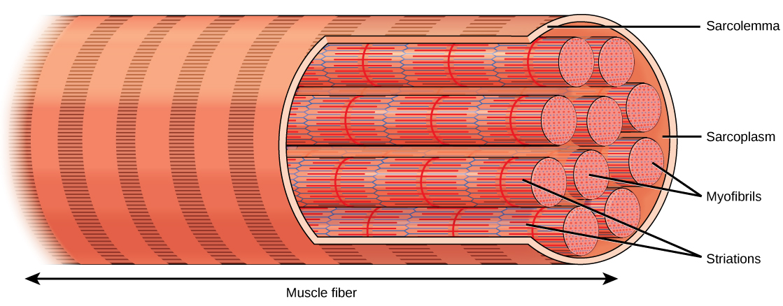
Figure 38.34図 38.34
A skeletal muscle cell is surrounded by a plasma membrane called the sarcolemma with a cytoplasm called the sarcoplasm. A muscle fiber is composed of many fibrils, packaged into orderly units.
骨格筋細胞は、筋鞘と呼ばれる細胞膜に囲まれ、筋形質という細胞質を含む。1本の筋線維は多数の筋原線維からなり、それらは規則正しい単位としてまとめられている。
The striated appearance of skeletal muscle tissue is a result of repeating bands of the proteins actin and myosin that are present along the length of myofibrils. Dark A bands and light I bands repeat along myofibrils, and the alignment of myofibrils in the cell causes the entire cell to appear striated or banded.
骨格筋組織の横紋は、筋原線維の長さ方向に沿って、アクチンとミオシンというタンパク質の帯が繰り返されることで生じる。暗いA帯と明るいI帯が筋原線維に沿って繰り返し並び、細胞内で筋原線維の位置がそろうことで、細胞全体に横紋、すなわち帯状の模様が現れる。
Each I band has a dense line running vertically through the middle called a Z disc or Z line. The Z discs mark the border of units called sarcomeres, which are the functional units of skeletal muscle. One sarcomere is the space between two consecutive Z discs and contains one entire A band and two halves of an I band, one on either side of the A band. A myofibril is composed of many sarcomeres running along its length, and as the sarcomeres individually contract, the myofibrils and muscle cells shorten (Figure 38.35).
英語のヒント
各I帯の中央を横切るように、Z板、またはZ線という密な線がある。Z板は、骨格筋の機能単位である筋節(サルコメア)の境界を示す。1つの筋節は、連続する2枚のZ板の間の区間であり、A帯全体を1本と、その両側にI帯の半分ずつを含む。1本の筋原線維は、長さ方向に連なった多数の筋節からなる。各筋節がそれぞれ収縮すると、筋原線維と筋細胞が短くなる(図38.35)。
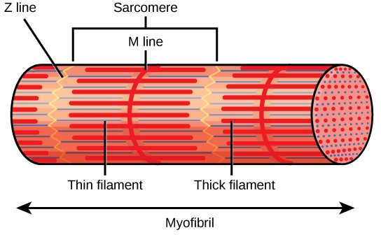


Figure 38.35図 38.35
A sarcomere is the region from one Z line to the next Z line. Many sarcomeres are present in a myofibril, resulting in the striation pattern characteristic of skeletal muscle.
筋節は、あるZ線から次のZ線までの領域である。1本の筋原線維には多数の筋節があり、それによって骨格筋に特有の横紋模様が生じる。
Myofibrils are composed of smaller structures called myofilaments. There are two main types of filaments: thick filaments and thin filaments; each has different compositions and locations. Thick filaments occur only in the A band of a myofibril. Thin filaments attach to a protein in the Z disc called alpha-actinin and occur across the entire length of the I band and partway into the A band. The region at which thick and thin filaments overlap has a dense appearance, as there is little space between the filaments. Thin filaments do not extend all the way into the A bands, leaving a central region of the A band that only contains thick filaments. This central region of the A band looks slightly lighter than the rest of the A band and is called the H zone. The middle of the H zone has a vertical line called the M line, at which accessory proteins hold together thick filaments. Both the Z disc and the M line hold myofilaments in place to maintain the structural arrangement and layering of the myofibril. Myofibrils are connected to each other by intermediate, or desmin, filaments that attach to the Z disc.
英語のヒント
筋原線維は、筋フィラメントという、さらに小さな構造からなる。フィラメントには、太いフィラメントと細いフィラメントの2種類があり、成分と位置が異なる。太いフィラメントは、筋原線維のA帯にだけ存在する。細いフィラメントは、Z板にあるαアクチニンというタンパク質に付着し、I帯の全長を通ってA帯の途中まで伸びる。太いフィラメントと細いフィラメントが重なる領域は、フィラメント間の隙間が少ないため、密に見える。細いフィラメントはA帯の奥まで伸びきらないので、A帯の中央には太いフィラメントだけの領域が残る。この部分はA帯のほかの部分よりわずかに明るく見え、H帯と呼ばれる。H帯の中央にはM線という縦の線があり、そこで補助タンパク質が太いフィラメントを束ねている。Z板とM線はいずれも筋フィラメントを所定の位置に保ち、筋原線維の構造的な配列と層の配置を維持する。筋原線維どうしは、Z板に付着する中間径フィラメント、すなわちデスミンフィラメントによってつながっている。
Thick and thin filaments are themselves composed of proteins. Thick filaments are composed of the protein myosin. The tail of a myosin molecule connects with other myosin molecules to form the central region of a thick filament near the M line, whereas the heads align on either side of the thick filament where the thin filaments overlap. The primary component of thin filaments is the actin protein. Two other components of the thin filament are tropomyosin and troponin. Actin has binding sites for myosin attachment. Strands of tropomyosin block the binding sites and prevent actin–myosin interactions when the muscles are at rest. Troponin consists of three globular subunits. One subunit binds to tropomyosin, one subunit binds to actin, and one subunit binds Ca2+ ions.
英語のヒント
太いフィラメントと細いフィラメント自体も、タンパク質からなる。太いフィラメントは、ミオシンというタンパク質でできている。ミオシン分子の尾部は、ほかのミオシン分子とつながり、M線の近くに太いフィラメントの中央部を形成する。一方、頭部は、細いフィラメントと重なる太いフィラメントの両側に並ぶ。細いフィラメントの主成分はアクチンである。ほかに、トロポミオシンとトロポニンという2つの成分がある。アクチンには、ミオシンが付着する結合部位がある。筋が静止しているときには、トロポミオシンのひも状の構造が結合部位をふさぎ、アクチンとミオシンの相互作用を防ぐ。トロポニンは、3つの球状サブユニットからなる。1つはトロポミオシンに、1つはアクチンに、1つはCa2+イオンに結合する。