第38章筋骨格系
The Skull頭蓋骨
The bones of the skull support the structures of the face and protect the brain. The skull consists of 22 bones, which are divided into two categories: cranial bones and facial bones. The cranial bones are eight bones that form the cranial cavity, which encloses the brain and serves as an attachment site for the muscles of the head and neck. The eight cranial bones are the frontal bone, two parietal bones, two temporal bones, occipital bone, sphenoid bone, and the ethmoid bone. Although the bones developed separately in the embryo and fetus, in the adult, they are tightly fused with connective tissue and adjoining bones do not move (Figure 38.6).
英語のヒント
頭蓋骨を構成する骨は、顔面の構造を支え、脳を保護する。頭蓋骨は22個の骨からなり、脳頭蓋の骨と顔面頭蓋の骨という2つの群に分けられる。脳頭蓋の骨は8個あり、頭蓋腔を形成する。頭蓋腔は脳を包み、頭や首の筋の付着部となる。8個の骨は、前頭骨、2個の頭頂骨、2個の側頭骨、後頭骨、蝶形骨、篩骨である。これらの骨は胚や胎児では別々に発達するが、成人では結合組織によって強くつながり、隣り合う骨は動かない(図38.6)。
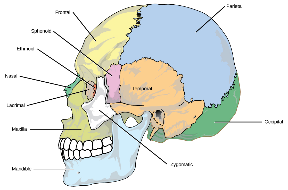
OpenStax, Rice University; Biology 2e. (credit: modification of work by Mariana Ruiz Villareal) · CC BY 4.0 · Source出典
Figure 38.6図 38.6
The bones of the skull support the structures of the face and protect the brain. (credit: modification of work by Mariana Ruiz Villareal)
頭蓋骨を構成する骨は、顔面の構造を支え、脳を保護する。(出典:Mariana Ruiz Villarealの作品を改変)
The auditory ossicles of the middle ear transmit sounds from the air as vibrations to the fluid-filled cochlea. The auditory ossicles consist of three bones each: the malleus, incus, and stapes. These are the smallest bones in the body and are unique to mammals.
英語のヒント
中耳の耳小骨は、空気中の音を振動として、液体で満たされた蝸牛へ伝える。耳小骨は左右それぞれ、ツチ骨、キヌタ骨、アブミ骨の3個からなる。これらは体内で最も小さい骨であり、哺乳類に特有である。
Fourteen facial bones form the face, provide cavities for the sense organs (eyes, mouth, and nose), protect the entrances to the digestive and respiratory tracts, and serve as attachment points for facial muscles. The 14 facial bones are the nasal bones, the maxillary bones, zygomatic bones, palatine, vomer, lacrimal bones, the inferior nasal conchae, and the mandible. All of these bones occur in pairs except for the mandible and the vomer (Figure 38.7).
英語のヒント
14個の顔面頭蓋の骨は、顔を形成し、感覚器(目・口・鼻)のための空所をつくり、消化管や気道の入口を保護し、顔面の筋の付着部となる。14個の骨は、鼻骨、上顎骨、頬骨、口蓋骨、鋤骨、涙骨、下鼻甲介、下顎骨である。下顎骨と鋤骨を除き、これらはすべて左右一対で存在する(図38.7)。
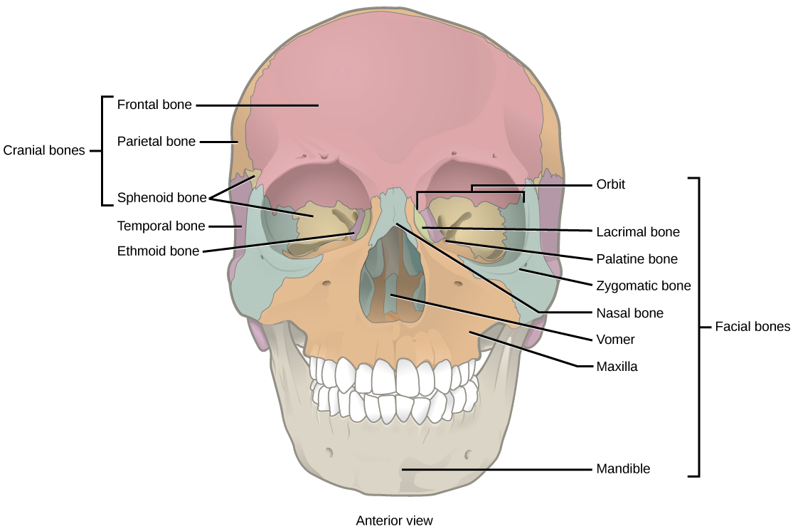


Figure 38.7図 38.7
The cranial bones, including the frontal, parietal, ethmoid, and sphenoid bones, cover the top of the head. The facial bones of the skull form the face and provide cavities for the eyes, nose, and mouth.
前頭骨、頭頂骨、篩骨、蝶形骨などの脳頭蓋の骨は、頭の上部を覆う。顔面頭蓋の骨は顔を形成し、目・鼻・口のための空所をつくる。
Although it is not found in the skull, the hyoid bone is considered a component of the axial skeleton. The hyoid bone lies below the mandible in the front of the neck. It acts as a movable base for the tongue and is connected to muscles of the jaw, larynx, and tongue. The mandible articulates with the base of the skull. The mandible controls the opening to the airway and gut. In animals with teeth, the mandible brings the surfaces of the teeth in contact with the maxillary teeth.
英語のヒント
舌骨は頭蓋骨に含まれないが、中軸骨格の構成要素とされる。舌骨は首の前側、下顎骨の下にある。舌の動く土台として働き、顎・喉頭・舌の筋につながっている。下顎骨は頭蓋底と関節を形成する。下顎骨は気道と消化管への入口を制御する。歯をもつ動物では、下顎骨によって下の歯の表面が上顎の歯と接触する。
The Vertebral Column脊柱
The vertebral column, or spinal column, surrounds and protects the spinal cord, supports the head, and acts as an attachment point for the ribs and muscles of the back and neck. The adult vertebral column comprises 26 bones: the 24 vertebrae, the sacrum, and the coccyx bones. In the adult, the sacrum is typically composed of five vertebrae that fuse into one. The coccyx is typically 3–4 vertebrae that fuse into one. Around the age of 70, the sacrum and the coccyx may fuse together. We begin life with approximately 33 vertebrae, but as we grow, several vertebrae fuse together. The adult vertebrae are further divided into the 7 cervical vertebrae, 12 thoracic vertebrae, and 5 lumbar vertebrae (Figure 38.8).
英語のヒント
脊柱は脊髄を囲んで保護し、頭を支え、肋骨や背中・首の筋の付着部となる。成人の脊柱は、24個の椎骨、仙骨、尾骨の計26個の骨からなる。成人の仙骨は、通常、5個の椎骨が融合して1つになったものである。尾骨は通常、3〜4個の椎骨が融合して1つになっている。70歳ごろには、仙骨と尾骨が互いに融合することもある。ヒトはおよそ33個の椎骨をもって生まれるが、成長につれて、いくつかの椎骨は互いに融合する。成人の椎骨はさらに、7個の頸椎、12個の胸椎、5個の腰椎に分けられる(図38.8)。
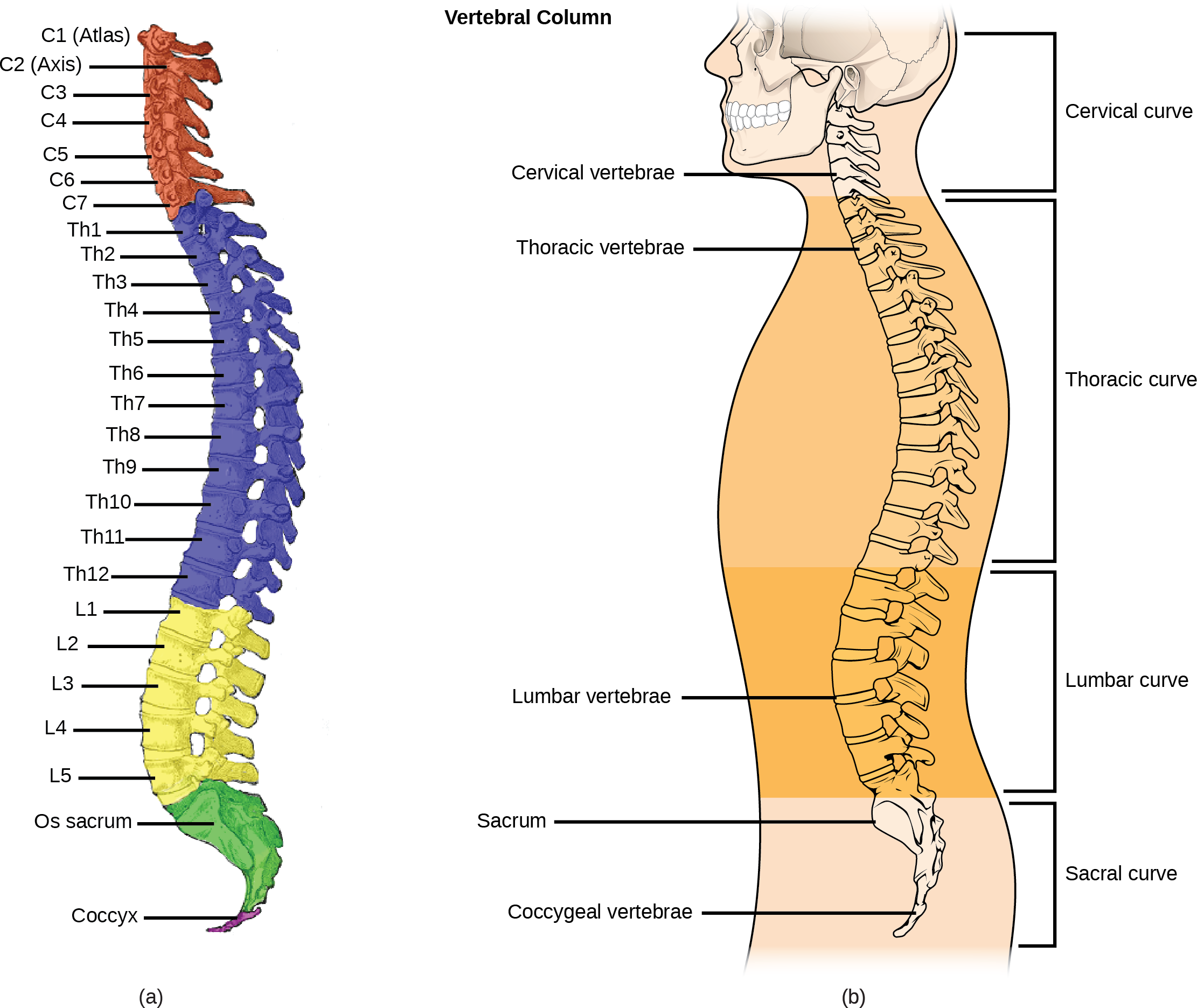
OpenStax, Rice University; Biology 2e. (credit a: modification of work by Uwe Gille based on original work by Gray's Anatomy; credit b: modification of work by NCI, NIH) · CC BY 4.0 · Source出典

Figure 38.8図 38.8
(a) The vertebral column consists of seven cervical vertebrae (C1–7) twelve thoracic vertebrae (Th1–12), five lumbar vertebrae (L1–5), the os sacrum, and the coccyx. (b) Spinal curves increase the strength and flexibility of the spine. (credit a: modification of work by Uwe Gille based on original work by Gray's Anatomy; credit b: modification of work by NCI, NIH)
(a)脊柱は、7個の頸椎(C1〜7)、12個の胸椎(Th1〜12)、5個の腰椎(L1〜5)、仙骨、尾骨からなる。(b)脊柱の弯曲は、脊柱の強度と柔軟性を高める。(出典a:Gray’s Anatomyの原図に基づくUwe Gilleの作品を改変;出典b:NCI、NIHの作品を改変)
Each vertebral body has a large hole in the center through which the nerves of the spinal cord pass. There is also a notch on each side through which the spinal nerves, which serve the body at that level, can exit from the spinal cord. The vertebral column is approximately 71 cm (28 inches) in adult male humans and is curved, which can be seen from a side view. The names of the spinal curves correspond to the region of the spine in which they occur. The thoracic and sacral curves are concave (curve inwards relative to the front of the body) and the cervical and lumbar curves are convex (curve outwards relative to the front of the body). The arched curvature of the vertebral column increases its strength and flexibility, allowing it to absorb shocks like a spring (Figure 38.8).
英語のヒント
各椎体の中央には大きな孔があり、そこを脊髄の神経が通る。両側には切痕もあり、その高さの体の領域に分布する脊髄神経が、そこを通って脊髄から出る。成人男性の脊柱は長さ約71cm(28インチ)で、横から見ると弯曲している。脊柱の弯曲には、その弯曲がある領域に対応した名前が付けられている。胸部と仙骨部の弯曲は凹型(体の前側に対して内向きに曲がる)であり、頸部と腰部の弯曲は凸型(体の前側に対して外向きに曲がる)である。脊柱の弓状の弯曲は、強度と柔軟性を高め、ばねのように衝撃を吸収できるようにする(図38.8)。
Intervertebral discs composed of fibrous cartilage lie between adjacent vertebral bodies from the second cervical vertebra to the sacrum. Each disc is part of a joint that allows for some movement of the spine and acts as a cushion to absorb shocks from movements such as walking and running. Intervertebral discs also act as ligaments to bind vertebrae together. The inner part of discs, the nucleus pulposus, hardens as people age and becomes less elastic. This loss of elasticity diminishes its ability to absorb shocks.
英語のヒント
第2頸椎から仙骨までの隣り合う椎体の間には、線維軟骨からなる椎間円板がある。各円板は、脊柱のある程度の動きを可能にする関節の一部であり、歩行や走行などの運動による衝撃を吸収するクッションとして働く。椎間円板はまた、椎骨どうしを結びつける靱帯としても働く。円板の内側にある髄核は、年齢とともに硬くなり、弾力性が低下する。この弾力性の低下によって、衝撃を吸収する能力が弱まる。