第38章筋骨格系
38.1 Types of Skeletal Systems38.1 骨格系の種類
Learning Objectives学習目標
By the end of this section, you will be able to do the following:
この節を終えると、次のことができるようになる。
- Discuss the different types of skeletal systemsさまざまな種類の骨格系を説明する。
- Explain the role of the human skeletal systemヒトの骨格系の役割を説明する。
- Compare and contrast different skeletal systems異なる骨格系の共通点と相違点を比較する。
A skeletal system is necessary to support the body, protect internal organs, and allow for the movement of an organism. There are three different skeleton designs that fulfill these functions: hydrostatic skeleton, exoskeleton, and endoskeleton.
骨格系は、体を支え、内臓を保護し、生物の運動を可能にするために必要である。これらの機能を果たす骨格には、静水骨格、外骨格、内骨格という3つの異なる形式がある。
Hydrostatic Skeleton静水骨格
A hydrostatic skeleton is a skeleton formed by a fluid-filled compartment within the body, called the coelom. The organs of the coelom are supported by the aqueous fluid, which also resists external compression. This compartment is under hydrostatic pressure because of the fluid and supports the other organs of the organism. This type of skeletal system is found in soft-bodied animals such as sea anemones, earthworms, Cnidaria, and other invertebrates (Figure 38.2).
英語のヒント
静水骨格は、体内にある、液体で満たされた区画によって形成される骨格である。この区画を体腔という。体腔内の器官は水性の液体によって支えられ、この液体は外部からの圧迫にも抵抗する。この区画には液体による静水圧がかかり、生物のほかの器官を支えている。このような骨格系は、イソギンチャク、ミミズ、刺胞動物、そのほかの無脊椎動物など、柔らかい体をもつ動物に見られる(図38.2)。
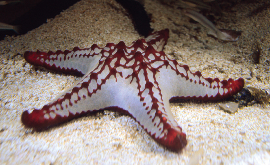
Figure 38.2図 38.2
The skeleton of the red-knobbed sea star (Protoreaster linckii) is an example of a hydrostatic skeleton. (credit: “Amada44”/Wikimedia Commons)
赤いこぶをもつヒトデ(Protoreaster linckii)の骨格は、静水骨格の一例である。(出典:“Amada44”/Wikimedia Commons)
Movement in a hydrostatic skeleton is provided by muscles that surround the coelom. The muscles in a hydrostatic skeleton contract to change the shape of the coelom; the pressure of the fluid in the coelom produces movement. For example, earthworms move by waves of muscular contractions of the skeletal muscle of the body wall hydrostatic skeleton, called peristalsis, which alternately shorten and lengthen the body. Lengthening the body extends the anterior end of the organism. Most organisms have a mechanism to fix themselves in the substrate. Shortening the muscles then draws the posterior portion of the body forward. Although a hydrostatic skeleton is well-suited to invertebrate organisms such as earthworms and some aquatic organisms, it is not an efficient skeleton for terrestrial animals.
英語のヒント
静水骨格での運動は、体腔を囲む筋によって生じる。静水骨格の筋が収縮すると体腔の形が変わり、体腔内の液体の圧力によって動きが生じる。たとえばミミズは、体壁の静水骨格にある骨格筋の収縮の波によって移動する。この波は蠕動と呼ばれ、体を交互に短くしたり長くしたりする。体を長くすると、生物の前端が伸びる。多くの生物は、基質に自分の体を固定する仕組みをもっている。続いて筋を短くすると、体の後部が前方へ引き寄せられる。静水骨格は、ミミズや一部の水生生物などの無脊椎動物には適しているが、陸生動物にとって効率的な骨格ではない。
Exoskeleton外骨格
An exoskeleton is an external skeleton that consists of a hard encasement on the surface of an organism. For example, the shells of crabs and insects are exoskeletons (Figure 38.3). This skeleton type provides defence against predators, supports the body, and allows for movement through the contraction of attached muscles. As with vertebrates, muscles must cross a joint inside the exoskeleton. Shortening of the muscle changes the relationship of the two segments of the exoskeleton. Arthropods such as crabs and lobsters have exoskeletons that consist of 30–50 percent chitin, a polysaccharide derivative of glucose that is a strong but flexible material. Chitin is secreted by the epidermal cells. The exoskeleton is further strengthened by the addition of calcium carbonate in organisms such as the lobster. Because the exoskeleton is acellular, arthropods must periodically shed their exoskeletons because the exoskeleton does not grow as the organism grows.
英語のヒント
外骨格は、生物の表面を覆う硬い殻からなる、体の外側の骨格である。たとえば、カニや昆虫の殻は外骨格である(図38.3)。この種類の骨格は、捕食者から身を守り、体を支え、付着した筋の収縮による運動を可能にする。脊椎動物の場合と同じく、筋は外骨格の内側で関節をまたがなければならない。筋が短くなると、外骨格の2つの節の位置関係が変わる。カニやロブスターなどの節足動物の外骨格は、30〜50%がキチンでできている。キチンは、グルコースから誘導される多糖で、強度と柔軟性を兼ね備えた材料である。キチンは表皮細胞から分泌される。ロブスターなどでは、炭酸カルシウムが加わることで外骨格がさらに強化される。外骨格には細胞がなく、生物の成長に合わせて成長しない。そのため、節足動物は定期的に外骨格を脱ぎ捨てなければならない。
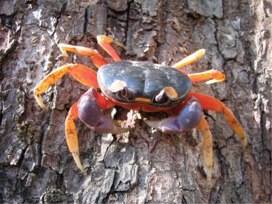
Figure 38.3図 38.3
Muscles attached to the exoskeleton of the Halloween crab (Gecarcinus quadratus) allow it to move.
ハロウィンクラブ(Gecarcinus quadratus)は、外骨格に付着する筋によって動くことができる。
Endoskeleton内骨格
An endoskeleton is a skeleton that consists of hard, mineralized structures located within the soft tissue of organisms. An example of a primitive endoskeletal structure is the spicules of sponges. The bones of vertebrates are composed of tissues, whereas sponges have no true tissues (Figure 38.4). Endoskeletons provide support for the body, protect internal organs, and allow for movement through contraction of muscles attached to the skeleton.
英語のヒント
内骨格は、生物の軟らかい組織の中にある、硬く無機質の沈着した構造からなる骨格である。原始的な内骨格構造の例として、海綿の骨片がある。脊椎動物の骨は組織でできているが、海綿には真の組織はない(図38.4)。内骨格は体を支え、内臓を保護し、骨格に付着した筋の収縮による運動を可能にする。
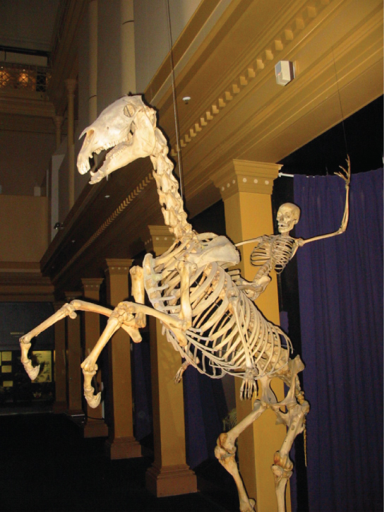


Figure 38.4図 38.4
The skeletons of humans and horses are examples of endoskeletons. (credit: Ross Murphy)
ヒトとウマの骨格は、内骨格の例である。(出典:Ross Murphy)
The human skeleton is an endoskeleton that consists of 206 bones in the adult. It has five main functions: providing support to the body, storing minerals and lipids, producing blood cells, protecting internal organs, and allowing for movement. The skeletal system in vertebrates is divided into the axial skeleton (which consists of the skull, vertebral column, and rib cage), and the appendicular skeleton (which consists of the shoulders, limb bones, the pectoral girdle, and the pelvic girdle).
英語のヒント
ヒトの骨格は内骨格であり、成人では206個の骨からなる。主な機能は5つある。体を支えること、無機質と脂質を蓄えること、血球をつくること、内臓を保護すること、そして運動を可能にすることである。脊椎動物の骨格系は、頭蓋骨・脊柱・胸郭からなる中軸骨格と、肩・四肢の骨・肩帯・骨盤帯からなる付属肢骨格に分けられる。
Human Axial Skeletonヒトの中軸骨格
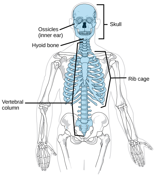
The axial skeleton forms the central axis of the body and includes the bones of the skull, ossicles of the middle ear, hyoid bone of the throat, vertebral column, and the thoracic cage (ribcage) (Figure 38.5). The function of the axial skeleton is to provide support and protection for the brain, the spinal cord, and the organs in the ventral body cavity. It provides a surface for the attachment of muscles that move the head, neck, and trunk, performs respiratory movements, and stabilizes parts of the appendicular skeleton.
中軸骨格は体の中心軸を形成し、頭蓋骨、中耳の耳小骨、喉にある舌骨、脊柱、胸郭を含む(図38.5)。中軸骨格は、脳、脊髄、腹側体腔内の器官を支え、保護する。また、頭・首・体幹を動かす筋の付着面となり、呼吸運動を行い、付属肢骨格の各部を安定させる。
OpenStax, Rice University; Biology 2e. (credit: modification of work by Mariana Ruiz Villareal) · CC BY 4.0 · Source出典

Figure 38.5図 38.5
The axial skeleton consists of the bones of the skull, ossicles of the middle ear, hyoid bone, vertebral column, and rib cage. (credit: modification of work by Mariana Ruiz Villareal)
中軸骨格は、頭蓋骨、中耳の耳小骨、舌骨、脊柱、胸郭からなる。(出典:Mariana Ruiz Villarealの作品を改変)