第38章筋骨格系
Sliding Filament Model of Contraction筋収縮の滑走フィラメントモデル
For a muscle cell to contract, the sarcomere must shorten. However, thick and thin filaments—the components of sarcomeres—do not shorten. Instead, they slide by one another, causing the sarcomere to shorten while the filaments remain the same length. The sliding filament theory of muscle contraction was developed to fit the differences observed in the named bands on the sarcomere at different degrees of muscle contraction and relaxation. The mechanism of contraction is the binding of myosin to actin, forming cross-bridges that generate filament movement (Figure 38.36).
英語のヒント
筋細胞が収縮するには、筋節が短くならなければならない。しかし、筋節の構成要素である太いフィラメントと細いフィラメント自体は短くならない。互いに滑り合うことで、フィラメントの長さを変えずに筋節が短くなる。筋収縮の滑走フィラメント説は、筋の収縮や弛緩の程度が変わったときに、筋節内の各帯で観察される違いを説明するために考えられた。収縮の仕組みは、ミオシンがアクチンに結合してクロスブリッジを形成し、それがフィラメントの運動を生み出すことである(図38.36)。
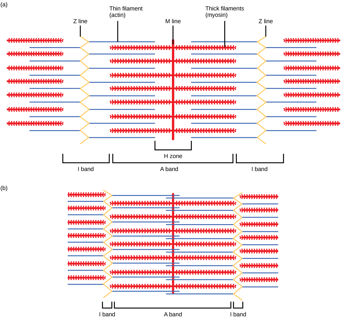

Figure 38.36図 38.36
When (a) a sarcomere (b) contracts, the Z lines move closer together and the I band gets smaller. The A band stays the same width and, at full contraction, the thin filaments overlap.
(a)筋節が(b)収縮すると、Z線どうしが近づき、I帯が小さくなる。A帯の幅は変わらず、完全に収縮すると、細いフィラメントどうしが重なる。
When a sarcomere shortens, some regions shorten whereas others stay the same length. A sarcomere is defined as the distance between two consecutive Z discs or Z lines; when a muscle contracts, the distance between the Z discs is reduced. The H zone—the central region of the A zone—contains only thick filaments and is shortened during contraction. The I band contains only thin filaments and also shortens. The A band does not shorten—it remains the same length—but A bands of different sarcomeres move closer together during contraction, eventually disappearing. Thin filaments are pulled by the thick filaments toward the center of the sarcomere until the Z discs approach the thick filaments. The zone of overlap, in which thin filaments and thick filaments occupy the same area, increases as the thin filaments move inward.
筋節が短くなるとき、短くなる領域もあれば、長さが変わらない領域もある。筋節は、連続する2枚のZ板、すなわちZ線の間の距離として定義される。筋が収縮すると、Z板間の距離が短くなる。A帯の中央部にあたるH帯は、太いフィラメントだけを含み、収縮時には短くなる。I帯は細いフィラメントだけを含み、これも短くなる。A帯自体は短くならず、同じ長さを保つが、異なる筋節のA帯どうしは収縮時に近づき、最終的には消失する。細いフィラメントは、Z板が太いフィラメントに近づくまで、太いフィラメントによって筋節の中心へ引かれる。細いフィラメントが内側へ動くにつれて、細いフィラメントと太いフィラメントが同じ領域を占める重なりの部分が増える。
ATP and Muscle ContractionATPと筋収縮
The motion of muscle shortening occurs as myosin heads bind to actin and pull the actin inwards. This action requires energy, which is provided by ATP. Myosin binds to actin at a binding site on the globular actin protein. Myosin has another binding site for ATP at which enzymatic activity hydrolyzes ATP to ADP, releasing an inorganic phosphate molecule and energy.
英語のヒント
ミオシン頭部がアクチンに結合し、それを内側へ引くことで、筋が短くなる。この動きにはエネルギーが必要であり、ATPがそれを供給する。ミオシンは、球状のアクチンタンパク質にある結合部位でアクチンと結合する。ミオシンにはATPのための別の結合部位もあり、そこで酵素活性によってATPがADPへ加水分解され、無機リン酸1分子とエネルギーが放出される。
ATP binding causes myosin to release actin, allowing actin and myosin to detach from each other. After this happens, the newly bound ATP is converted to ADP and inorganic phosphate, Pi. The enzyme at the binding site on myosin is called ATPase. The energy released during ATP hydrolysis changes the angle of the myosin head into a “cocked” position. The myosin head is then in a position for further movement, possessing potential energy, but ADP and Pi are still attached. If actin binding sites are covered and unavailable, the myosin will remain in the high energy configuration with ATP hydrolyzed, but still attached.
英語のヒント
ATPが結合すると、ミオシンはアクチンを放し、両者が離れる。その後、新しく結合したATPはADPと無機リン酸Piに変換される。ミオシンのこの結合部位にある酵素をATPアーゼという。ATPの加水分解で放出されたエネルギーは、ミオシン頭部の角度を変え、引き金を引く準備をしたような「起き上がった」位置にする。頭部は次の動きを行える位置になり、位置エネルギーを蓄えるが、ADPとPiはまだ付着している。アクチンの結合部位が覆われていて利用できない場合、ミオシンは、ATPが加水分解されたものの、まだ結合している高エネルギー状態の配置にとどまる。
If the actin binding sites are uncovered, a cross-bridge will form; that is, the myosin head spans the distance between the actin and myosin molecules. Pi is then released, allowing myosin to expend the stored energy as a conformational change. The myosin head moves toward the M line, pulling the actin along with it. As the actin is pulled, the filaments move approximately 10 nm toward the M line. This movement is called the power stroke, as it is the step at which force is produced. As the actin is pulled toward the M line, the sarcomere shortens and the muscle contracts.
英語のヒント
アクチンの結合部位が露出していれば、クロスブリッジが形成される。つまり、ミオシン頭部がアクチン分子とミオシン分子の間を橋渡しする。その後、Piが放出され、ミオシンは蓄えたエネルギーを立体構造の変化として使えるようになる。ミオシン頭部はM線へ向かって動き、アクチンを一緒に引く。アクチンが引かれると、フィラメントはM線に向かって約10nm動く。この動きをパワーストロークという。力が生み出される段階だからである。アクチンがM線へ引かれるにつれて、筋節が短くなり、筋が収縮する。
When the myosin head is “cocked,” it contains energy and is in a high-energy configuration. This energy is expended as the myosin head moves through the power stroke; at the end of the power stroke, the myosin head is in a low-energy position. After the power stroke, ADP is released; however, the cross-bridge formed is still in place, and actin and myosin are bound together. ATP can then attach to myosin, which allows the cross-bridge cycle to start again and further muscle contraction can occur (Figure 38.37).
ミオシン頭部が「起き上がった」状態では、エネルギーを蓄えた高エネルギーの配置にある。このエネルギーは、頭部がパワーストロークを行う際に使われ、パワーストロークの終わりには頭部は低エネルギーの位置になる。パワーストロークの後、ADPは放出されるが、形成されたクロスブリッジは残り、アクチンとミオシンは結合したままである。そこへATPがミオシンに結合すると、クロスブリッジのサイクルが再び始まり、筋はさらに収縮できる(図38.37)。