第38章筋骨格系
The Pelvic Girdle骨盤帯
The pelvic girdle bones provide the points of attachment of the lower limbs to the axial skeleton. Because it is responsible for bearing the weight of the body and for locomotion, the pelvic girdle is securely attached to the axial skeleton by strong ligaments. It also has deep sockets with robust ligaments to securely attach the femur to the body. The pelvic girdle is further strengthened by two large hip bones. In adults, the hip bones, or coxal bones, are formed by the fusion of three pairs of bones: the ilium, ischium, and pubis. The pelvis joins together in the anterior of the body at a joint called the pubic symphysis and with the bones of the sacrum at the posterior of the body.
英語のヒント
骨盤帯の骨は、下肢を中軸骨格につなぐ付着部となる。体重を支え、移動を担うため、骨盤帯は強い靱帯によって中軸骨格にしっかりと固定されている。また、強固な靱帯を伴う深いくぼみがあり、大腿骨を体にしっかりとつないでいる。骨盤帯は、2つの大きな寛骨によってさらに強化される。成人の寛骨は、腸骨、坐骨、恥骨という3対の骨が融合してできる。骨盤は体の前側で恥骨結合という関節によってつながり、後ろ側では仙骨とつながる。
The female pelvis is slightly different from the male pelvis. Over generations of evolution, females with a wider pubic angle and larger diameter pelvic canal reproduced more successfully. Therefore, their offspring also had pelvic anatomy that enabled successful childbirth (Figure 38.13).
英語のヒント
女性の骨盤は男性の骨盤とわずかに異なる。何世代にもわたる進化の過程で、恥骨下角が広く、骨盤腔の直径が大きい女性は、よりよく子孫を残した。その結果、その子孫も、出産を成功させやすい骨盤の構造をもつようになった(図38.13)。
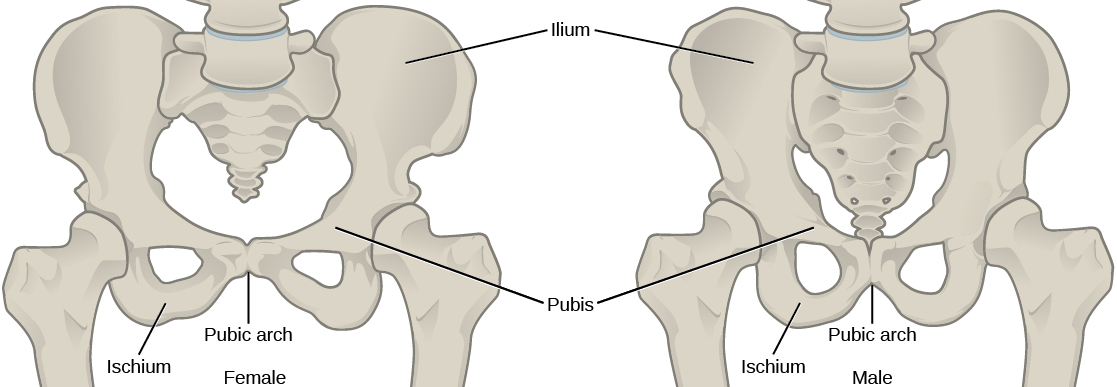
Figure 38.13図 38.13
To adapt to reproductive fitness, the (a) female pelvis is lighter, wider, shallower, and has a broader angle between the pubic bones than (b) the male pelvis.
繁殖上の適応度を高めるように、(a)女性の骨盤は(b)男性の骨盤より軽く、幅が広く、浅く、恥骨の間の角度も広い。
The Lower Limb下肢
The lower limb consists of the thigh, the leg, and the foot. The bones of the lower limb are the femur (thigh bone), patella (kneecap), tibia and fibula (bones of the leg), tarsals (bones of the ankle), and metatarsals and phalanges (bones of the foot) (Figure 38.14). The bones of the lower limbs are thicker and stronger than the bones of the upper limbs because of the need to support the entire weight of the body and the resulting forces from locomotion. In addition to evolutionary fitness, the bones of an individual will respond to forces exerted upon them.
英語のヒント
下肢は大腿、下腿、足からなる。下肢の骨には、大腿骨、膝蓋骨、脛骨と腓骨(下腿の骨)、足根骨(足首の骨)、中足骨と趾骨(足の骨)がある(図38.14)。下肢の骨は、体重全体と移動によって生じる力を支える必要があるため、上肢の骨より太く強い。進化における適応度に加え、個々の人の骨も、それに加わる力に応じて変化する。
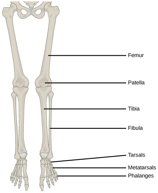
Figure 38.14図 38.14
The lower limb consists of the thigh (femur), kneecap (patella), leg (tibia and fibula), ankle (tarsals), and foot (metatarsals and phalanges) bones.
下肢は、大腿(大腿骨)、膝頭(膝蓋骨)、下腿(脛骨と腓骨)、足首(足根骨)、足(中足骨と趾骨)の骨からなる。
The femur, or thighbone, is the longest, heaviest, and strongest bone in the body. The femur and pelvis form the hip joint at the proximal end. At the distal end, the femur, tibia, and patella form the knee joint. The patella, or kneecap, is a triangular bone that lies anterior to the knee joint. The patella is embedded in the tendon of the femoral extensors (quadriceps). It improves knee extension by reducing friction. The tibia, or shinbone, is a large bone of the leg that is located directly below the knee. The tibia articulates with the femur at its proximal end, with the fibula and the tarsal bones at its distal end. It is the second largest bone in the human body and is responsible for transmitting the weight of the body from the femur to the foot. The fibula, or calf bone, parallels and articulates with the tibia. It does not articulate with the femur and does not bear weight. The fibula acts as a site for muscle attachment and forms the lateral part of the ankle joint.
英語のヒント
大腿骨は、体内で最も長く、重く、強い骨である。近位端では、大腿骨と骨盤が股関節を形成する。遠位端では、大腿骨、脛骨、膝蓋骨が膝関節を形成する。膝蓋骨は、膝関節の前側にある三角形の骨である。大腿の伸筋である大腿四頭筋の腱の中に埋め込まれている。摩擦を減らすことによって、膝を伸ばす働きを助ける。脛骨は、膝のすぐ下にある、下腿の大きな骨である。近位端で大腿骨と、遠位端で腓骨および足根骨と関節を形成する。ヒトの体で2番目に大きな骨であり、大腿骨から足へ体重を伝える役割を担う。腓骨は脛骨に平行に並び、脛骨と関節を形成する。大腿骨とは関節を形成せず、体重も支えない。腓骨は筋の付着部として働き、足関節の外側部分を形成する。
The tarsals are the seven bones of the ankle. The ankle transmits the weight of the body from the tibia and the fibula to the foot. The metatarsals are the five bones of the foot. The phalanges are the 14 bones of the toes. Each toe consists of three phalanges, except for the big toe that has only two (Figure 38.15). Variations exist in other species; for example, the horse’s metacarpals and metatarsals are oriented vertically and do not make contact with the substrate.
英語のヒント
足根骨は、足首を構成する7個の骨である。足首は、脛骨と腓骨から足へ体重を伝える。中足骨は、足を構成する5個の骨である。趾骨は、足の指を構成する14個の骨である。各趾は3個の趾骨からなるが、母趾だけは2個である(図38.15)。ほかの種では違いが見られる。たとえばウマでは、中手骨と中足骨は垂直に向き、基質には接触しない。
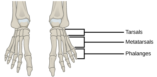


Figure 38.15図 38.15
This drawing shows the bones of the human foot and ankle, including the metatarsals and the phalanges.
この図は、中足骨や趾骨など、ヒトの足と足首の骨を示している。