第38章筋骨格系
Spongy Bone Tissue海綿骨組織
Whereas compact bone tissue forms the outer layer of all bones, spongy bone or cancellous bone forms the inner layer of all bones. Spongy bone tissue does not contain osteons that constitute compact bone tissue. Instead, it consists of trabeculae, which are lamellae that are arranged as rods or plates. Red bone marrow is found between the trabeculae. Blood vessels within this tissue deliver nutrients to osteocytes and remove waste. The red bone marrow of the femur and the interior of other large bones, such as the ilium, forms blood cells.
英語のヒント
緻密骨組織がすべての骨の外層をつくるのに対し、海綿骨はすべての骨の内層をつくる。海綿骨組織には、緻密骨組織を構成するオステオンは含まれない。代わりに、骨層板が棒状や板状に配列した骨梁からなる。骨梁の間には赤色骨髄がある。この組織の血管は、骨細胞に栄養を届け、老廃物を取り除く。大腿骨や、腸骨などのほかの大きな骨の内部にある赤色骨髄は、血球をつくる。
Spongy bone reduces the density of bone and allows the ends of long bones to compress as the result of stresses applied to the bone. Spongy bone is prominent in areas of bones that are not heavily stressed or where stresses arrive from many directions. The epiphyses of bones, such as the neck of the femur, are subject to stress from many directions. Imagine laying a heavy framed picture flat on the floor. You could hold up one side of the picture with a toothpick if the toothpick was perpendicular to the floor and the picture. Now drill a hole and stick the toothpick into the wall to hang up the picture. In this case, the function of the toothpick is to transmit the downward pressure of the picture to the wall. The force on the picture is straight down to the floor, but the force on the toothpick is both the picture wire pulling down and the bottom of the hole in the wall pushing up. The toothpick will break off right at the wall.
英語のヒント
海綿骨は骨の密度を下げ、骨に応力が加わったときに長骨の端が圧縮されるようにする。海綿骨は、大きな応力がかからない領域や、多方向から応力が加わる領域で発達している。大腿骨頸部などの骨端には、多方向から応力がかかる。重い額入りの絵を、床の上に平らに置くことを想像してみよう。つまようじが床と絵に対して垂直であれば、それで絵の片側を支えることができる。次に、壁に穴を開けてつまようじを差し込み、そこへ絵を掛けるとしよう。この場合、つまようじの役割は、絵が下向きに加える力を壁へ伝えることである。絵にかかる力は床へまっすぐ下向きだが、つまようじには、絵をつるすワイヤーが下へ引く力と、壁の穴の底面が上へ押す力の両方がかかる。つまようじは、壁のすぐそばで折れる。
The neck of the femur is horizontal like the toothpick in the wall. The weight of the body pushes it down near the joint, but the vertical diaphysis of the femur pushes it up at the other end. The neck of the femur must be strong enough to transfer the downward force of the body weight horizontally to the vertical shaft of the femur (Figure 38.20).
大腿骨頸部は、壁に差したつまようじのように水平である。体重は関節に近い側を下へ押すが、垂直な大腿骨の骨幹は、反対側を上へ押す。大腿骨頸部は、体重による下向きの力を、水平方向に垂直な大腿骨の骨幹へ伝えられるだけの強さをもたなければならない(図38.20)。
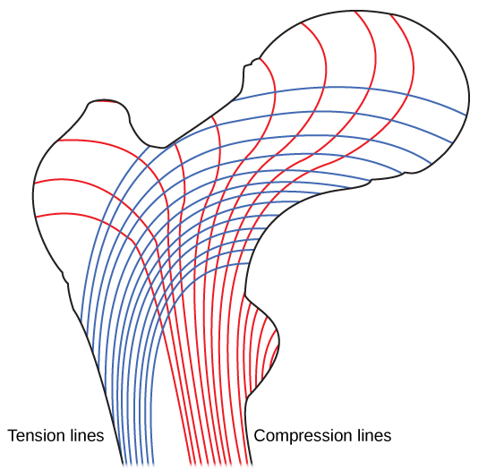

Figure 38.20図 38.20
Trabeculae in spongy bone are arranged such that one side of the bone bears tension and the other withstands compression.
海綿骨の骨梁は、骨の一方の側が張力を受け、もう一方の側が圧縮に耐えるように配列している。
Cell Types in Bones骨の細胞の種類
Bone consists of four types of cells: osteoblasts, osteoclasts, osteocytes, and osteoprogenitor cells. Osteoblasts are bone cells that are responsible for bone formation. Osteoblasts synthesize and secrete the organic part and inorganic part of the extracellular matrix of bone tissue, and collagen fibers. Osteoblasts become trapped in these secretions and differentiate into less active osteocytes. Osteoclasts are large bone cells with up to 50 nuclei. They remove bone structure by releasing lysosomal enzymes and acids that dissolve the bony matrix. These minerals, released from bones into the blood, help regulate calcium concentrations in body fluids. Bone may also be resorbed for remodeling, if the applied stresses have changed. Osteocytes are mature bone cells and are the main cells in bony connective tissue; these cells cannot divide. Osteocytes maintain normal bone structure by recycling the mineral salts in the bony matrix. Osteoprogenitor cells are squamous stem cells that divide to produce daughter cells that differentiate into osteoblasts. Osteoprogenitor cells are important in the repair of fractures.
英語のヒント
骨は、骨芽細胞、破骨細胞、骨細胞、骨前駆細胞という4種類の細胞からなる。骨芽細胞は、骨形成を担う細胞である。骨組織の細胞外基質の有機成分と無機成分、およびコラーゲン線維を合成して分泌する。骨芽細胞は、自らの分泌物に囲まれると、活動性の低い骨細胞へ分化する。破骨細胞は、最大50個もの核をもつ大きな骨の細胞である。骨基質を溶かすリソソーム酵素や酸を放出することで、骨の構造を取り除く。骨から血液中へ放出された無機質は、体液のカルシウム濃度の調節を助ける。加わる応力が変化した場合には、骨の再構築のために骨吸収が起こることもある。骨細胞は成熟した骨の細胞で、骨性の結合組織の主要な細胞である。これらの細胞は分裂できない。骨基質の無機塩類を再利用することで、正常な骨の構造を維持する。骨前駆細胞は、扁平な幹細胞である。分裂によって娘細胞を生み、その娘細胞は骨芽細胞に分化する。骨前駆細胞は、骨折の修復で重要な役割を果たす。
Development of Bone骨の発達
Ossification, or osteogenesis, is the process of bone formation by osteoblasts. Ossification is distinct from the process of calcification; whereas calcification takes place during the ossification of bones, it can also occur in other tissues. Ossification begins approximately six weeks after fertilization in an embryo. Before this time, the embryonic skeleton consists entirely of fibrous membranes and hyaline cartilage. The development of bone from fibrous membranes is called intramembranous ossification; development from hyaline cartilage is called endochondral ossification. Bone growth continues until approximately age 25. Bones can grow in thickness throughout life, but after age 25, ossification functions primarily in bone remodeling and repair.
英語のヒント
骨化、すなわち骨形成とは、骨芽細胞によって骨が形成される過程である。骨化は石灰化とは異なる。石灰化は骨化の途中に起こるが、ほかの組織でも起こりうる。胚では、受精後約6週で骨化が始まる。それ以前の胚の骨格は、すべて線維性の膜と硝子軟骨からなる。線維性の膜から骨が発達することを膜内骨化といい、硝子軟骨から発達することを軟骨内骨化という。骨の成長はおよそ25歳まで続く。骨は一生を通じて厚くなりうるが、25歳以降の骨化は、主に骨の再構築と修復に働く。
Intramembranous Ossification膜内骨化
Intramembranous ossification is the process of bone development from fibrous membranes. It is involved in the formation of the flat bones of the skull, the mandible, and the clavicles. Ossification begins as mesenchymal cells form a template of the future bone. They then differentiate into osteoblasts at the ossification center. Osteoblasts secrete the extracellular matrix and deposit calcium, which hardens the matrix. The non-mineralized portion of the bone or osteoid continues to form around blood vessels, forming spongy bone. Connective tissue in the matrix differentiates into red bone marrow in the fetus. The spongy bone is remodeled into a thin layer of compact bone on the surface of the spongy bone.
英語のヒント
膜内骨化は、線維性の膜から骨が発達する過程である。頭蓋の扁平骨、下顎骨、鎖骨の形成に関与する。間葉系細胞が将来の骨の原型をつくると、骨化が始まる。その後、骨化中心で骨芽細胞に分化する。骨芽細胞は細胞外基質を分泌し、カルシウムを沈着させる。カルシウムによって基質は硬くなる。骨のうち、まだ無機質の沈着していない部分である類骨は、血管の周囲で形成され続け、海綿骨となる。胎児では、基質内の結合組織が赤色骨髄に分化する。海綿骨の表面は再構築され、薄い緻密骨の層になる。