第38章筋骨格系
Endochondral Ossification軟骨内骨化
Endochondral ossification is the process of bone development from hyaline cartilage. All of the bones of the body, except for the flat bones of the skull, mandible, and clavicles, are formed through endochondral ossification.
軟骨内骨化は、硝子軟骨から骨が発達する過程である。頭蓋の扁平骨、下顎骨、鎖骨を除き、体のすべての骨は軟骨内骨化によって形成される。
In long bones, chondrocytes form a template of the hyaline cartilage diaphysis. Responding to complex developmental signals, the matrix begins to calcify. This calcification prevents diffusion of nutrients into the matrix, resulting in chondrocytes dying and the opening up of cavities in the diaphysis cartilage. Blood vessels invade the cavities, and osteoblasts and osteoclasts modify the calcified cartilage matrix into spongy bone. Osteoclasts then break down some of the spongy bone to create a medullary, or marrow, cavity in the center of the diaphysis. Dense, irregular connective tissue forms a sheath (periosteum) around the bones. The periosteum assists in attaching the bone to surrounding tissues, tendons, and ligaments. The bone continues to grow and elongate as the cartilage cells at the epiphyses divide.
英語のヒント
長骨では、軟骨細胞が硝子軟骨からなる骨幹の原型をつくる。複雑な発生シグナルに応答して、基質の石灰化が始まる。この石灰化は基質への栄養分の拡散を妨げるため、軟骨細胞が死に、骨幹の軟骨に空洞が生じる。空洞に血管が侵入し、骨芽細胞と破骨細胞が、石灰化した軟骨基質を海綿骨へと変える。続いて、破骨細胞が海綿骨の一部を分解し、骨幹の中央に骨髄腔をつくる。密で不規則な結合組織が、骨の周囲に鞘状の骨膜を形成する。骨膜は、骨を周囲の組織や腱、靱帯へつなぐのを助ける。骨端の軟骨細胞が分裂するにつれて、骨は成長し、長くなり続ける。
In the last stage of prenatal bone development, the centers of the epiphyses begin to calcify. Secondary ossification centers form in the epiphyses as blood vessels and osteoblasts enter these areas and convert hyaline cartilage into spongy bone. Until adolescence, hyaline cartilage persists at the epiphyseal plate (growth plate), which is the region between the diaphysis and epiphysis that is responsible for the lengthwise growth of long bones (Figure 38.21).
英語のヒント
出生前の骨の発達の最終段階では、骨端の中心が石灰化し始める。血管と骨芽細胞がこの領域へ入り、硝子軟骨を海綿骨へ変えるにつれて、骨端に二次骨化中心が形成される。思春期までは、骨端板(成長板)に硝子軟骨が残る。骨端板は、骨幹と骨端の間の領域であり、長骨の長さ方向の成長を担う(図38.21)。
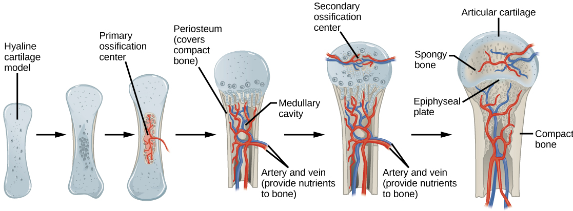
Figure 38.21図 38.21
Endochondral ossification is the process of bone development from hyaline cartilage. The periosteum is the connective tissue on the outside of bone that acts as the interface between bone, blood vessels, tendons, and ligaments.
軟骨内骨化は、硝子軟骨から骨が発達する過程である。骨膜は骨の外側にある結合組織で、骨、血管、腱、靱帯の間をつなぐ境界面として働く。
Growth of Bone骨の成長
Long bones continue to lengthen, potentially until adolescence, through the addition of bone tissue at the epiphyseal plate. They also increase in width through appositional growth.
長骨は、骨端板に骨組織が加わることによって、思春期まで長さ方向に成長し続ける場合がある。また、付加成長によって幅も増す。
Lengthening of Long Bones長骨が長くなる過程
Chondrocytes on the epiphyseal side of the epiphyseal plate divide; one cell remains undifferentiated near the epiphysis, and one cell moves toward the diaphysis. The cells, which are pushed from the epiphysis, mature and are destroyed by calcification. This process replaces cartilage with bone on the diaphyseal side of the plate, resulting in a lengthening of the bone.
骨端板の骨端側にある軟骨細胞が分裂すると、一方の細胞は骨端の近くで未分化のまま残り、もう一方は骨幹の方向へ移動する。骨端から押し出された細胞は成熟し、石灰化によって破壊される。この過程によって、骨端板の骨幹側で軟骨が骨に置き換えられ、骨が長くなる。
Long bones stop growing at around the age of 18 in females and the age of 21 in males in a process called epiphyseal plate closure. During this process, cartilage cells stop dividing and all of the cartilage is replaced by bone. The epiphyseal plate fades, leaving a structure called the epiphyseal line or epiphyseal remnant, and the epiphysis and diaphysis fuse.
英語のヒント
長骨は、女性ではおよそ18歳、男性ではおよそ21歳で、骨端板閉鎖という過程によって成長を止める。この過程では、軟骨細胞が分裂を停止し、すべての軟骨が骨に置き換わる。骨端板は消失して、骨端線、または骨端板の遺残と呼ばれる構造を残し、骨端と骨幹が融合する。
Thickening of Long Bones長骨が太くなる過程
Appositional growth is the increase in the diameter of bones by the addition of bony tissue at the surface of bones. Osteoblasts at the bone surface secrete bone matrix, and osteoclasts on the inner surface break down bone. The osteoblasts differentiate into osteocytes. A balance between these two processes allows the bone to thicken without becoming too heavy.
英語のヒント
付加成長とは、骨の表面に骨組織が加わることで、骨の直径が増すことである。骨の表面では骨芽細胞が骨基質を分泌し、内面では破骨細胞が骨を分解する。骨芽細胞は骨細胞に分化する。この2つの過程の釣り合いにより、骨は重くなりすぎずに太くなることができる。
Bone Remodeling and Repair骨の再構築と修復
Bone renewal continues after birth into adulthood. Bone remodeling is the replacement of old bone tissue by new bone tissue. It involves the processes of bone deposition by osteoblasts and bone resorption by osteoclasts. Normal bone growth requires vitamins D, C, and A, plus minerals such as calcium, phosphorous, and magnesium. Hormones such as parathyroid hormone, growth hormone, and calcitonin are also required for proper bone growth and maintenance.
英語のヒント
骨の更新は、出生後も成人期まで続く。骨の再構築(リモデリング)とは、古い骨組織が新しい骨組織に置き換わることである。骨芽細胞による骨形成と、破骨細胞による骨吸収の過程を伴う。正常な骨の成長には、ビタミンD・C・Aに加え、カルシウム、リン、マグネシウムなどの無機質が必要である。適切な骨の成長と維持には、副甲状腺ホルモン、成長ホルモン、カルシトニンなどのホルモンも必要となる。
Bone turnover rates are quite high, with five to seven percent of bone mass being recycled every week. Differences in turnover rate exist in different areas of the skeleton and in different areas of a bone. For example, the bone in the head of the femur may be fully replaced every six months, whereas the bone along the shaft is altered much more slowly.
英語のヒント
骨の代謝回転はかなり速く、毎週、骨量の5〜7%が入れ替わる。代謝回転の速さは、骨格の部位や、同じ骨の中の部位によって異なる。たとえば、大腿骨頭の骨は6か月ごとに完全に置き換わることがある一方、骨幹に沿った部分の骨の入れ替わりは、はるかに遅い。
Bone remodeling allows bones to adapt to stresses by becoming thicker and stronger when subjected to stress. Bones that are not subject to normal stress, for example when a limb is in a cast, will begin to lose mass. A fractured or broken bone undergoes repair through four stages:
骨の再構築によって、骨は応力を受けると太く強くなり、応力に適応できる。たとえば、四肢をギプスで固定したときのように、通常の応力を受けない骨は、骨量が減少し始める。骨折した骨は、次の4段階を経て修復される。
- Blood vessels in the broken bone tear and hemorrhage, resulting in the formation of clotted blood, or a hematoma, at the site of the break. The severed blood vessels at the broken ends of the bone are sealed by the clotting process, and bone cells that are deprived of nutrients begin to die.
英語のヒント
骨折した骨の血管が裂けて出血し、骨折部位に血液の凝固塊、すなわち血腫が形成される。骨折端で切断された血管は、血液凝固によってふさがれ、栄養分を失った骨の細胞は死に始める。 - Within days of the fracture, capillaries grow into the hematoma, and phagocytic cells begin to clear away the dead cells. Though fragments of the blood clot may remain, fibroblasts and osteoblasts enter the area and begin to reform bone. Fibroblasts produce collagen fibers that connect the broken bone ends, and osteoblasts start to form spongy bone. The repair tissue between the broken bone ends is called the fibrocartilaginous callus, as it is composed of both hyaline and fibrocartilage (Figure 38.22). Some bone spicules may also appear at this point.
英語のヒント
骨折後数日以内に、毛細血管が血腫の中へ伸び、食細胞が死んだ細胞を取り除き始める。血餅の断片が残ることもあるが、線維芽細胞と骨芽細胞がこの領域に入り、骨の再形成を始める。線維芽細胞は、折れた骨の両端をつなぐコラーゲン線維をつくり、骨芽細胞は海綿骨をつくり始める。骨折端の間の修復組織は、硝子軟骨と線維軟骨の両方からなるため、線維軟骨性仮骨と呼ばれる(図38.22)。この時点で、骨片が一部現れることもある。 - The fibrocartilaginous callus is converted into a bony callus of spongy bone. It takes about two months for the broken bone ends to be firmly joined together after the fracture. This is similar to the endochondral formation of bone, as cartilage becomes ossified; osteoblasts, osteoclasts, and bone matrix are present.
英語のヒント
線維軟骨性仮骨は、海綿骨からなる骨性仮骨へ変わる。骨折後、折れた骨の両端がしっかり結合するまでには、およそ2か月かかる。軟骨が骨化し、骨芽細胞、破骨細胞、骨基質が存在するという点で、この過程は軟骨内骨化による骨形成に似ている。 - The bony callus is then remodelled by osteoclasts and osteoblasts, with excess material on the exterior of the bone and within the medullary cavity being removed. Compact bone is added to create bone tissue that is similar to the original, unbroken bone. This remodeling can take many months, and the bone may remain uneven for years.続いて、骨性仮骨は破骨細胞と骨芽細胞によって再構築される。このとき、骨の外側と骨髄腔の中にある余分な物質が取り除かれる。緻密骨が加わることで、骨折前の元の骨に似た骨組織がつくられる。この再構築には何か月もかかることがあり、何年も骨の表面に凹凸が残る場合もある。
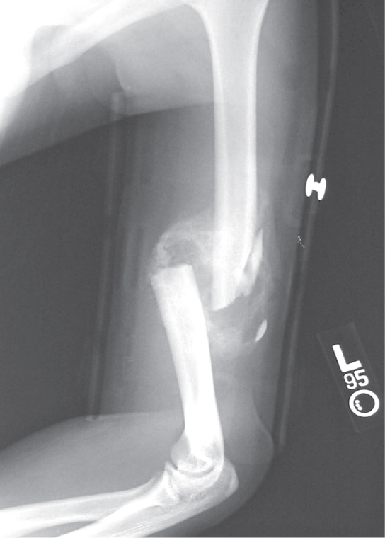


Figure 38.22図 38.22
After this bone is set, a callus will knit the two ends together. (credit: Bill Rhodes)
この骨の位置が整えられた後、仮骨が両端をつなぎ合わせる。(出典:Bill Rhodes)