第34章動物の栄養と消化器系
Small Intestine小腸
Chyme moves from the stomach to the small intestine. The small intestine is the organ where the digestion of protein, fats, and carbohydrates is completed. The small intestine is a long tube-like organ with a highly folded surface containing finger-like projections called the villi. The apical surface of each villus has many microscopic projections called microvilli. These structures, illustrated in Figure 34.12, are lined with epithelial cells on the luminal side and allow for the nutrients to be absorbed from the digested food and absorbed into the bloodstream on the other side. The villi and microvilli, with their many folds, increase the surface area of the intestine and increase absorption efficiency of the nutrients. Absorbed nutrients in the blood are carried into the hepatic portal vein, which leads to the liver. There, the liver regulates the distribution of nutrients to the rest of the body and removes toxic substances, including drugs, alcohol, and some pathogens.
英語のヒント
キームは胃から小腸へ移動する。小腸は、タンパク質、脂肪、炭水化物の消化が完了する器官である。長い管状の器官で、その表面は複雑に折りたたまれ、絨毛という指状の突起を持つ。各絨毛の頂端面には、微絨毛という顕微鏡的な突起が多数ある。図34.12に示すこれらの構造は、内腔側が上皮細胞で覆われており、消化した食物から栄養素を吸収し、反対側の血流へ取り込めるようにする。絨毛と微絨毛は、多くの折りたたみによって腸の表面積を増やし、栄養吸収の効率を高める。血中へ吸収された栄養素は、肝臓へ通じる肝門脈へ運ばれる。肝臓は、体の他の部分への栄養素の分配を調節し、薬物、アルコール、一部の病原体などの有害物質を除く。
The human small intestine is over 6m long and is divided into three parts: the duodenum, the jejunum, and the ileum. The “C-shaped,” fixed part of the small intestine is called the duodenum and is shown in Figure 34.11. The duodenum is separated from the stomach by the pyloric sphincter which opens to allow chyme to move from the stomach to the duodenum. In the duodenum, chyme is mixed with pancreatic juices in an alkaline solution rich in bicarbonate that neutralizes the acidity of chyme and acts as a buffer. Pancreatic juices also contain several digestive enzymes. Digestive juices from the pancreas, liver, and gallbladder, as well as from gland cells of the intestinal wall itself, enter the duodenum. Bile is produced in the liver and stored and concentrated in the gallbladder. Bile contains bile salts which emulsify lipids while the pancreas produces enzymes that catabolize starches, disaccharides, proteins, and fats. These digestive juices break down the food particles in the chyme into glucose, triglycerides, and amino acids. The bulk of chemical digestion of food takes place in the duodenum. Absorption of fatty acids also takes place in the duodenum.
英語のヒント
ヒトの小腸は長さが6mを超え、十二指腸、空腸、回腸の三つに分かれる。図34.11の、C字形で固定されている部分が十二指腸である。十二指腸と胃は幽門括約筋で隔てられ、この筋が開くとキームが胃から十二指腸へ移れる。十二指腸では、キームは炭酸水素イオンに富むアルカリ性の溶液の中で膵液と混ざる。この溶液はキームの酸性を中和し、緩衝液として働く。膵液には複数の消化酵素も含まれる。膵臓、肝臓、胆嚢、さらに腸壁そのものの腺細胞からの消化液が十二指腸へ入る。胆汁は肝臓でつくられ、胆嚢で貯蔵・濃縮される。胆汁には脂質を乳化する胆汁酸塩が含まれ、膵臓はデンプン、二糖、タンパク質、脂肪を分解する酵素をつくる。これらの消化液は、キーム内の食物粒子をグルコース、トリグリセリド、アミノ酸へ分解する。食物の化学的消化の大半は十二指腸で行われる。脂肪酸の吸収もここで起こる。
The second part of the small intestine is called the jejunum, shown in Figure 34.11. Here, hydrolysis of nutrients is continued while most of the carbohydrates and amino acids are absorbed through the intestinal lining. Some chemical digestion and the bulk of nutrient absorption occurs in the jejunum.
小腸の二番目の部分は空腸と呼ばれ、図34.11に示されている。ここでは栄養素の加水分解が続くとともに、炭水化物とアミノ酸の大半が腸の内面を通して吸収される。空腸では一部の化学的消化と、栄養吸収の大半が行われる。
The ileum, also illustrated in Figure 34.11 is the last part of the small intestine and here the bile salts and vitamins are absorbed into the bloodstream. The undigested food is sent to the colon from the ileum via peristaltic movements of the muscle. The ileum ends and the large intestine begins at the ileocecal valve. The vermiform, “worm-like,” appendix is located at the ileocecal valve. The appendix of humans secretes no enzymes and has an insignificant role in immunity.
英語のヒント
図34.11の回腸は小腸の最後の部分で、胆汁酸塩とビタミンがここで血流へ吸収される。未消化の食物は筋の蠕動によって回腸から結腸へ送られる。回盲弁の位置で回腸が終わり、大腸が始まる。蠕虫状、すなわち「虫のような形」の虫垂は回盲弁の位置にある。ヒトの虫垂は酵素を分泌せず、免疫で果たす役割もわずかである。
Large Intestine大腸
The large intestine, illustrated in Figure 34.13, reabsorbs the water from the undigested food material and processes the waste material. The human large intestine is much smaller in length compared to the small intestine but larger in diameter. It has three parts: the cecum, the colon, and the rectum. The cecum joins the ileum to the colon and is the receiving pouch for the waste matter. The colon is home to many bacteria or “intestinal flora” that aid in the digestive processes. The colon can be divided into four regions, the ascending colon, the transverse colon, the descending colon, and the sigmoid colon. The main functions of the colon are to extract the water and mineral salts from undigested food, and to store waste material. Carnivorous mammals have a shorter large intestine compared to herbivorous mammals due to their diet.
英語のヒント
図34.13に示す大腸は、未消化の食物から水分を再吸収し、不要物を処理する。ヒトの大腸は小腸よりはるかに短いが、直径は大きい。盲腸、結腸、直腸の三つの部分からなる。盲腸は回腸と結腸をつなぐ、不要物を受け取る袋である。結腸には、消化過程を助ける多数の細菌、すなわち「腸内細菌叢」が生息する。結腸は、上行結腸、横行結腸、下行結腸、S状結腸の四領域に分けられる。主な機能は、未消化の食物から水分と無機塩類を取り出し、不要物を貯蔵することである。食性の違いにより、肉食哺乳類の大腸は草食哺乳類より短い。
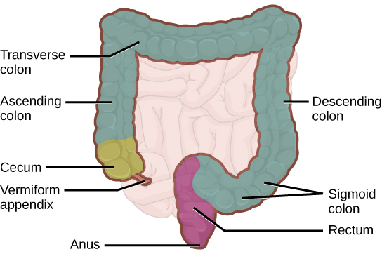

Figure 34.13図 34.13
The large intestine reabsorbs water from undigested food and stores waste material until it is eliminated.
大腸は未消化の食物から水分を再吸収し、不要物を排出まで貯蔵する。