第34章動物の栄養と消化器系
Esophagus食道
The esophagus is a tubular organ that connects the mouth to the stomach. The chewed and softened food passes through the esophagus after being swallowed. The smooth muscles of the esophagus undergo a series of wave like movements called peristalsis that push the food toward the stomach, as illustrated in Figure 34.10. The peristalsis wave is unidirectional—it moves food from the mouth to the stomach, and reverse movement is not possible. The peristaltic movement of the esophagus is an involuntary reflex; it takes place in response to the act of swallowing.
食道は口と胃をつなぐ管状の器官である。かまれて柔らかくなった食物は、飲み込まれた後に食道を通る。食道の平滑筋は、図34.10に示す蠕動という一連の波状運動を起こし、食物を胃へ押し進める。蠕動の波は一方向性で、食物を口から胃へ動かし、逆向きには動かせない。食道の蠕動は不随意的な反射で、嚥下に応じて起こる。
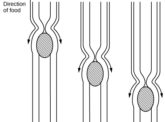

Figure 34.10図 34.10
The esophagus transfers food from the mouth to the stomach through peristaltic movements.
食道は蠕動によって、食物を口から胃へ運ぶ。
A ring-like muscle called a sphincter forms valves in the digestive system. The gastro-esophageal sphincter is located at the stomach end of the esophagus. In response to swallowing and the pressure exerted by the bolus of food, this sphincter opens, and the bolus enters the stomach. When there is no swallowing action, this sphincter is shut and prevents the contents of the stomach from traveling up the esophagus. Many animals have a true sphincter; however, in humans, there is no true sphincter, but the esophagus remains closed when there is no swallowing action. Acid reflux or “heartburn” occurs when the acidic digestive juices escape into the esophagus.
英語のヒント
括約筋と呼ばれる輪状の筋が、消化系で弁を形成する。胃食道括約筋は、食道の胃に近い端にある。嚥下と食塊が加える圧力に応じてこの括約筋が開き、食塊が胃に入る。嚥下していないときは閉じて、胃の内容物が食道を上がるのを防ぐ。多くの動物には真の括約筋がある。しかしヒトには真の括約筋はなく、嚥下していないときに食道が閉じたまま保たれる。酸性の消化液が食道へ漏れ出すと、胃酸逆流、すなわち「胸焼け」が起こる。
Stomach胃
A large part of digestion occurs in the stomach, shown in Figure 34.11. The stomach is a saclike organ that secretes gastric digestive juices. The pH in the stomach is between 1.5 and 2.5. This highly acidic environment is required for the chemical breakdown of food and the extraction of nutrients. When empty, the stomach is a rather small organ; however, it can expand to up to 20 times its resting size when filled with food. This characteristic is particularly useful for animals that need to eat when food is available.
英語のヒント
消化のかなりの部分は、図34.11に示す胃で行われる。胃は胃液を分泌する袋状の器官である。胃内のpHは1.5〜2.5で、この強い酸性環境が、食物の化学的分解と栄養素の取り出しに必要となる。空の胃はかなり小さいが、食物で満たされると、空のときの最大20倍にまで拡張できる。この特徴は、食物が手に入ったときに食べる必要のある動物にとって、特に有用である。
The stomach is also the major site for protein digestion in animals other than ruminants. Protein digestion is mediated by an enzyme called pepsin in the stomach chamber. Pepsin is secreted by the chief cells in the stomach in an inactive form called pepsinogen. Pepsin breaks peptide bonds and cleaves proteins into smaller polypeptides; it also helps activate more pepsinogen, starting a positive feedback mechanism that generates more pepsin. Another cell type—parietal cells—secrete hydrogen and chloride ions, which combine in the lumen to form hydrochloric acid, the primary acidic component of the stomach juices. Hydrochloric acid helps to convert the inactive pepsinogen to pepsin. The highly acidic environment also kills many microorganisms in the food and, combined with the action of the enzyme pepsin, results in the hydrolysis of protein in the food. Chemical digestion is facilitated by the churning action of the stomach. Contraction and relaxation of smooth muscles mixes the stomach contents about every 20 minutes. The partially digested food and gastric juice mixture is called chyme. Chyme passes from the stomach to the small intestine. Further protein digestion takes place in the small intestine. Gastric emptying occurs within two to six hours after a meal. Only a small amount of chyme is released into the small intestine at a time. The movement of chyme from the stomach into the small intestine is regulated by the pyloric sphincter.
英語のヒント
反芻動物以外の動物では、胃はタンパク質消化の主な場所でもある。胃内のタンパク質消化は、ペプシンという酵素が担う。ペプシンは胃の主細胞から、ペプシノーゲンという不活性な形で分泌される。ペプシンはペプチド結合を切り、タンパク質をより小さなポリペプチドへ分解する。また、さらに多くのペプシノーゲンの活性化を助け、ペプシンを増やす正のフィードバック機構を始動させる。別の種類の細胞である壁細胞は、水素イオンと塩化物イオンを分泌する。これらは内腔で合わさり、胃液の主な酸性成分である塩酸となる。塩酸は、不活性なペプシノーゲンをペプシンへ変えるのを助ける。強い酸性環境は、食物中の多くの微生物を殺すとともに、ペプシンの作用と相まって食物中のタンパク質を加水分解する。胃が内容物をかき混ぜる作用によって、化学的消化が促される。平滑筋の収縮と弛緩により、胃の内容物は約20分ごとに混合される。部分的に消化された食物と胃液の混合物をキームという。キームは胃から小腸へ進み、そこでタンパク質がさらに消化される。胃内容物の排出は食後2〜6時間以内に起こる。一度に小腸へ放出されるキームは少量だけである。胃から小腸へのキームの移動は、幽門括約筋によって調節される。
When digesting protein and some fats, the stomach lining must be protected from getting digested by pepsin. There are two points to consider when describing how the stomach lining is protected. First, as previously mentioned, the enzyme pepsin is synthesized in the inactive form. This protects the chief cells, because pepsinogen does not have the same enzyme functionality of pepsin. Second, the stomach has a thick mucus lining that protects the underlying tissue from the action of the digestive juices. When this mucus lining is ruptured, ulcers can form in the stomach. Ulcers are open wounds in or on an organ caused by bacteria (Helicobacter pylori) when the mucus lining is ruptured and fails to reform.
英語のヒント
タンパク質や一部の脂肪を消化する際、胃の内面はペプシンによって消化されないよう守られなければならない。胃の内面の保護を説明するうえで、考慮すべき点は二つある。第一に、すでに述べたように、ペプシンは不活性な形で合成される。ペプシノーゲンはペプシンと同じ酵素機能を持たないため、これによって主細胞が守られる。第二に、胃の内面には厚い粘液の層があり、その下の組織を消化液の作用から守る。この粘液層が破れると、胃に潰瘍ができることがある。潰瘍とは、粘液層が破れて再生しなくなったとき、細菌(ヘリコバクター・ピロリ)によって器官の内部や表面にできる、開いた傷である。