第23章原生生物
Excavataエクスカバータ
Many of the protist species classified into the supergroup Excavata are asymmetrical, single-celled organisms with a feeding groove “excavated” from one side. This supergroup includes heterotrophic predators, photosynthetic species, and parasites. Its subgroups are the diplomonads, parabasalids, and euglenozoans. The group includes a variety of modified mitochondria, as well as chloroplasts derived from green algae by secondary endosymbiosis. Many of the euglenozoans are free-living, but most diplomonads and parabasalids are symbionts or parasites.
英語のヒント
エクスカバータに分類される多くの原生生物は、片側に「掘り込まれた」摂食溝をもつ、非対称な単細胞生物である。このスーパーグループには、従属栄養性の捕食者、光合成種、寄生者が含まれる。下位群は、ディプロモナス類、パラバサリア、ユーグレノゾアである。この群には、多様な変型ミトコンドリアと、緑藻との二次的な細胞内共生に由来する葉緑体が見られる。ユーグレノゾアの多くは独立生活するが、ディプロモナス類とパラバサリアの大部分は、共生者または寄生者である。
Diplomonadsディプロモナス類
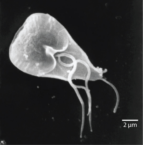
Among the Excavata are the diplomonads, which include the intestinal parasite, Giardia lamblia (Figure 23.30). Until recently, these protists were believed to lack mitochondria. Mitochondrial remnant organelles, called mitosomes, have since been identified in diplomonads, but although these mitosomes are essentially nonfunctional as respiratory organelles, they do function in iron and sulfur metabolism. Diplomonads exist in anaerobic environments and use alternative pathways, such as glycolysis, to generate energy. Each diplomonad cell has two similar, but not identical haploid nuclei. Diplomonads have four pairs of locomotor flagella that are fairly deeply rooted in basal bodies that lie between the two nuclei.
英語のヒント
エクスカバータには、腸内寄生者Giardia lamblia(図23.30)を含むディプロモナス類がある。最近まで、これらの原生生物はミトコンドリアを欠くと考えられていた。その後、ミトソームと呼ばれる、ミトコンドリアの名残の細胞小器官が確認された。ミトソームは、呼吸小器官としては基本的に機能しないが、鉄と硫黄の代謝では確かに機能する。ディプロモナス類は嫌気的環境に存在し、解糖などの別の経路を用いてエネルギーを生じる。各細胞には、似ているが同一ではない二つの半数体核がある。移動用の鞭毛を4対もち、それらは、二つの核の間にある基底小体にかなり深く根づいている。
OpenStax, Rice University; Biology 2e. (credit: modification of work by Janice Carr, CDC; scale-bar data from Matt Russell) · CC BY 4.0 · Source出典
Figure 23.30図 23.30
Giardia. The mammalian intestinal parasite Giardia lamblia, visualized here using scanning electron microscopy, is a waterborne protist that causes severe diarrhea when ingested. (credit: modification of work by Janice Carr, CDC; scale-bar data from Matt Russell)
Giardia。走査電子顕微鏡で観察した、哺乳類の腸内寄生者Giardia lamblia。水を介して伝播する原生生物で、摂取すると激しい下痢を引き起こす。(クレジット:CDCのJanice Carrによる作品を改変。スケールバーのデータ:Matt Russell)
Parabasalidsパラバサリア
A second Excavata subgroup, the parabasalids, are named for the parabasal apparatus, which consists of a Golgi complex associated with cytoskeletal fibers. Other cytoskeletal features include an axostyle, a bundle of fibers that runs the length of the cell and may even extend beyond it. Parabasalids move with flagella and membrane rippling, and these and other cytoskeletal modifications may assist locomotion. Like the diplomonads, the parabasalids exhibit modified mitochondria. In parabasalids these structures function anaerobically and are called hydrogenosomes because they produce hydrogen gas as a byproduct.
英語のヒント
エクスカバータの第2の下位群であるパラバサリアは、細胞骨格繊維と結びついたゴルジ体からなる、副基底装置にちなんで名づけられた。他の細胞骨格の特徴としては、細胞の長さ方向を走り、細胞の外へ伸びることもある繊維束、軸柱がある。パラバサリアは、鞭毛と膜の波打ちによって動き、これらや他の細胞骨格の変型が、移動を助けているのかもしれない。ディプロモナス類と同様に、パラバサリアも変型したミトコンドリアをもつ。この構造は嫌気的に機能し、副産物として水素ガスを生じるため、ハイドロジェノソームと呼ばれる。
The parabasalid Trichomonas vaginalis causes trichomoniasis, a sexually transmitted disease in humans, which appears in an estimated 180 million cases worldwide each year. Whereas males rarely exhibit symptoms during an infection with this protist, infected females may become more susceptible to secondary infection with human immunodeficiency virus (HIV) and may be more likely to develop cervical cancer. Pregnant people infected with T. vaginalis are at an increased risk of serious complications, such as pre-term delivery.
英語のヒント
パラバサリアのTrichomonas vaginalisは、ヒトの性感染症であるトリコモナス症を引き起こし、世界では年間推定1億8,000万例が発生する。男性はこの原生生物に感染しても、症状を示すことが少ない。一方、感染した女性は、ヒト免疫不全ウイルス(HIV)の二次感染を受けやすくなり、子宮頸がんを発症する可能性も高くなることがある。T. vaginalisに感染した妊娠中の人は、早産などの重大な合併症のリスクが高まる。
Some of the most complex of the parabasalids are those that colonize the rumen of ruminant animals and the guts of termites. These organisms can digest cellulose, a metabolic talent that is unusual among eukaryotic cells. They have multiple flagella arranged in complex patterns and some additionally recruit spirochetes that attach to their surface to act as accessory locomotor structures.
英語のヒント
パラバサリアの中でも特に複雑なものには、反芻動物の第一胃や、シロアリの腸に定着するものがある。これらの生物は、真核細胞では珍しい代謝能力として、セルロースを消化できる。複雑な配列をとる複数の鞭毛をもち、さらに一部は、表面に付着するスピロヘータを取り込み、補助的な運動構造として働かせる。
Euglenozoansユーグレノゾア
Euglenozoans includes parasites, heterotrophs, autotrophs, and mixotrophs, ranging in size from 10 to 500 µm. Euglenoids move through their aquatic habitats using two long flagella that guide them toward light sources sensed by a primitive ocular organ called an eyespot. The familiar genus, Euglena, encompasses some mixotrophic species that display a photosynthetic capability only when light is present. The chloroplast of Euglena descends from a green alga by secondary endosymbiosis. In the dark, the chloroplasts of Euglena shrink up and temporarily cease functioning, and the cells instead take up organic nutrients from their environment. Euglena has a tough pellicle composed of bands of protein attached to the cytoskeleton. The bands spiral around the cell and give Euglena its exceptional flexibility.
英語のヒント
ユーグレノゾアには、寄生者、従属栄養生物、独立栄養生物、混合栄養生物が含まれ、大きさは10〜500 µmである。ユーグレナ類は、眼点という原始的な視覚器官で感知した光源へ導く2本の長い鞭毛を使って、水中の生息環境を移動する。よく知られたミドリムシ属Euglenaには、光があるときだけ光合成能力を示す混合栄養性の種が含まれる。Euglenaの葉緑体は、緑藻との二次的な細胞内共生に由来する。暗所では、葉緑体は縮小して一時的に機能を停止し、細胞は代わりに環境から有機栄養物を取り込む。Euglenaは、細胞骨格に付着したタンパク質の帯からなる、丈夫なペリクルをもつ。その帯は細胞の周りを螺旋状に巻き、極めて高い柔軟性を与える。
The human parasite, Trypanosoma brucei, belongs to a different subgroup of Euglenozoa, the kinetoplastids. The kinetoplastid subgroup is named after the kinetoplast, a large modified mitochondrion carrying multiple circular DNAs. This subgroup includes several parasites, collectively called trypanosomes, which cause devastating human diseases and infect an insect species during a portion of their life cycle. T. brucei develops in the gut of the tsetse fly after the fly bites an infected human or other mammalian host. The parasite then travels to the insect salivary glands to be transmitted to another human or other mammal when the infected tsetse fly consumes another blood meal. T. brucei is common in central Africa and is the causative agent of African sleeping sickness, a disease associated with severe chronic fatigue, coma, and can be fatal if left untreated since it leads to progressive decline of the function of the central nervous system.
英語のヒント
ヒトの寄生者Trypanosoma bruceiは、ユーグレノゾアの別の下位群であるキネトプラスト類に属する。この下位群の名は、複数の環状DNAをもつ、大きく変型したミトコンドリアであるキネトプラストに由来する。この群には、まとめてトリパノソーマと呼ばれる複数の寄生者が含まれ、ヒトに深刻な病気を起こし、生活環の一部で昆虫種に感染する。T. bruceiは、ツェツェバエが感染したヒトや他の哺乳類宿主を刺した後、その腸内で発育する。その後、昆虫の唾液腺へ移り、感染したツェツェバエが次に吸血するとき、別のヒトや哺乳類へ伝播する。T. bruceiは中央アフリカによく見られ、アフリカ睡眠病を引き起こす。この病気は、重い慢性疲労や昏睡を伴い、中枢神経系の機能を進行性に低下させるため、治療せず放置すると死に至ることがある。
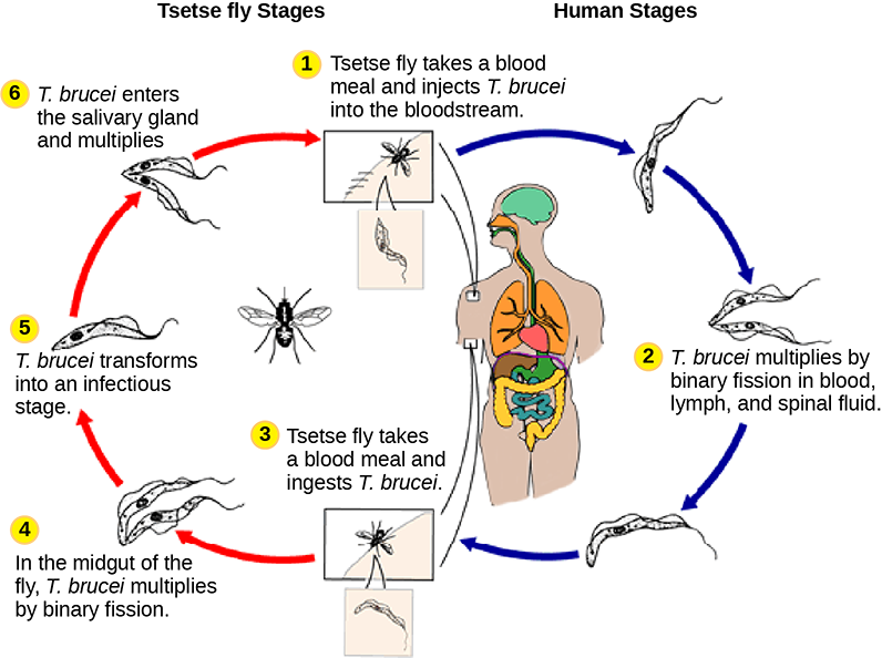


Figure 23.31図 23.31
Sleeping sickness. Trypanosoma brucei, the causative agent of sleeping sickness, spends part of its life cycle in the tsetse fly and part in humans. (credit: modification of work by CDC)
睡眠病。病原体Trypanosoma bruceiは、生活環の一部をツェツェバエの中で、別の一部をヒトの中で過ごす。(クレジット:CDCによる作品を改変)