第23章原生生物
Slime Molds粘菌
A subset of the amoebozoans, the slime molds, has several morphological similarities to fungi that are thought to be the result of convergent evolution. For instance, during times of stress, some slime molds develop into spore-generating fruiting bodies, much like fungi.
英語のヒント
アメーボゾアの一群である粘菌には、菌類との形態的類似がいくつか見られ、収斂進化の結果だと考えられている。たとえば、一部の粘菌は、ストレスのある時期に、菌類とよく似た胞子を作る子実体へ発達する。
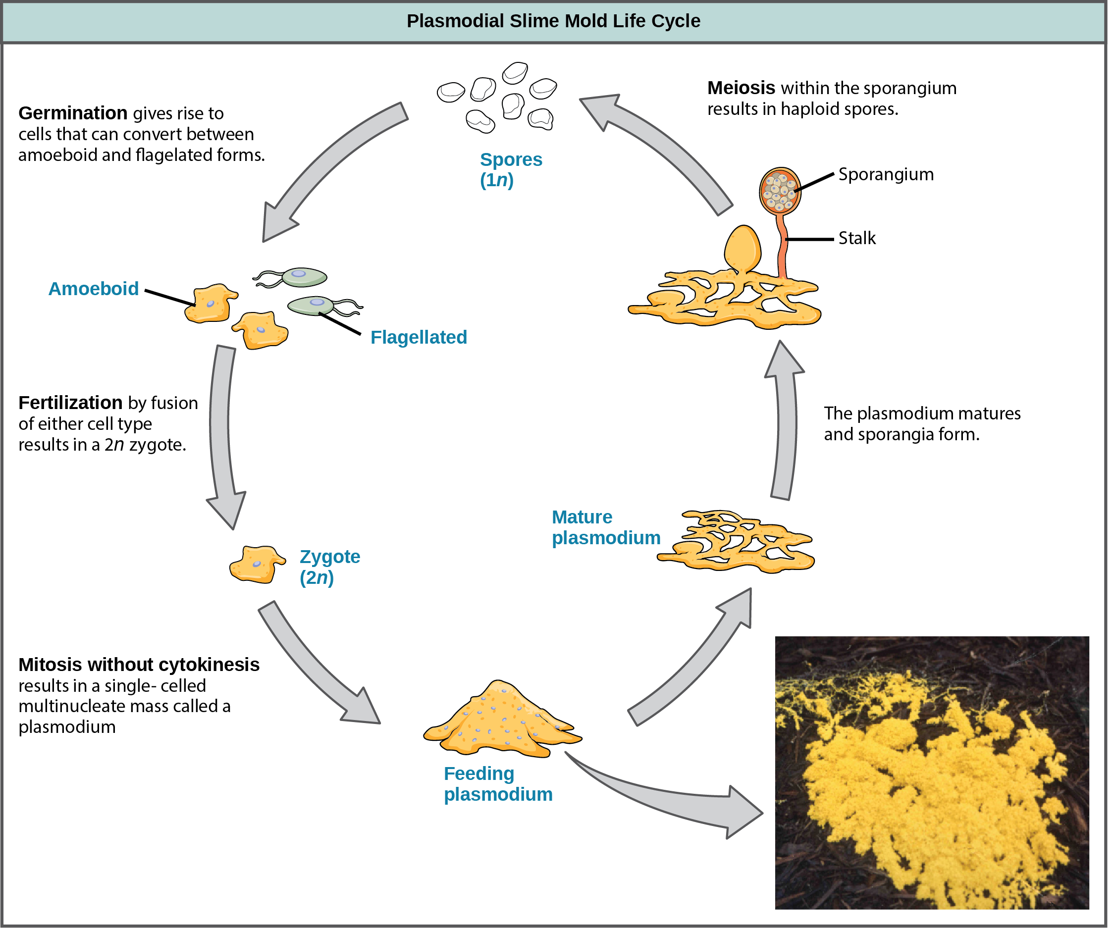
The slime molds are categorized on the basis of their life cycles into plasmodial or cellular types. Plasmodial slime molds are composed of large, multinucleate cells and move along surfaces like an amorphous blob of slime during their feeding stage (Figure 23.14). Food particles are lifted and engulfed into the slime mold as it glides along. The "dog vomit" slime mold seen in Figure 23.14 is a particularly colorful specimen and its ability to creep about might well trigger suspicion of alien invasion. Upon maturation, the plasmodium takes on a net-like appearance with the ability to form fruiting bodies, or sporangia, during times of stress. Haploid spores are produced by meiosis within the sporangia, and spores can be disseminated through the air or water to potentially land in more favorable environments. If this occurs, the spores germinate to form ameboid or flagellate haploid cells that can combine with each other and produce a diploid zygotic slime mold to complete the life cycle.
英語のヒント
粘菌は、生活環に基づいて変形体性と細胞性の二つに分類される。変形体性粘菌は、大きな多核細胞からなり、摂食段階では不定形の粘液の塊のように表面を移動する(図23.14)。粘菌は滑るように進みながら、食物粒子を持ち上げて取り込む。図23.14に示す「犬の吐瀉物」粘菌は、特に色鮮やかな標本で、はい回る能力は、宇宙人の侵入を疑わせても不思議ではない。成熟すると、変形体は網状の外観になり、ストレスのある時期には子実体、すなわち胞子嚢を形成できる。胞子嚢内では減数分裂によって半数体の胞子が作られ、胞子は空気や水で散布され、より好適な環境に到達する可能性がある。そうなると、胞子は発芽してアメーバ状または鞭毛をもつ半数体細胞となる。これらは互いに融合し、二倍体の接合子段階の粘菌を生じて、生活環を完結できる。
OpenStax, Rice University; Biology 2e. (credit: modification of work by Dr. Jonatha Gott and the Center for RNA Molecular Biology, Case Western Reserve University) · CC BY 4.0 · Source出典

Figure 23.14図 23.14
Plasmodial slime molds. The life cycle of the plasmodial slime mold is shown. The brightly colored plasmodium in the inset photo is a single-celled, multinucleate mass. (credit: modification of work by Dr. Jonatha Gott and the Center for RNA Molecular Biology, Case Western Reserve University)
変形体性粘菌。変形体性粘菌の生活環を示す。挿入写真の鮮やかな変形体は、単細胞の多核の塊である。(クレジット:Dr. Jonatha GottおよびCase Western Reserve UniversityのCenter for RNA Molecular Biologyによる作品を改変)
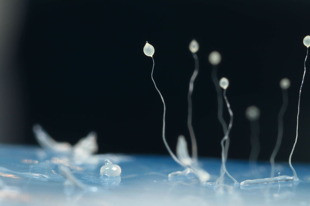
The cellular slime molds function as independent amoeboid cells when nutrients are abundant. When food is depleted, cellular slime molds aggregate into a mass of cells that behaves as a single unit, called a slug. Some cells in the slug contribute to a 2–3-millimeter stalk, drying up and dying in the process. Cells atop the stalk form an asexual fruiting body that contains haploid spores (Figure 23.15). As with plasmodial slime molds, the spores are disseminated and can germinate if they land in a moist environment. One representative genus of the cellular slime molds is Dictyostelium, which commonly exists in the damp soil of forests.
英語のヒント
細胞性粘菌は、栄養分が豊富なとき、独立したアメーバ状細胞として働く。食物が枯渇すると、細胞が集合して、ナメクジ体と呼ばれる、一つの単位として振る舞う細胞塊となる。ナメクジ体の一部の細胞は、長さ2〜3ミリメートルの柄を作り、その過程で乾燥して死ぬ。柄の先端の細胞は、半数体の胞子を含む無性生殖の子実体を作る(図23.15)。変形体性粘菌と同様に、胞子は散布され、湿った環境に到達すれば発芽できる。細胞性粘菌の代表的な属の一つはDictyosteliumで、森林の湿った土によく生息する。
Usman Bashir, Queller/Strassmann Lab; Dictyostelium discoideum. OpenStax, Biology 2e adaptation. · CC BY-SA 4.0 · Source出典

Figure 23.15図 23.15
Cellular Slime Mold. The image shows several stages in the life cycle of Dictyostelium discoideum, including aggregated cells, mobile slugs and their transformation into fruiting bodies with a cluster of spores supported by a stalk. (credit: By Usman Bashir (Own work) [CC BY-SA 4.0 (http://creativecommons.org/licenses/by-sa/4.0)], via Wikimedia Commons)
細胞性粘菌。Dictyostelium discoideumの生活環のいくつかの段階を示す。集合した細胞、移動するナメクジ体、そして柄に支えられた胞子の塊をもつ子実体への変化が見える。(クレジット:Usman Bashir、本人の作品、CC BY-SA 4.0(http://creativecommons.org/licenses/by-sa/4.0)、Wikimedia Commons経由)
Opisthokontaオピストコンタ
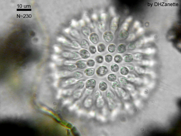
The Opisthokonts are named for the single posterior flagellum seen in flagellated cells of the group. The flagella of other protists are anterior and their movement pulls the cells along, while the opisthokonts are pushed. Protist members of the opisthokonts include the animal-like choanoflagellates, which are believed to resemble the common ancestor of sponges and perhaps, all animals. Choanoflagellates include unicellular and colonial forms (Figure 23.16), and number about 244 described species. In these organisms, the single, apical flagellum is surrounded by a contractile collar composed of microvilli. The collar is used to filter and collect bacteria for ingestion by the protist. A similar feeding mechanism is seen in the collar cells of sponges, which suggests a possible connection between choanoflagellates and animals.
英語のヒント
オピストコンタという名は、この群の鞭毛をもつ細胞に見られる、後方の1本の鞭毛に由来する。他の原生生物では鞭毛は前方にあり、その運動によって細胞が引かれるのに対し、オピストコンタは押される。この群の原生生物には、動物に似た襟鞭毛虫類が含まれ、海綿動物、そしておそらく全動物の共通祖先に似ていると考えられる。襟鞭毛虫類には単細胞と群体の形があり(図23.16)、記載済みの種は約244種である。これらの生物では、頂端にある1本の鞭毛を、微絨毛からなる収縮性の襟が囲む。襟は細菌を濾し集め、原生生物が摂取するために用いられる。似た摂食機構は海綿動物の襟細胞にも見られ、襟鞭毛虫類と動物のつながりの可能性を示唆している。
The Mesomycetozoa form a small group of parasites, primarily of fish, and at least one form that can parasitize humans. Their life cycles are poorly understood. These organisms are of special interest, because they appear to be so closely related to animals. In the past, they were grouped with fungi and other protists based on their morphology.
英語のヒント
メソミケトゾアは、主に魚類に寄生する小さな群で、少なくとも一つはヒトにも寄生できる。その生活環は、よく分かっていない。これらの生物は、動物に非常に近縁なように見えるため、特に興味深い。かつては、その形態から、菌類や他の原生生物と同じ群にまとめられていた。
Dhzanette; colonial choanoflagellate. Public-domain status explicitly supplied in the source caption. OpenStax, Biology 2e adaptation. · Public domain · Source出典
Figure 23.16図 23.16
A Colonial Choanoflagellate. (credit: By Dhzanette (http://en.wikipedia.org/wiki/Choanoflagellate) [Public domain], via Wikimedia Commons)
群体を作る襟鞭毛虫。(クレジット:Dhzanette(http://en.wikipedia.org/wiki/Choanoflagellate)、パブリックドメイン、Wikimedia Commons経由)
The previous supergroups are all the products of primary endosymbiontic events and their organelles—nucleus, mitochondria, and chloroplasts—are what would be considered "typical," i.e., matching the diagrams you would find in an introductory biology book. The next three supergroups all contain at least some photosynthetic members whose chloroplasts were derived by secondary endosymbiosis. They also show some interesting variations in nuclear structure, and modification of mitochondria or chloroplasts.
英語のヒント
ここまでのスーパーグループは、すべて一次的な細胞内共生の産物であり、核、ミトコンドリア、葉緑体といった細胞小器官は、「典型的」とされるもの、すなわち生物学の入門書で見る図と一致するものである。次の三つのスーパーグループはすべて、二次的な細胞内共生によって葉緑体を得た光合成生物を、少なくとも一部に含む。また、核構造の興味深い変型や、ミトコンドリアや葉緑体の変型も示す。