第23章原生生物
Alveolates: Dinoflagellates, Apicomplexians, and Ciliatesアルベオラータ:渦鞭毛藻、アピコンプレクサ、繊毛虫
A large body of data supports that the alveolates are derived from a shared common ancestor. The alveolates are named for the presence of an alveolus, or membrane-enclosed sac, beneath the cell membrane. The exact function of the alveolus is unknown, but it may be involved in osmoregulation. The alveolates are further categorized into some of the better-known protists: the dinoflagellates, the apicomplexans, and the ciliates.
英語のヒント
多くのデータが、アルベオラータは一つの共通祖先に由来することを支持している。その名称は、細胞膜の下に、アルベオールという膜で囲まれた袋があることに由来する。アルベオールの正確な機能は不明だが、浸透圧調節に関わる可能性がある。アルベオラータはさらに、比較的よく知られた原生生物である渦鞭毛藻、アピコンプレクサ、繊毛虫に分類される。
Dinoflagellates exhibit extensive morphological diversity and can be photosynthetic, heterotrophic, or mixotrophic. The chloroplast of photosynthetic dinoflagellates was derived by secondary endosymbiosis of a red alga. Many dinoflagellates are encased in interlocking plates of cellulose. Two perpendicular flagella fit into the grooves between the cellulose plates, with one flagellum extending longitudinally and a second encircling the dinoflagellate (Figure 23.21). Together, the flagella contribute to the characteristic spinning motion of dinoflagellates. These protists exist in freshwater and marine habitats, and are a component of plankton, the typically microscopic organisms that drift through the water and serve as a crucial food source for larger aquatic organisms.
英語のヒント
渦鞭毛藻は形態的に幅広い多様性を示し、光合成性、従属栄養性、混合栄養性のいずれの場合もある。光合成性渦鞭毛藻の葉緑体は、紅藻との二次的な細胞内共生に由来する。多くの渦鞭毛藻は、互いにかみ合うセルロースの板に包まれる。互いに直交する2本の鞭毛が、セルロース板の間の溝に収まり、1本は縦方向へ伸び、もう1本は細胞を取り巻く(図23.21)。この2本が一緒に働き、渦鞭毛藻特有の回転運動を生む。これらの原生生物は、淡水と海水に生息し、プランクトンの一部を構成する。プランクトンは、通常は微小な、水中を漂う生物で、より大きな水生生物の極めて重要な食物源となる。
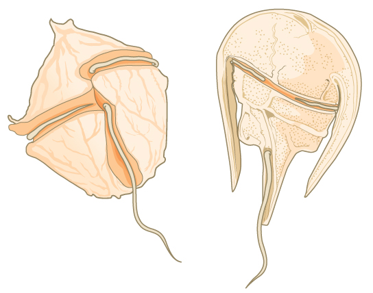
Figure 23.21図 23.21
Dinoflagellates. The dinoflagellates exhibit great diversity in shape. Many are encased in cellulose armor and have two flagella that fit in grooves between the plates. Movement of these two perpendicular flagella causes a spinning motion.
渦鞭毛藻。渦鞭毛藻の形は、非常に多様である。多くはセルロースのよろいに包まれ、板の間の溝に収まる2本の鞭毛をもつ。互いに直交する2本の鞭毛の運動が、回転運動を引き起こす。
Dinoflagellates have a nuclear variant called a dinokaryon. The chromosomes in the dinokaryon are highly condensed throughout the cell cycle and do not have typical histones. Mitosis in dinoflagellates is closed, that is, the spindle separates the chromosomes from outside of the nucleus without breakdown of the nuclear envelope.
英語のヒント
渦鞭毛藻は、ダイノカリオンと呼ばれる特殊な核をもつ。この核の染色体は、細胞周期全体を通じて強く凝縮しており、典型的なヒストンをもたない。渦鞭毛藻の有糸分裂は閉鎖型である。つまり、核膜を崩壊させずに、紡錘体が核の外側から染色体を分離する。
Some dinoflagellates generate light, called bioluminescence, when they are jarred or stressed. Large numbers of marine dinoflagellates (billions or trillions of cells per wave) can emit light and cause an entire breaking wave to twinkle or take on a brilliant blue color (Figure 23.22). For approximately 20 species of marine dinoflagellates, population explosions (also called blooms) during the summer months can tint the ocean with a muddy red color. This phenomenon is called a red tide, and it results from the abundant red pigments present in dinoflagellate plastids. In large quantities, these dinoflagellate species secrete an asphyxiating toxin that can kill fish, birds, and marine mammals. Red tides can be massively detrimental to commercial fisheries, and humans who consume these protists may become poisoned.
英語のヒント
一部の渦鞭毛藻は、揺さぶられたりストレスを受けたりすると、生物発光と呼ばれる光を生じる。海産渦鞭毛藻が大量に存在すると、1波あたり数十億〜数兆個の細胞が発光し、砕ける波全体がきらめいたり、鮮やかな青色になったりする(図23.22)。海産渦鞭毛藻の約20種では、夏の個体数の爆発的増加、いわゆるブルームによって、海が濁った赤色に染まることがある。この現象を赤潮といい、色素体内の豊富な赤色色素によって生じる。これらの渦鞭毛藻が大量にいると、魚、鳥、海生哺乳類を殺しうる窒息性の毒素を分泌する。赤潮は商業漁業に甚大な損害を与えることがあり、これらの原生生物を摂取したヒトも、中毒になる可能性がある。
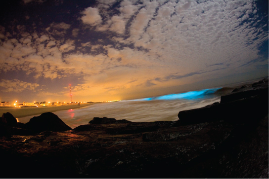


Figure 23.22図 23.22
Dinoflagellate bioluminescence. Bioluminescence is emitted from dinoflagellates in a breaking wave, as seen from the New Jersey coast. (credit: “catalano82”/Flickr)
渦鞭毛藻の生物発光。ニュージャージー沿岸から見た、砕ける波の中で渦鞭毛藻が放つ生物発光。(クレジット:“catalano82”/Flickr)
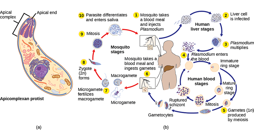
The apicomplexan protists are named for a structure called an apical complex (Figure 23.23), which appears to be a highly modified secondary chloroplast. The apicoplast genome is similar to those of dinoflagellate chloroplasts. The apical complex is specialized for entry and infection of host cells. Indeed, all apicomplexans are parasitic. This group includes the genus Plasmodium, which causes malaria in humans. Apicomplexan life cycles are complex, involving multiple hosts and stages of sexual and asexual reproduction.
英語のヒント
アピコンプレクサという原生生物の名は、アピカルコンプレックスという構造(図23.23)に由来する。これは、高度に変型した二次葉緑体のように見える。アピコプラストのゲノムは、渦鞭毛藻の葉緑体ゲノムに似ている。アピカルコンプレックスは、宿主細胞への侵入と感染に特化している。実際、アピコンプレクサはすべて寄生性である。この群には、ヒトにマラリアを引き起こすPlasmodium属が含まれる。アピコンプレクサの生活環は複雑で、複数の宿主と、有性生殖・無性生殖の段階が関わる。
OpenStax, Rice University; Biology 2e. (credit b: modification of work by CDC) · CC BY 4.0 · Source出典

Figure 23.23図 23.23
Apicomplexa. (a) Apicomplexans are parasitic protists. They have a characteristic apical complex that enables them to infect host cells. (b) Plasmodium, the causative agent of malaria, has a complex life cycle typical of apicomplexans. (credit b: modification of work by CDC)
アピコンプレクサ。(a)アピコンプレクサは、寄生性原生生物である。特徴的なアピカルコンプレックスをもち、これによって宿主細胞に感染する。(b)マラリアの病原体Plasmodiumは、アピコンプレクサに典型的な複雑な生活環をもつ。(bのクレジット:CDCによる作品を改変)
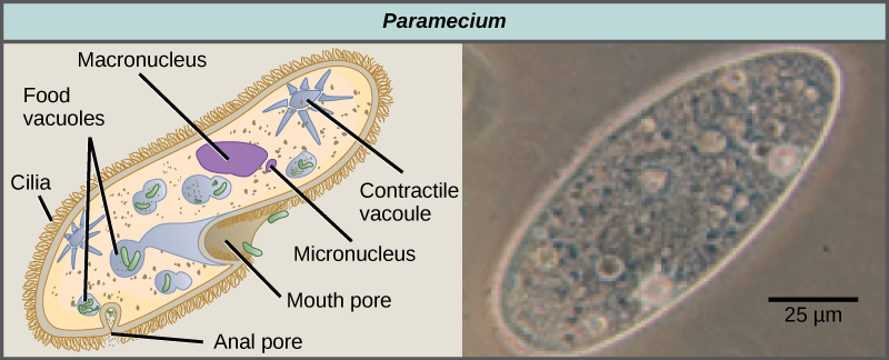
The ciliates, which include Paramecium and Tetrahymena, are a group of protists 10 to 3,000 micrometers in length that are covered in rows, tufts, or spirals of tiny cilia. By beating their cilia synchronously or in waves, ciliates can coordinate directed movements and ingest food particles. Certain ciliates have fused cilia-based structures that function like paddles, funnels, or fins. Ciliates also are surrounded by a pellicle, providing protection without compromising agility. The genus Paramecium includes protists that have organized their cilia into a plate-like primitive mouth, called an oral groove, which is used to capture and digest bacteria (Figure 23.24). Food captured in the oral groove enters a food vacuole, where it combines with digestive enzymes. Waste particles are expelled by an exocytic vesicle that fuses at a specific region on the cell membrane, called the anal pore. In addition to a vacuole-based digestive system, Paramecium also uses contractile vacuoles, which are osmoregulatory vesicles that fill with water as it enters the cell by osmosis and then contract to squeeze water from the cell. Ciliates therefore exhibit considerable structural complexity without having achieved multicellularity.
英語のヒント
ゾウリムシやテトラヒメナを含む繊毛虫は、長さ10〜3,000マイクロメートルの原生生物の群で、小さな繊毛の列、束、螺旋に覆われる。繊毛を同期させて、または波状に打ち動かすことで、方向性のある運動を調整し、食物粒子を摂取できる。ある種の繊毛虫では、繊毛が融合した構造が、櫂、漏斗、ひれのように働く。繊毛虫はペリクルにも囲まれ、敏捷さを損なわずに保護される。ゾウリムシ属には、繊毛を板状の原始的な口へまとめた原生生物が含まれる。この口溝は、細菌の捕獲と消化に用いられる(図23.24)。口溝で捕らえた食物は食胞へ入り、消化酵素と混ざる。老廃物粒子は、細胞膜の細胞肛門という特定の領域で融合するエキソサイトーシス小胞によって排出される。液胞を基盤とする消化系に加えて、ゾウリムシは収縮胞も用いる。収縮胞は浸透圧調節を行う小胞で、浸透によって細胞へ入った水で満たされた後、収縮して水を細胞外へ絞り出す。したがって、繊毛虫は多細胞化を達成しなくても、かなり複雑な構造を示す。
OpenStax, Rice University; Biology 2e. (credit “paramecium micrograph”: modification of work by NIH; scale-bar data from Matt Russell) · CC BY 4.0 · Source出典

Figure 23.24図 23.24
Paramecium. Paramecium has a primitive mouth (called an oral groove) to ingest food, and an anal pore to eliminate waste. Contractile vacuoles allow the organism to excrete excess water. Cilia enable the organism to move. (credit “paramecium micrograph”: modification of work by NIH; scale-bar data from Matt Russell)
ゾウリムシ。ゾウリムシは、食物を摂取する原始的な口(口溝)と、老廃物を除く細胞肛門をもつ。収縮胞は余分な水の排出を可能にし、繊毛は移動を可能にする。(ゾウリムシ顕微鏡像のクレジット:NIHによる作品を改変。スケールバーのデータ:Matt Russell)
Paramecium has two nuclei, a macronucleus and a micronucleus, in each cell. The micronucleus is essential for sexual reproduction, and is in many ways a typical eukaryotic nucleus, except that its genes are not transcribed. The transcribed nucleus is the macronucleus, which directs asexual binary fission and all other biological functions. The macronucleus is a multiploid nucleus constructed from the micronucleus during sexual reproduction. Periodic reconstruction of the macronucleus is necessary because the macronucleus divides amitotically, and thus becomes genetically unbalanced over a period of successive cell replications. Paramecium and most other ciliates reproduce sexually by conjugation. This process begins when two different mating types of Paramecium make physical contact and join with a cytoplasmic bridge (Figure 23.25). The diploid micronucleus in each cell then undergoes meiosis to produce four haploid micronuclei. Three of these degenerate in each cell, leaving one micronucleus that then undergoes mitosis, generating two haploid micronuclei. The cells each exchange one of these haploid nuclei and move away from each other. Fusion of the haploid micronuclei generates a completely novel diploid pre-micronucleus in each conjugative cell. This pre-micronucleus undergoes three rounds of mitosis to produce eight copies, and the original macronucleus disintegrates. Four of the eight pre-micronuclei become full-fledged micronuclei, whereas the other four perform multiple rounds of DNA replication. The copies of the micronuclear chromosomes are severely edited to form hundreds of smaller chromosomes that contain only the protein coding genes. Each of these smaller chromosomes gets new telomeres as the macronucleus differentiates. Two cycles of cell division then yield four new Paramecia from each original conjugative cell.
英語のヒント
ゾウリムシは各細胞に、大核と小核という二つの核をもつ。小核は有性生殖に不可欠であり、その遺伝子が転写されないことを除けば、多くの点で典型的な真核細胞の核である。転写が行われる核は大核で、無性の二分裂と、他のすべての生命機能を指揮する。大核は、有性生殖の際に小核から作られる多倍性の核である。大核は無糸分裂で分かれ、細胞の複製を繰り返すうちに遺伝的な不均衡が生じるため、定期的に作り直す必要がある。ゾウリムシと大部分の繊毛虫は、接合によって有性生殖する。この過程は、異なる接合型のゾウリムシ2個体が接触し、細胞質の橋でつながると始まる(図23.25)。各細胞の二倍体小核は減数分裂して、四つの半数体小核を作る。各細胞でそのうち三つが退化し、残る一つが有糸分裂して二つの半数体小核となる。各細胞は、この半数体核の一つを交換し、互いに離れる。半数体小核が融合すると、接合した各細胞に、まったく新しい二倍体の小核前駆体ができる。これは有糸分裂を3回行って八つになり、元の大核は崩壊する。八つの小核前駆体のうち四つは完全な小核となり、残りの四つはDNA複製を何度も行う。小核の染色体のコピーには大幅な編集が加わり、タンパク質をコードする遺伝子だけを含む、数百本のより小さな染色体となる。大核が分化する際、これらの小さな染色体には、それぞれ新しいテロメアが付く。その後、細胞分裂が2回起こり、元の各接合細胞から四つの新しいゾウリムシが生じる。