第3章生体高分子
Secondary Structure二次構造
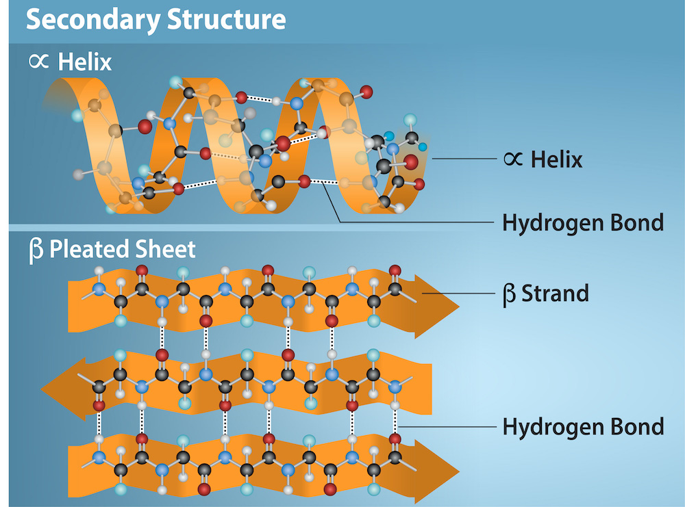
The local folding of the polypeptide in some regions gives rise to the secondary structure of the protein. The most common are the α-helix and β-pleated sheet structures (Figure 3.28). Both structures are held in shape by hydrogen bonds. The hydrogen bonds form between the oxygen atom in the carbonyl group in one amino acid and another amino acid that is four amino acids farther along the chain.
英語のヒント
ポリペプチドの一部の領域が局所的に折りたたまれることで、タンパク質の二次構造が生じる。最も一般的なのは、αヘリックスとβシートである(図3.28)。どちらも水素結合によって形が保たれる。水素結合は、あるアミノ酸のカルボニル基の酸素原子と、鎖に沿って四つ先にある別のアミノ酸との間に形成される。
OpenStax, Rice University; Biology 2e. Credit: Rao, A., Ryan, K. Fletcher, S. and Tag, A. Department of Biology, Texas A&M University. · CC BY 4.0 · Source出典
Figure 3.28図 3.28
The α-helix and β-pleated sheet are secondary protein structures formed when hydrogen bonds form between the carbonyl oxygen and the amino hydrogen in the peptide backbone. Certain amino acids have a propensity to form an α-helix while others favor β-pleated sheet formation. Black = carbon, White = hydrogen, Blue = nitrogen, and Red = oxygen. Credit: Rao, A., Ryan, K. Fletcher, S. and Tag, A. Department of Biology, Texas A&M University.
αヘリックスとβシートは、ペプチド骨格のカルボニル基の酸素とアミノ基の水素との間に水素結合ができて形成される、タンパク質の二次構造である。アミノ酸の種類によって、αヘリックスを作りやすいものとβシートを作りやすいものがある。黒は炭素、白は水素、青は窒素、赤は酸素を表す。出典:Rao, A., Ryan, K., Fletcher, S. and Tag, A.(テキサスA&M大学生物学部)
Every helical turn in an alpha helix has 3.6 amino acid residues. The polypeptide's R groups (the variant groups) protrude out from the α-helix chain. In the β-pleated sheet, hydrogen bonding between atoms on the polypeptide chain's backbone form the "pleats". The R groups are attached to the carbons and extend above and below the pleat's folds. The pleated segments align parallel or antiparallel to each other, and hydrogen bonds form between the partially positive hydrogen atom in the amino group and the partially negative oxygen atom in the peptide backbone's carbonyl group. The α-helix and β-pleated sheet structures are in most globular and fibrous proteins and they play an important structural role.
英語のヒント
αヘリックスのらせん一回転には、3.6個のアミノ酸残基が含まれる。ポリペプチドのR基(可変の基)は、αヘリックスの鎖から外へ突き出している。βシートでは、ポリペプチド鎖の骨格の原子間の水素結合が、「ひだ」を作る。R基は炭素に結合し、ひだの折れ曲がりの上と下へ伸びている。ひだ状の部分は、互いに平行または逆平行に並ぶ。アミノ基の部分的に正に帯電した水素原子と、ペプチド骨格のカルボニル基の部分的に負に帯電した酸素原子との間に、水素結合が形成される。αヘリックスとβシートは、大部分の球状・繊維状タンパク質に存在し、構造上重要な役割を果たす。
Tertiary Structure三次構造
The polypeptide's unique three-dimensional structure is its tertiary structure (Figure 3.29). This structure is in part due to chemical interactions at work on the polypeptide chain. Primarily, the interactions among R groups create the protein's complex three-dimensional tertiary structure. The nature of the R groups in the amino acids involved can counteract forming the hydrogen bonds we described for standard secondary structures. For example, R groups with like charges repel each other and those with unlike charges are attracted to each other (ionic bonds). When protein folding takes place, the nonpolar amino acids' hydrophobic R groups lie in the protein's interior; whereas, the hydrophilic R groups lie on the outside. Scientists also call the former interaction types hydrophobic interactions. Interaction between cysteine side chains forms disulfide linkages in the presence of oxygen, the only covalent bond that forms during protein folding.
英語のヒント
ポリペプチドに固有の三次元構造が、三次構造である(図3.29)。この構造は、一部にはポリペプチド鎖に働く化学的相互作用によって生じる。主にR基同士の相互作用が、タンパク質の複雑な三次元の三次構造を作る。関与するアミノ酸のR基の性質によって、通常の二次構造で述べた水素結合の形成が妨げられる場合がある。例えば、同符号の電荷を持つR基は反発し、逆符号の電荷を持つR基は引き合う(イオン結合)。タンパク質が折りたたまれるとき、無極性アミノ酸の疎水性R基は内部に、親水性R基は外側に位置する。前者の相互作用は、疎水性相互作用とも呼ばれる。酸素の存在下では、システインの側鎖同士の相互作用によってジスルフィド結合ができる。これは、タンパク質の折りたたみの際に形成される唯一の共有結合である。
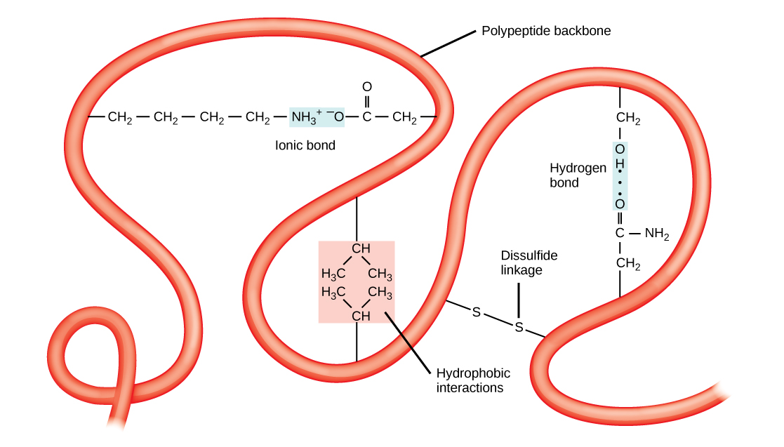


Figure 3.29図 3.29
A variety of chemical interactions determine the proteins' tertiary structure. These include hydrophobic interactions, ionic bonding, hydrogen bonding, and disulfide linkages.
さまざまな化学的相互作用がタンパク質の三次構造を決める。疎水性相互作用、イオン結合、水素結合、ジスルフィド結合などが含まれる。
All of these interactions, weak and strong, determine the protein's final three-dimensional shape. When a protein loses its three-dimensional shape, it may no longer be functional.
英語のヒント
弱いものも強いものも、これらすべての相互作用が、タンパク質の最終的な三次元の形を決める。三次元の形を失うと、タンパク質は機能しなくなる場合がある。
Quaternary Structure四次構造
In nature, some proteins form from several polypeptides, or subunits, and the interaction of these subunits forms the quaternary structure. Weak interactions between the subunits help to stabilize the overall structure. For example, insulin (a globular protein) has a combination of hydrogen and disulfide bonds that cause it to mostly clump into a ball shape. Insulin starts out as a single polypeptide and loses some internal sequences in the presence of post-translational modification after forming the disulfide linkages that hold the remaining chains together. Silk (a fibrous protein), however, has a β-pleated sheet structure that is the result of hydrogen bonding between different chains.
英語のヒント
自然界には、複数のポリペプチド、すなわちサブユニットからなるタンパク質があり、サブユニット同士の相互作用が四次構造を作る。サブユニット間の弱い相互作用が、全体の構造を安定させる。例えば、球状タンパク質のインスリンは、水素結合とジスルフィド結合の組み合わせによって、ほぼ球状にまとまる。インスリンは最初は一本のポリペプチドで、残る鎖をつなぐジスルフィド結合ができた後、翻訳後修飾によって内部の一部の配列を失う。一方、繊維状タンパク質の絹は、異なる鎖同士の水素結合によって生じるβシート構造を持つ。
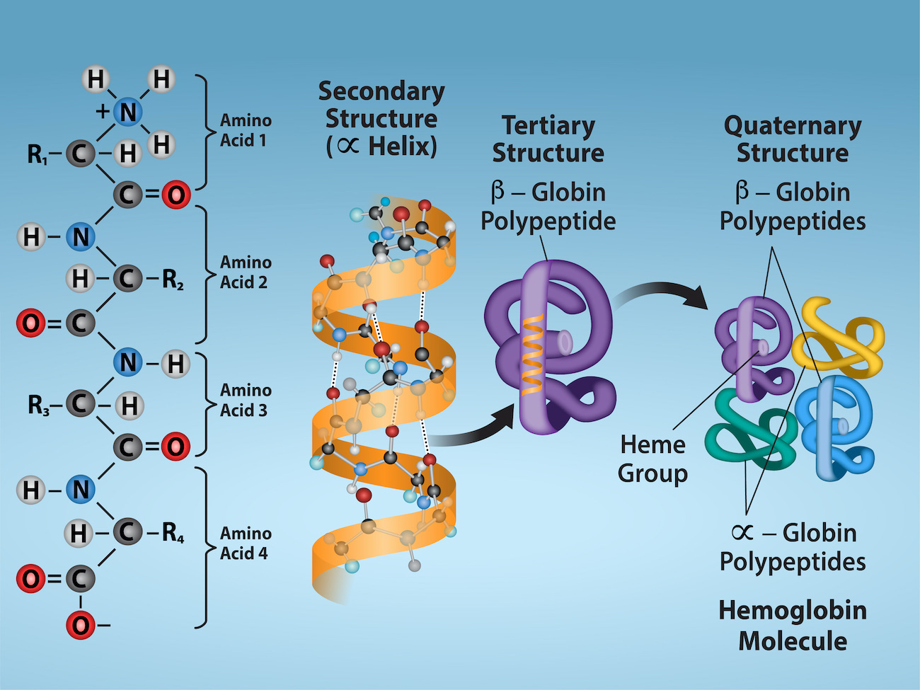
Figure 3.30 illustrates the four levels of protein structure (primary, secondary, tertiary, and quaternary).
図3.30に、タンパク質の四つの構造段階、すなわち一次、二次、三次、四次構造を示す。
OpenStax, Rice University; Biology 2e. Credit: Rao, A. Ryan, K. and Tag, A. Department of Biology, Texas A&M University. · CC BY 4.0 · Source出典

Figure 3.30図 3.30
Observe the four levels of protein structure in these illustrations. Credit: Rao, A. Ryan, K. and Tag, A. Department of Biology, Texas A&M University.
これらの図で、タンパク質の四つの構造段階を観察しよう。出典:Rao, A., Ryan, K. and Tag, A.(テキサスA&M大学生物学部)
Denaturation and Protein Folding変性とタンパク質の折りたたみ
Each protein has its own unique sequence and shape that chemical interactions hold together. If the protein is subject to changes in temperature, pH, or exposure to chemicals, the protein structure may change, losing its shape without losing its primary sequence in what scientists call denaturation. Denaturation is often reversible because the polypeptide's primary structure is conserved in the process if the denaturing agent is removed, allowing the protein to resume its function. Sometimes denaturation is irreversible, leading to loss of function. One example of irreversible protein denaturation is frying an egg. The albumin protein in the liquid egg white denatures when placed in a hot pan. Not all proteins denature at high temperatures. For instance, bacteria that survive in hot springs have proteins that function at temperatures close to boiling. The stomach is also very acidic, has a low pH, and denatures proteins as part of the digestion process; however, the stomach's digestive enzymes retain their activity under these conditions.
英語のヒント
各タンパク質は固有の配列と形を持ち、その形は化学的相互作用によって保たれている。温度やpHが変わったり、化学物質にさらされたりすると、タンパク質の構造が変わる場合がある。一次配列は失わずに形を失う、この現象を変性という。変性ではポリペプチドの一次構造が保たれるため、変性を引き起こす作用を取り除けば元に戻り、タンパク質が機能を回復できることが多い。変性が不可逆で、機能が失われる場合もある。卵を焼くことは、不可逆的なタンパク質変性の一例である。液状の卵白にあるアルブミンは、熱いフライパンに入れると変性する。すべてのタンパク質が高温で変性するわけではない。例えば、温泉で生きる細菌には、沸点近くでも働くタンパク質がある。胃も強い酸性、つまり低いpHを持ち、消化過程の一部としてタンパク質を変性させる。しかし、胃の消化酵素は、この条件でも活性を保つ。
Protein folding is critical to its function. Scientists originally thought that the proteins themselves were responsible for the folding process. Only recently researchers discovered that often they receive assistance in the folding process from protein helpers, or chaperones (or chaperonins) that associate with the target protein during the folding process. They act by preventing polypeptide aggregation that comprise the complete protein structure, and they disassociate from the protein once the target protein is folded.
英語のヒント
タンパク質の折りたたみは、その機能に不可欠である。当初は、タンパク質自身が折りたたみを担うと考えられていた。しかし、折りたたみの際に、タンパク質の補助役であるシャペロン(またはシャペロニン)の助けを受けることが多いと分かったのは、つい最近のことである。これらは、折りたたみの過程で標的タンパク質に結合する。シャペロンは、完全なタンパク質構造を構成するポリペプチドの凝集を防ぐことで働き、標的タンパク質が折りたたまれると、そこから離れる。