第3章生体高分子
Protein Structureタンパク質の構造
As we discussed earlier, a protein's shape is critical to its function. For example, an enzyme can bind to a specific substrate at an active site. If this active site is altered because of local changes or changes in overall protein structure, the enzyme may be unable to bind to the substrate. To understand how the protein gets its final shape or conformation, we need to understand the four levels of protein structure: primary, secondary, tertiary, and quaternary.
英語のヒント
先に述べたように、タンパク質の形は機能に極めて重要である。例えば酵素は、活性部位で特定の基質に結合できる。この活性部位が局所的な変化やタンパク質全体の構造変化によって変わると、基質に結合できなくなることがある。タンパク質が最終的な形、すなわち立体配座を得る仕組みを理解するには、一次、二次、三次、四次という四つの構造段階を理解する必要がある。
Primary Structure一次構造
Amino acids' unique sequence in a polypeptide chain is its primary structure. For example, the pancreatic hormone insulin has two polypeptide chains, A and B, and they are linked together by disulfide bonds. The N terminal amino acid of the A chain is glycine; whereas, the C terminal amino acid is asparagine (Figure 3.25). The amino acid sequences in the A and B chains are unique to insulin.
英語のヒント
ポリペプチド鎖内の固有のアミノ酸配列が、その一次構造である。例えば膵臓のホルモンであるインスリンには、A鎖とB鎖の二本のポリペプチド鎖があり、ジスルフィド結合でつながっている。A鎖のN末端アミノ酸はグリシン、C末端アミノ酸はアスパラギンである(図3.25)。A鎖とB鎖のアミノ酸配列は、インスリンに固有のものである。
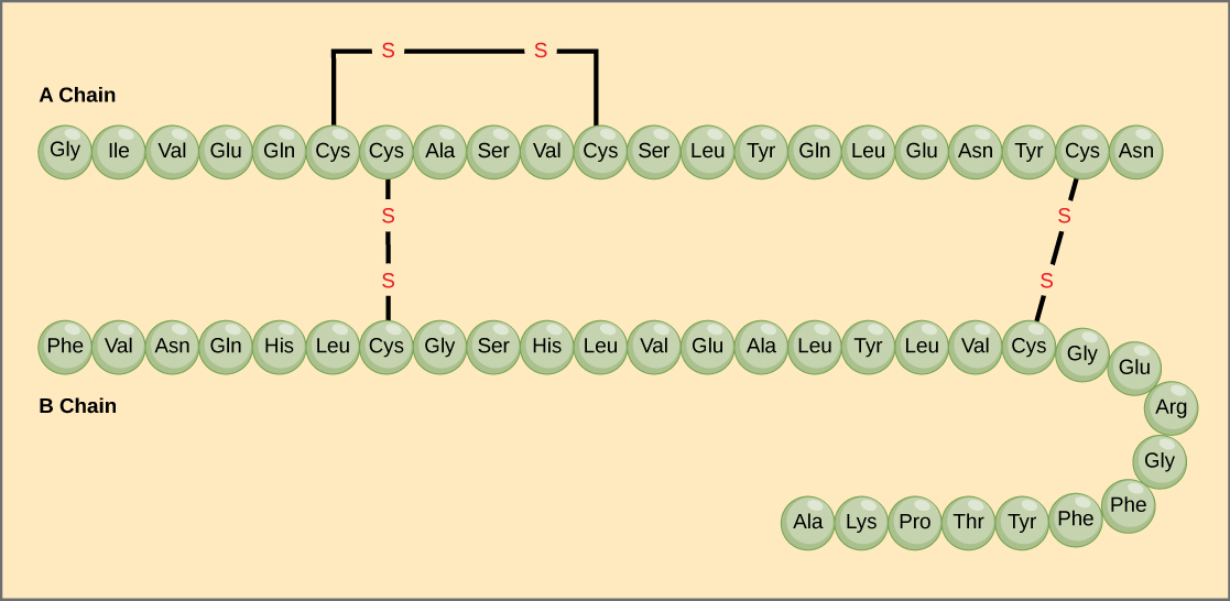

Figure 3.25図 3.25
Bovine serum insulin is a protein hormone comprised of two peptide chains, A (21 amino acids long) and B (30 amino acids long). In each chain, three-letter abbreviations that represent the amino acids' names in the order they are present indicate primary structure. The amino acid cysteine (cys) has a sulfhydryl (SH) group as a side chain. Two sulfhydryl groups can react in the presence of oxygen to form a disulfide (S-S) bond. Two disulfide bonds connect the A and B chains together, and a third helps the A chain fold into the correct shape. Note that all disulfide bonds are the same length, but we have drawn them different sizes for clarity.
英語のヒント
ウシ血清インスリンは、A鎖(アミノ酸21個)とB鎖(アミノ酸30個)の二本のペプチド鎖からなるタンパク質ホルモンである。各鎖の一次構造は、アミノ酸名を表す三文字略号を、実際に存在する順に並べて示している。システイン(cys)は、側鎖にスルフヒドリル基(SH)を持つ。酸素があると、二つのスルフヒドリル基が反応してジスルフィド結合(S–S)を形成できる。二つのジスルフィド結合がA鎖とB鎖をつなぎ、三つ目はA鎖が正しい形に折りたたまれるのを助ける。すべてのジスルフィド結合の長さは同じだが、図を見やすくするため、異なる長さで描いてある。
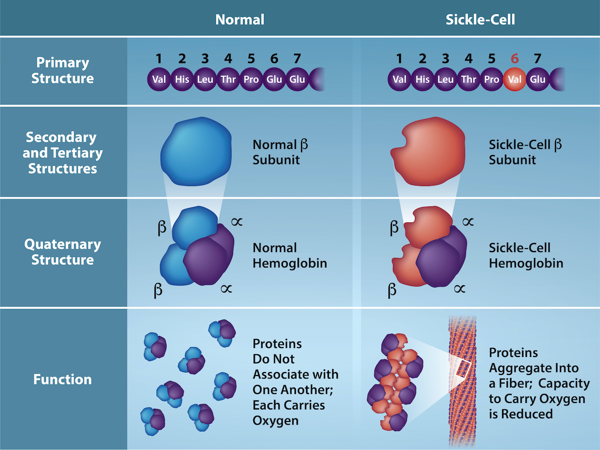
The gene encoding the protein ultimately determines the unique sequence for every protein. A change in nucleotide sequence of the gene’s coding region may lead to adding a different amino acid to the growing polypeptide chain, causing a change in protein structure and function. In sickle cell anemia, the hemoglobin β chain (a small portion of which we show in Figure 3.26) has a single amino acid substitution, causing a change in protein structure and function. Specifically, valine in the β chain substitutes the amino acid glutamic. What is most remarkable to consider is that a hemoglobin molecule is comprised of two alpha and two beta chains that each consist of about 150 amino acids. The molecule, therefore, has about 600 amino acids. The structural difference between a normal hemoglobin molecule and a sickle cell molecule—which dramatically decreases life expectancy—is a single amino acid of the 600. What is even more remarkable is that three nucleotides each encode those 600 amino acids, and a single base change (point mutation), 1 in 1800 bases causes the mutation.
英語のヒント
各タンパク質の固有の配列は、最終的にはそのタンパク質をコードする遺伝子によって決まる。遺伝子のコード領域の塩基配列が変わると、伸長中のポリペプチド鎖に異なるアミノ酸が加わり、タンパク質の構造や機能が変わる場合がある。鎌状赤血球貧血では、ヘモグロビンのβ鎖(その一部を図3.26に示す)のアミノ酸が一つ置換され、タンパク質の構造と機能が変わる。具体的には、β鎖のグルタミン酸がバリンに置き換わる。特に注目すべきなのは、ヘモグロビン分子が二本のα鎖と二本のβ鎖からなり、それぞれが約150個のアミノ酸を含むという点である。したがって、分子全体には約600個のアミノ酸がある。正常なヘモグロビン分子と鎌状赤血球のヘモグロビン分子との構造上の違いは、この600個のうち一つのアミノ酸にすぎないが、それが平均余命を大きく縮める。さらに驚くべきことに、600個のアミノ酸はそれぞれ三つのヌクレオチドでコードされており、1800個の塩基のうち、ただ一つの塩基の変化、すなわち点突然変異がこの変異を引き起こす。
OpenStax, Rice University; Biology 2e. (credit: Rao, A., Tag, A. Ryan, K. and Fletcher, S. Department of Biology, Texas A&M University) · CC BY 4.0 · Source出典

Figure 3.26図 3.26
Because of this change of one amino acid in the chain, hemoglobin molecules form long fibers that distort the biconcave, or disc-shaped, red blood cells and causes them to assume a crescent or “sickle” shape, which clogs blood vessels (Figure 3.27). The beta (β)- chain of hemoglobin is 147 amino acids in length, yet a single amino acid substitution in the primary sequence leads to changes in secondary, tertiary, and quaternary structures and sickle cell anemia. In normal hemoglobin, the amino acid at position six is glutamate. In sickle cell hemoglobin glutamate is replaced by valine. (credit: Rao, A., Tag, A. Ryan, K. and Fletcher, S. Department of Biology, Texas A&M University)
鎖の一つのアミノ酸が変わることで、ヘモグロビン分子は長い繊維を作り、両凹形、つまり円盤状の赤血球を変形させる。赤血球は三日月形、すなわち「鎌形」となり、血管を詰まらせる(図3.27)。ヘモグロビンのβ鎖は147個のアミノ酸からなるが、一次配列中のアミノ酸一つの置換だけで二次・三次・四次構造が変わり、鎌状赤血球貧血になる。正常なヘモグロビンでは6番目のアミノ酸はグルタミン酸であり、鎌状赤血球のヘモグロビンではバリンに置き換わっている。(出典:Rao, A., Tag, A., Ryan, K. and Fletcher, S.、テキサスA&M大学生物学部)
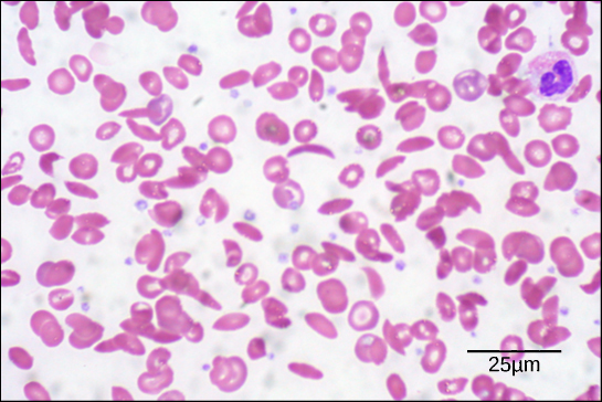
Because of this change of one amino acid in the chain, hemoglobin molecules form long fibers that distort the biconcave, or disc-shaped, red blood cells and causes them to assume a crescent or “sickle” shape, which clogs blood vessels (Figure 3.27). This can lead to myriad serious health problems such as breathlessness, dizziness, headaches, and abdominal pain for those affected by this disease. William Warrick Cardozo showed that sickle-cell anemia is an inherited disorder, meaning that the difference in the specific gene's encoding region is passed down from parents to children. As you will learn in the genetics unit, the inheritance of such traits is determined by a combination of genes from both parents, and these very small differences can have significant impacts on organisms.
英語のヒント
鎖のアミノ酸一つが変わることで、ヘモグロビン分子は長い繊維を作り、両凹形、つまり円盤状の赤血球を変形させる。赤血球は三日月形、すなわち「鎌形」となり、血管を詰まらせる(図3.27)。この病気の人には、息切れ、めまい、頭痛、腹痛など、数多くの深刻な健康問題が起こりうる。ウィリアム・ウォリック・カルドーゾは、鎌状赤血球貧血が遺伝性の疾患であることを示した。つまり、特定の遺伝子のコード領域の違いが、親から子へ伝わるのである。遺伝学の部で学ぶように、こうした形質の遺伝は両親の遺伝子の組み合わせで決まる。ごく小さな違いでも、生物に大きな影響を及ぼすことがある。
OpenStax, Rice University; Biology 2e. (credit: modification of work by Ed Uthman; scale-bar data from Matt Russell) · CC BY 4.0 · Source出典

Figure 3.27図 3.27
In this blood smear, visualized at 535x magnification using bright field microscopy, sickle cells are crescent shaped, while normal cells are disc-shaped. (credit: modification of work by Ed Uthman; scale-bar data from Matt Russell)
この血液塗抹標本は、明視野顕微鏡で535倍に拡大している。鎌状赤血球は三日月形で、正常な赤血球は円盤状である。(出典:Ed Uthmanの作品を改変。スケールバーのデータ:Matt Russell)