第32章植物の生殖
Male Gametophyte (The Pollen Grain)雄性配偶体(花粉粒)
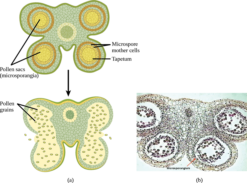
The male gametophyte develops and reaches maturity in an immature anther. In a plant’s male reproductive organs, development of pollen takes place in a structure known as the microsporangium (Figure 32.6). The microsporangia, which are usually bilobed, are pollen sacs in which the microspores develop into pollen grains. These are found in the anther, which is at the end of the stamen—the long filament that supports the anther.
英語のヒント
雄性配偶体は、未成熟な葯の中で発達して成熟する。植物の雄の生殖器官では、小胞子嚢と呼ばれる構造で花粉が発達する(図32.6)。小胞子嚢は通常二裂した花粉嚢で、その中で小胞子が花粉粒へ発達する。小胞子嚢は葯の中にあり、葯は、それを支える長い花糸である雄しべの先端にある。
OpenStax, Rice University; Biology 2e. (credit a: modification of work by LibreTexts; b: modification of work by Robert R. Wise; scale-bar data from Matt Russell) · CC BY 4.0 · Source出典

Figure 32.6図 32.6
Shown is (a) a cross section of an anther at two developmental stages. The immature anther (top) contains four microsporangia, or pollen sacs. Each microsporangium contains hundreds of microspore mother cells that will each give rise to four pollen grains. The tapetum supports the development and maturation of the pollen grains. Upon maturation of the pollen (bottom), the pollen sac walls split open and the pollen grains (male gametophytes) are released, as shown in the (b) micrograph of an immature lily anther. In these scanning electron micrographs, pollen sacs are ready to burst, releasing their grains. (credit a: modification of work by LibreTexts; b: modification of work by Robert R. Wise; scale-bar data from Matt Russell)
(a)二つの発達段階における葯の横断面。未成熟な葯(上)には、小胞子嚢、すなわち花粉嚢が四つある。それぞれの小胞子嚢には数百個の小胞子母細胞があり、各母細胞から4個の花粉粒が生じる。タペート層は花粉粒の発達と成熟を支える。花粉が成熟すると(下)、花粉嚢の壁が裂けて開き、雄性配偶体である花粉粒が放出される。その様子は、(b)未成熟なユリの葯の顕微鏡写真に示されている。これらの走査型電子顕微鏡写真では、花粉嚢が破れて粒を放出する直前にある。(クレジットa:LibreTextsの作品を改変;b:Robert R. Wiseの作品を改変;スケールバーのデータ:Matt Russell)
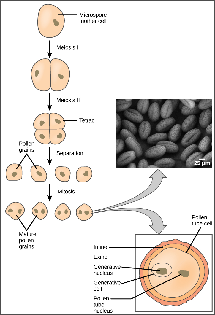
Within the microsporangium, each of the microspore mother cells divides by meiosis to give rise to four microspores, each of which will ultimately form a pollen grain (Figure 32.7). An inner layer of cells, known as the tapetum, provides nutrition to the developing microspores and contributes key components to the pollen wall. Mature pollen grains contain two cells: a generative cell and a pollen tube cell. The generative cell is contained within the larger pollen tube cell. Upon germination, the tube cell forms the pollen tube through which the generative cell migrates to enter the ovary. During its transit inside the pollen tube, the generative cell divides to form two male gametes (sperm cells). Upon maturity, the microsporangia burst, releasing the pollen grains from the anther.
英語のヒント
小胞子嚢の中では、それぞれの小胞子母細胞が減数分裂して4個の小胞子を生じ、各小胞子が最終的に花粉粒となる(図32.7)。タペート層と呼ばれる内側の細胞層は、発達中の小胞子へ栄養を供給し、花粉壁の主要な成分を与える。成熟した花粉粒には、雄原細胞と花粉管細胞の二つの細胞が含まれる。雄原細胞は、大きな花粉管細胞の内側にある。発芽すると、花粉管細胞が花粉管を作り、その中を雄原細胞が移動して子房へ入る。雄原細胞は花粉管内を移動する間に分裂し、二つの雄性配偶子である精細胞を作る。成熟すると小胞子嚢が破れ、葯から花粉粒が放出される。
OpenStax, Rice University; Biology 2e. (credit “pollen micrograph”: modification of work by Robert R. Wise; scale-bar data from Matt Russell) · CC BY 4.0 · Source出典

Figure 32.7図 32.7
Pollen develops from the microspore mother cells. The mature pollen grain is composed of two cells: the pollen tube cell and the generative cell, which is inside the tube cell. The pollen grain has two coverings: an inner layer (intine) and an outer layer (exine). The inset scanning electron micrograph shows Arabidopsis lyrata pollen grains. (credit “pollen micrograph”: modification of work by Robert R. Wise; scale-bar data from Matt Russell)
花粉は小胞子母細胞から発達する。成熟した花粉粒は、花粉管細胞と、その内側にある雄原細胞の二つの細胞からなる。花粉粒には、内側の内壁と外側の外壁という二つの覆いがある。挿入された走査型電子顕微鏡写真は、Arabidopsis lyrataの花粉粒を示す。(花粉顕微鏡写真のクレジット:Robert R. Wiseの作品を改変;スケールバーのデータ:Matt Russell)
Each pollen grain has two coverings: the exine (thicker, outer layer) and the intine (Figure 32.7). The exine contains sporopollenin, a complex waterproofing substance supplied by the tapetal cells. Sporopollenin allows the pollen to survive under unfavorable conditions and to be carried by wind, water, or biological agents without undergoing damage.
それぞれの花粉粒には、厚い外側の層である外壁と、内壁という二つの覆いがある(図32.7)。外壁には、タペート細胞が供給する複雑な防水性物質、スポロポレニンが含まれる。スポロポレニンによって、花粉は不利な条件下でも生き延び、風、水、生物に運ばれても損傷を受けずにすむ。
Female Gametophyte (The Embryo Sac)雌性配偶体(胚嚢)
While the details may vary between species, the development of the female gametophyte occurs in a two-phase process called megasporogenesis. In the first phase, a single cell in the diploid megasporangium—an area of tissue in the ovules—undergoes meiosis to produce four megaspores, only one of which survives. During the second phase, megagametogenesis, the surviving haploid megaspore undergoes mitosis to produce an eight-nucleate, seven-cell female gametophyte, also known as the megagametophyte or embryo sac. Two of the nuclei—the polar nuclei—move to the equator and fuse, forming a single, diploid central cell. This central cell later fuses with a sperm to form the triploid endosperm. Three nuclei position themselves on the end of the embryo sac opposite the micropyle and develop into the antipodal cells, which later degenerate. The nucleus closest to the micropyle becomes the female gamete, or egg cell, and the two adjacent nuclei develop into synergid cells (Figure 32.8). The synergids help guide the pollen tube for successful fertilization, after which they disintegrate. Once fertilization is complete, the resulting diploid zygote develops into the embryo, and the fertilized ovule forms the other tissues of the seed.
英語のヒント
細部は種によって異なることがあるが、雌性配偶体の発達は、大胞子形成と呼ばれる二段階の過程で起こる。第1段階では、胚珠内の組織領域である二倍体の大胞子嚢の中で、一つの細胞が減数分裂して4個の大胞子を作り、そのうち1個だけが生き残る。第2段階の大配偶子形成では、生き残った半数体の大胞子が有糸分裂を行い、8核7細胞の雌性配偶体を作る。これは大配偶体、または胚嚢とも呼ばれる。二つの核である極核は赤道面へ移動して融合し、一つの二倍体の中央細胞を作る。この中央細胞は後に精細胞と融合して、三倍体の胚乳を作る。三つの核は、胚嚢の珠孔とは反対側の端へ移動し、反足細胞へ発達する。反足細胞は後に退化する。珠孔にもっとも近い核は雌性配偶子である卵細胞となり、その隣の二つの核は助細胞になる(図32.8)。助細胞は、受精が成功するよう花粉管を誘導し、その後に崩壊する。受精が完了すると、生じた二倍体の接合子は胚へ発達し、受精した胚珠は種子のその他の組織を作る。
A double-layered integument protects the megasporangium and, later, the embryo sac. The integument will develop into the seed coat after fertilization and protect the entire seed. The ovule wall will become part of the fruit. The integuments, while protecting the megasporangium, do not enclose it completely, but leave an opening called the micropyle. The micropyle allows the pollen tube to enter the female gametophyte for fertilization.
英語のヒント
二重の珠皮が大胞子嚢を保護し、その後は胚嚢を保護する。珠皮は受精後に種皮へ発達し、種子全体を守る。胚珠の壁は果実の一部となる。珠皮は大胞子嚢を保護するが、完全には囲まず、珠孔という開口部を残す。珠孔を通して、花粉管は受精のために雌性配偶体へ入ることができる。