プリンキピア

第22章原核生物:細菌と古細菌

22.2 Structure of Prokaryotes: Bacteria and Archaea22.2 原核生物の構造:細菌と古細菌

Learning Objectives学習目標

By the end of this section, you will be able to do the following:
この節を終えると、次のことができるようになる。
  • Describe the basic structure of a typical prokaryote
    典型的な原核生物の基本構造を説明する。
  • Describe important differences in structure between Archaea and Bacteria
    古細菌と細菌の重要な構造上の違いを説明する。
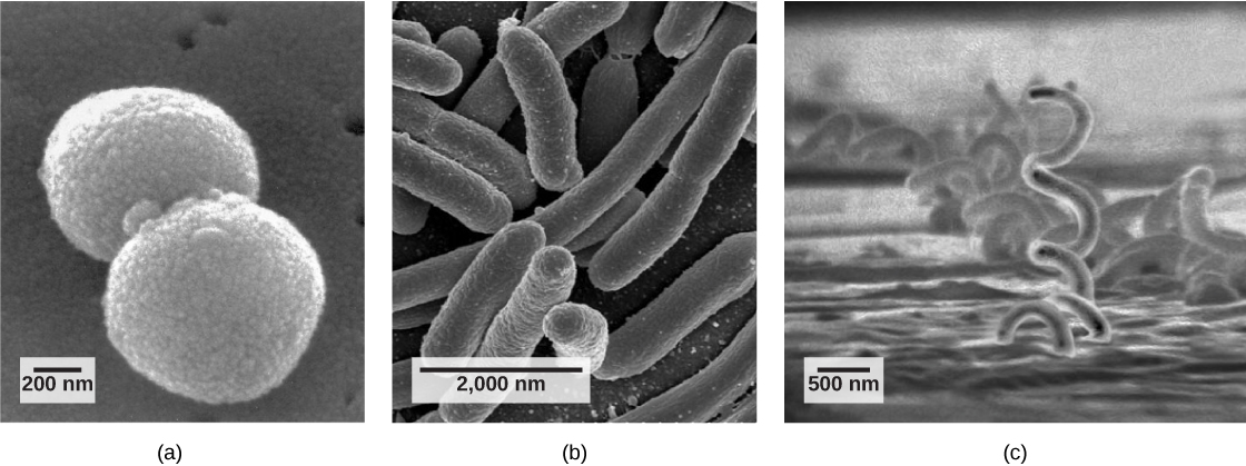
There are many differences between prokaryotic and eukaryotic cells. The name "prokaryote" suggests that prokaryotes are defined by exclusion—they are not eukaryotes, or organisms whose cells contain a nucleus and other internal membrane-bound organelles. However, all cells have four common structures: the plasma membrane, which functions as a barrier for the cell and separates the cell from its environment; the cytoplasm, a complex solution of organic molecules and salts inside the cell; a double-stranded DNA genome, the informational archive of the cell; and ribosomes, where protein synthesis takes place. Prokaryotes come in various shapes, but many fall into three categories: cocci (spherical), bacilli (rod-shaped), and spirilli (spiral-shaped) (Figure 22.9).
英語のヒント
原核細胞と真核細胞には、多くの違いがある。「原核生物」という名前は、それが除外によって定義されていることを示唆する。つまり、核や、膜に囲まれたほかの内部細胞小器官をもつ真核生物ではない、という定義である。しかし、すべての細胞には四つの共通構造がある。細胞の障壁として働き、周囲の環境と細胞を隔てる細胞膜。有機分子と塩類からなる、細胞内の複雑な溶液である細胞質。細胞の情報の保管庫となる二本鎖DNAゲノム。そして、タンパク質合成が起こるリボソームである。原核生物にはさまざまな形があるが、多くは球菌(球形)、桿菌(棒状)、らせん菌(らせん状)の三つに分類される(図22.9)。
a:直径約800ナノメートルの球状の球菌の顕微鏡写真。b:長さ約2000ナノメートルの、細長い楕円形の桿菌の顕微鏡写真。c:直径500ナノメートルでかなり長い、コルク抜き状のらせん菌の顕微鏡写真。

OpenStax, Rice University; Biology 2e. (credit a: modification of work by Janice Haney Carr, Dr. Richard Facklam, CDC; credit c: modification of work by Dr. David Cox; scale-bar data from Matt Russell) · CC BY 4.0 · Source出典

Figure 22.9図 22.9
Common prokaryotic cell types. Prokaryotes fall into three basic categories based on their shape, visualized here using scanning electron microscopy: (a) cocci, or spherical (a pair is shown); (b) bacilli, or rod-shaped; and (c) spirilli, or spiral-shaped. (credit a: modification of work by Janice Haney Carr, Dr. Richard Facklam, CDC; credit c: modification of work by Dr. David Cox; scale-bar data from Matt Russell)
一般的な原核細胞の型。原核生物は形に基づき、三つの基本的な群に分かれる。走査型電子顕微鏡で示したのは、(a)球形の球菌(ここでは一対)、(b)棒状の桿菌、(c)らせん状のらせん菌である。(クレジットa:Janice Haney Carr、Dr. Richard Facklam、CDCの作品を改変。クレジットc:Dr. David Coxの作品を改変。スケールバーのデータ:Matt Russell)

The Prokaryotic Cell原核細胞

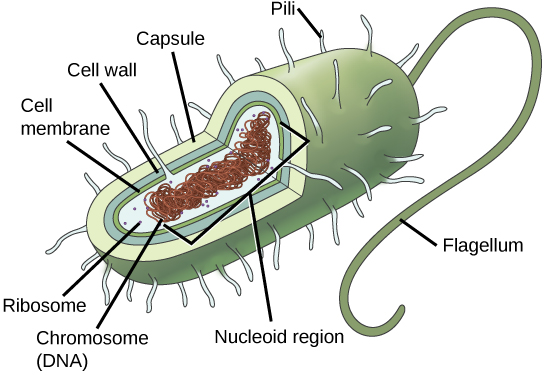
Recall that prokaryotes are unicellular organisms that lack membrane-bound organelles or other internal membrane-bound structures (Figure 22.10). Their chromosome—usually single—consists of a piece of circular, double-stranded DNA located in an area of the cell called the nucleoid. Most prokaryotes have a cell wall outside the plasma membrane. The cell wall functions as a protective layer, and it is responsible for the organism’s shape. Some bacterial species have a capsule outside the cell wall. The capsule enables the organism to attach to surfaces, protects it from dehydration and attack by phagocytic cells, and makes pathogens more resistant to our immune responses. Some species also have flagella (singular, flagellum) used for locomotion, and pili (singular, pilus) used for attachment to surfaces including the surfaces of other cells. Plasmids, which consist of extra-chromosomal DNA, are also present in many species of bacteria and archaea.
英語のヒント
原核生物は単細胞生物で、膜に囲まれた細胞小器官や、ほかの膜に囲まれた内部構造をもたないことを思い出してほしい(図22.10)。通常一つの染色体は、環状の二本鎖DNAからなり、細胞内の核様体と呼ばれる領域に位置する。大半の原核生物は、細胞膜の外側に細胞壁をもつ。細胞壁は保護層として働き、生物の形を決める。一部の細菌種には、細胞壁の外側に莢膜がある。莢膜は、表面への付着を可能にし、乾燥や食細胞の攻撃から生物を守り、病原体の免疫応答への耐性を高める。種によっては、移動に使う鞭毛(英語の複数形flagella、単数形flagellum)や、ほかの細胞の表面などへ付着するための線毛(複数形pili、単数形pilus)ももつ。染色体外DNAからなるプラスミドも、多くの細菌種や古細菌種に存在する。
この図の原核細胞は棒状である。環状染色体は、核様体と呼ばれる領域にまとまっている。細胞内の液体は細胞質と呼ばれる。小さな円で描かれたリボソームが、細胞質の中に浮かぶ。 細胞質は細胞膜に囲まれ、細胞膜はさらに細胞壁に囲まれる。細胞壁の周囲には莢膜がある。描かれた細菌では、細い端の一方から鞭毛が突き出ている。線毛は、細菌全体の莢膜から毛のように伸びる小さな突起である。

OpenStax, Rice University; Biology 2e. · CC BY 4.0 · Source出典

Figure 22.10図 22.10
The features of a typical prokaryotic cell. Flagella, capsules, and pili are not found in all prokaryotes.
典型的な原核細胞の特徴。鞭毛、莢膜、線毛は、すべての原核生物にあるわけではない。
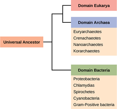
Recall that prokaryotes are divided into two different domains, Bacteria and Archaea, which together with Eukarya, comprise the three domains of life (Figure 22.11).
原核生物は細菌と古細菌という二つの異なるドメインに分かれ、真核生物と合わせて生命の三つのドメインを構成することを思い出してほしい(図22.11)。
系統樹の幹は、全生物の共通祖先を示す。木は二本の枝に分かれる。一方は細菌ドメインにつながり、プロテオバクテリア、クラミジア、スピロヘータ、シアノバクテリア、グラム陽性細菌の各門を含む。 もう一方はさらに分岐し、真核生物ドメインと古細菌ドメインになる。古細菌ドメインは、ユーリアーキオータ、クレンアーキオータ、ナノアーキオータ、コルアーキオータの各門を含む。

OpenStax, Rice University; Biology 2e. · CC BY 4.0 · Source出典

Figure 22.11図 22.11
The three domains of living organisms. Bacteria and Archaea are both prokaryotes but differ enough to be placed in separate domains. An ancestor of modern Archaea is believed to have given rise to Eukarya, the third domain of life. Major groups of Archaea and Bacteria are shown.
生物の三つのドメイン。細菌と古細菌はどちらも原核生物だが、別々のドメインに置くほどの違いがある。現生古細菌の祖先から、生命の第三のドメインである真核生物が生じたと考えられている。古細菌と細菌の主な群を示す。
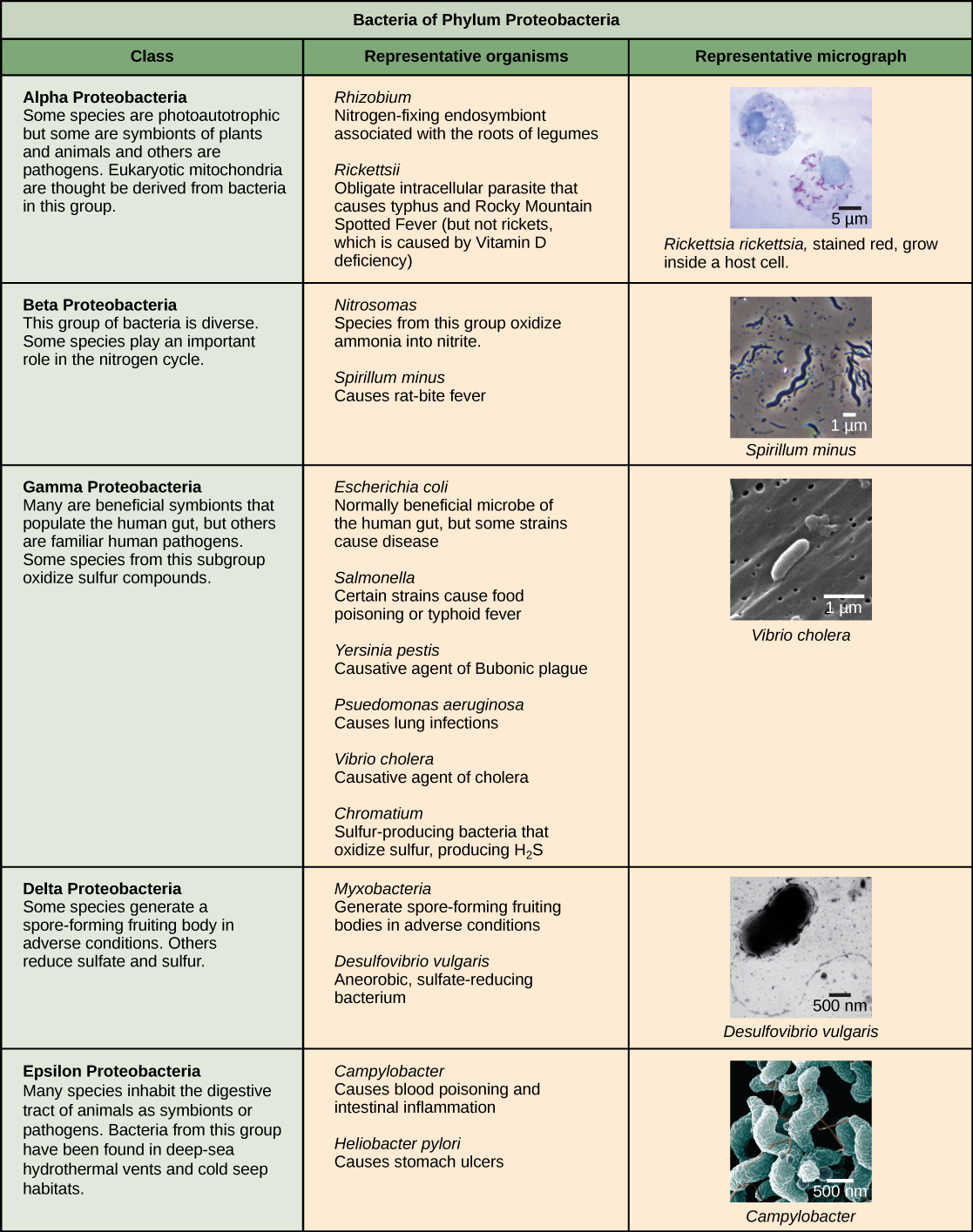
Characteristics of bacterial phyla are described in Figure 22.12 and Figure 22.13. Major bacterial phyla include the Proteobacteria, the Chlamydias, the Spirochaetes, the photosynthetic Cyanobacteria, and the Gram-positive bacteria. The Proteobacteria are in turn subdivided into several classes, from the Alpha- to the Epsilon proteobacteria. Eukaryotic mitochondria are thought to be the descendants of alphaproteobacteria, while eukaryotic chloroplasts are derived from cyanobacteria. Archaeal phyla are described in Figure 22.14.
英語のヒント
細菌の各門の特徴を、図22.12と図22.13に示す。主な門には、プロテオバクテリア、クラミジア、スピロヘータ、光合成を行うシアノバクテリア、グラム陽性細菌がある。プロテオバクテリアは、さらにアルファからイプシロンまで、複数の綱に分けられる。真核生物のミトコンドリアはアルファプロテオバクテリアの子孫と考えられ、葉緑体はシアノバクテリアに由来する。古細菌の各門は、図22.14に示す。
細菌のプロテオバクテリア門に属する五つの綱、アルファ、ベータ、ガンマ、デルタ、イプシロンの特徴を示す。アルファプロテオバクテリアの大半は光独立栄養性だが、植物や動物の共生体も、病原体もいる。真核生物のミトコンドリアは、この群の細菌に由来すると考えられる。 代表的な生物には、マメ科植物の根に共生して窒素を固定する細胞内共生体Rhizobiumと、発疹チフスやロッキー山紅斑熱を起こす偏性細胞内寄生体Rickettsiaがある。ただし、ビタミンD欠乏が原因のくる病、英語のricketsを起こすわけではない。顕微鏡写真には、はるかに大きい真核細胞内にいる、棒状のRickettsia rickettsiiが示される。 ベータプロテオバクテリアは、多様な細菌群である。一部の種は窒素循環に重要な役割をもつ。代表例は、アンモニアを亜硝酸へ酸化するNitrosomasと、鼠咬症を起こすSpirillum minusである。らせん状のSpirillum minusの顕微鏡写真を示す。 ガンマプロテオバクテリアには、ヒトの腸に住む多くの有益な共生体と、よく知られたヒトの病原体が含まれる。この亜群の一部は硫黄化合物を酸化する。代表例には、通常は腸内の有益な微生物だが、一部の株は病気を起こすEscherichia coli、特定の株が食中毒や腸チフスを起こすSalmonella、腺ペストの病原体Yersinia pestis、肺感染症を起こすPsuedomonas aeruganosa、コレラの病原体Vibrio cholera、硫黄を酸化してH2Sを作る硫黄生成細菌Chromatiumがある。顕微鏡写真は、長さ約1マイクロメートルの棒状のVibrio choleraを示す。 デルタプロテオバクテリアには、条件が悪化すると胞子を作る子実体を形成する種がいる。硫酸塩や硫黄を還元する種もいる。代表例は、悪条件で胞子形成性の子実体を作る粘液細菌と、嫌気性の硫酸塩還元細菌Desulfovibrio vulgarisである。顕微鏡写真は、長い鞭毛をもつ、曲がった棒状のDesulfovibrio vulgarisを示す。 イプシロンプロテオバクテリアには、動物の消化管に共生体や病原体として住む多くの種が含まれる。この群の細菌は、深海の熱水噴出孔や冷水湧出域で見つかっている。代表例には、敗血症や腸の炎症を起こすCampylobacterと、胃潰瘍を起こすHeliobacter pyloriがある。顕微鏡写真は、短いらせん状、またはS字形のCampylobacterを示す。

OpenStax, Rice University; Biology 2e. (credit “Rickettsia rickettsii”: modification of work by CDC; credit “Spirillum minus”: modification of work by Wolframm Adlassnig; credit “Vibrio cholera”: modification of work by Janice Haney Carr, CDC; credit “Desulfovibrio vulgaris”: modification of work by Graham Bradley; credit “Campylobacter”: modification of work by De Wood, Pooley, USDA, ARS, EMU; scale-bar data from Matt Russell) · CC BY 4.0 · Source出典

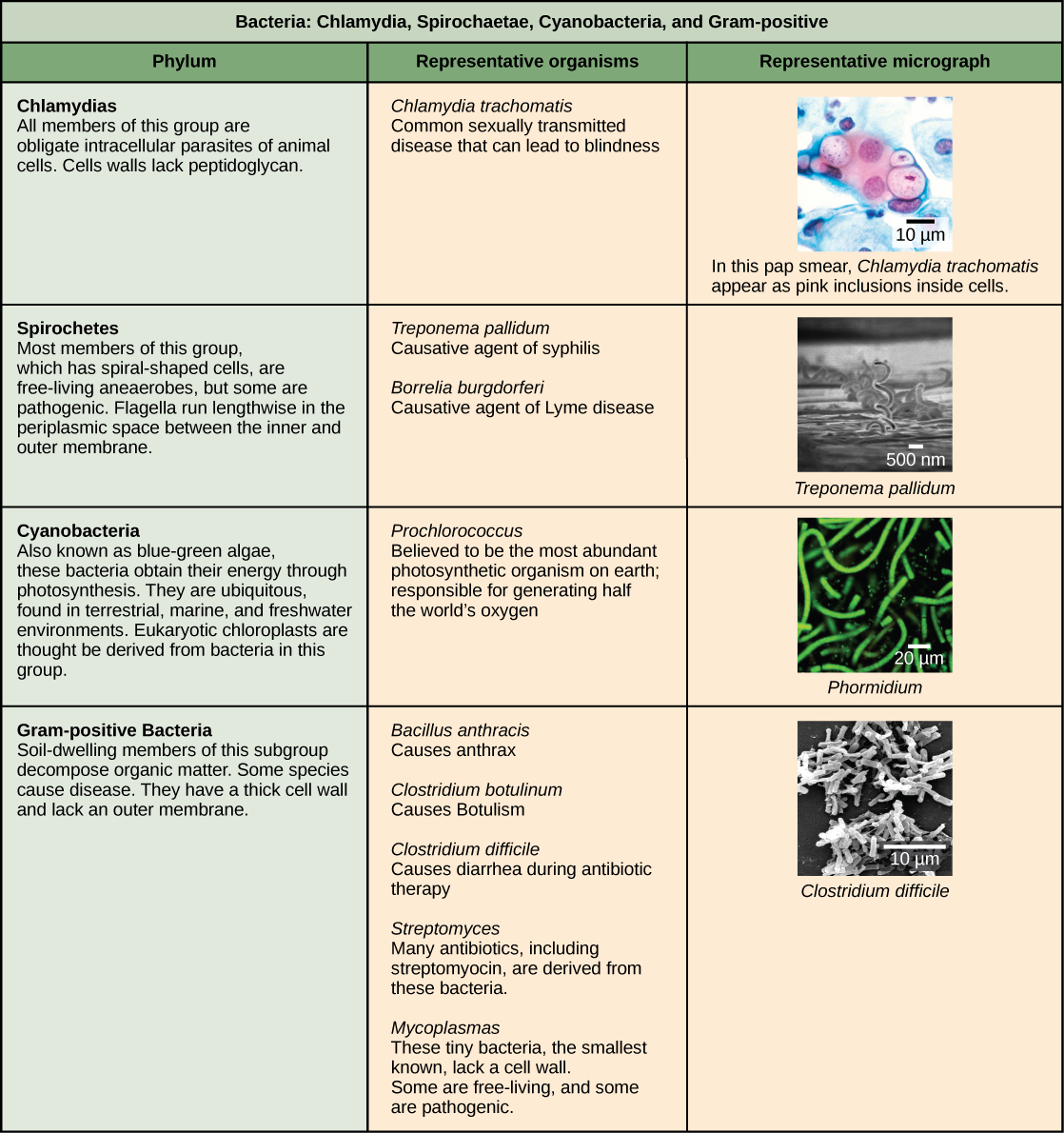
Figure 22.12図 22.12
The Proteobacteria. Phylum Proteobacteria is one of up to 52 bacteria phyla. Proteobacteria is further subdivided into five classes, Alpha through Epsilon. (credit “Rickettsia rickettsii”: modification of work by CDC; credit “Spirillum minus”: modification of work by Wolframm Adlassnig; credit “Vibrio cholera”: modification of work by Janice Haney Carr, CDC; credit “Desulfovibrio vulgaris”: modification of work by Graham Bradley; credit “Campylobacter”: modification of work by De Wood, Pooley, USDA, ARS, EMU; scale-bar data from Matt Russell)
プロテオバクテリア。プロテオバクテリア門は、最大52ある細菌の門の一つである。さらに、アルファからイプシロンまでの五つの綱に分けられる。(クレジット「Rickettsia rickettsii」:CDCの作品を改変。「Spirillum minus」:Wolframm Adlassnigの作品を改変。「Vibrio cholera」:Janice Haney Carr、CDCの作品を改変。「Desulfovibrio vulgaris」:Graham Bradleyの作品を改変。「Campylobacter」:De Wood、Pooley、USDA、ARS、EMUの作品を改変。スケールバーのデータ:Matt Russell)
この表は、ほかの四つの細菌の門を説明する。最初はクラミジア門である。この群の全メンバーは、動物細胞の偏性細胞内寄生体である。細胞壁にペプチドグリカンはない。顕微鏡写真は、Chlamydia trachomatisに感染した細胞のパップスメア標本。クラミジア感染は最も一般的な性感染症で、失明につながることもある。 スピロヘータ門の細胞はすべてらせん状である。大半は自由生活性の嫌気性生物だが、病原性のものもいる。鞭毛は、内膜と外膜の間のペリプラズム空間を、長さ方向に走る。代表例は、梅毒の病原体Treponema pallidumと、ライム病の病原体Borrelia burgdorferi。顕微鏡写真は、幅約1マイクロメートルのコルク抜き状のTrepanema pallidumを示す。 シアノバクテリア門の細菌は、藍藻とも呼ばれ、光合成でエネルギーを得る。陸、海、淡水と、至るところに存在する。真核生物の葉緑体は、この群の細菌に由来すると考えられる。シアノバクテリアのProchlorococcusは、地球上で最も豊富な光合成生物であり、世界の酸素の半分を作ると考えられている。顕微鏡写真は、Phormidiumという細長い棒状の種。 グラム陽性細菌は厚い細胞壁をもち、外膜がない。この亜群の土壌に住むものは、有機物を分解する。病気を起こす種もいる。代表例には、炭疽を起こすBacillus anthracis、ボツリヌス症を起こすClostridium botulinum、抗生物質治療中の下痢を起こすClostridium difficile、ストレプトマイシンを含む多くの抗生物質の由来となるStreptomyces、既知で最小の細菌で細胞壁をもたないマイコプラズマがある。自由生活性のものも病原性のものもいる。顕微鏡写真は、長さ約3マイクロメートルの棒状のClostridium difficileを示す。

OpenStax, Rice University; Biology 2e. (credit “Chlamydia trachomatis”: modification of work by Dr. Lance Liotta Laboratory, NCI; credit “Treponema pallidum”: modification of work by Dr. David Cox, CDC; credit “Phormidium”: modification of work by USGS; credit “Clostridium difficile”: modification of work by Lois S. Wiggs, CDC; scale-bar data from Matt Russell) · CC BY 4.0 · Source出典

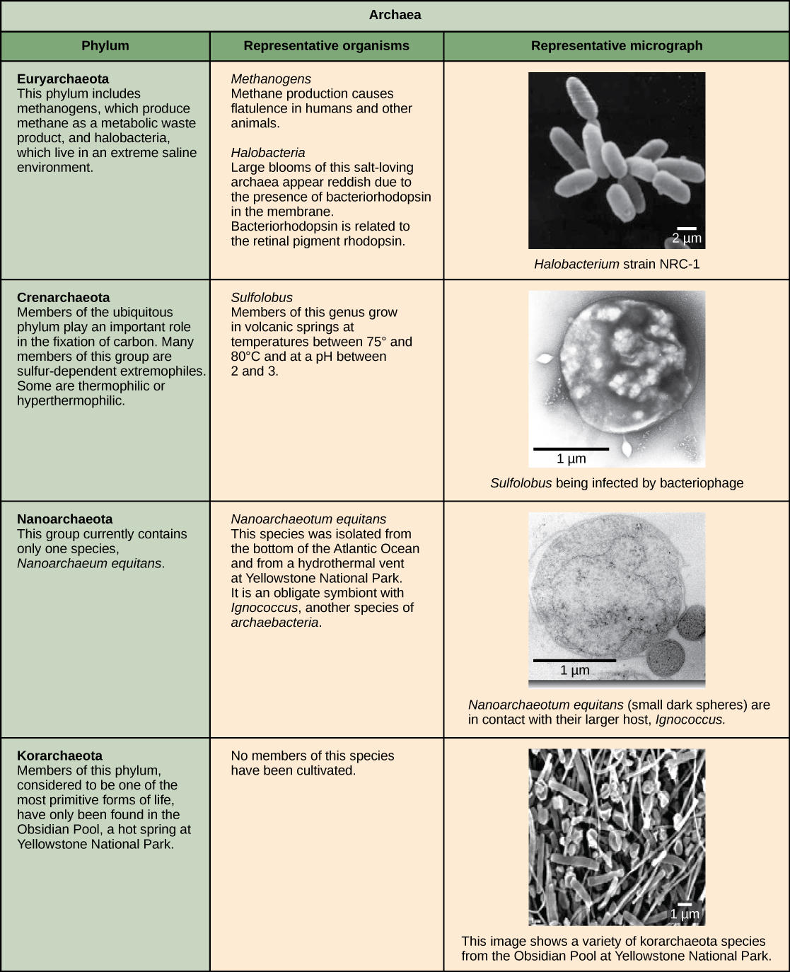
Figure 22.13図 22.13
Other bacterial phyla. Chlamydia, Spirochetes, Cyanobacteria, and Gram-positive bacteria are described in this table. Note that bacterial shape is not phylum-dependent; bacteria within a phylum may be cocci, rod-shaped, or spiral. (credit “Chlamydia trachomatis”: modification of work by Dr. Lance Liotta Laboratory, NCI; credit “Treponema pallidum”: modification of work by Dr. David Cox, CDC; credit “Phormidium”: modification of work by USGS; credit “Clostridium difficile”: modification of work by Lois S. Wiggs, CDC; scale-bar data from Matt Russell)
ほかの細菌の門。この表は、クラミジア、スピロヘータ、シアノバクテリア、グラム陽性細菌を説明する。細菌の形は門によって決まるわけではない。同じ門の中に、球菌、棒状の細菌、らせん状の細菌が含まれることがある。(クレジット「Chlamydia trachomatis」:Dr. Lance Liotta Laboratory、NCIの作品を改変。「Treponema pallidum」:Dr. David Cox、CDCの作品を改変。「Phormidium」:USGSの作品を改変。「Clostridium difficile」:Lois S. Wiggs、CDCの作品を改変。スケールバーのデータ:Matt Russell)
古細菌の四つの門の特徴を示す。ユーリアーキオータには、代謝廃物としてメタンを作るメタン生成菌と、極端な高塩分環境に住むハロバクテリアが含まれる。メタン生成菌は、ヒトやほかの動物の放屁の原因となる。ハロバクテリアは大量発生することがあり、膜にバクテリオロドプシンがあるため、赤みを帯びて見える。バクテリオロドプシンは、網膜色素ロドプシンと関連する。顕微鏡写真は棒状のHalobacterium。 至るところにあるクレンアーキオータ門の生物は、炭素固定に重要な役割をもつ。この群の多くは、硫黄に依存する極限環境生物である。好熱性や超好熱性のものもいる。顕微鏡写真は球菌状のSulfolobusで、この属は75〜80 °C、pH 2〜3の火山性温泉に生育する。 ナノアーキオータ門は現在、Nanoarchaeum equitansの1種だけを含む。これは大西洋の海底や、イエローストーン国立公園の熱水噴出孔から分離されている。別の古細菌種Ignicoccusとの偏性共生体である。顕微鏡写真には、大きなIgnicoccus細胞に付着した、小さく丸い2個のN. equitans細胞が示される。 コルアーキオータは、最も原始的な生命の形の一つと考えられ、これまでイエローストーン国立公園の温泉、オブシディアン・プールでしか見つかっていない。顕微鏡写真は、この群のさまざまな形をした標本を示す。

OpenStax, Rice University; Biology 2e. (credit “Halobacterium”: modification of work by NASA; credit “Nanoarchaeotum equitans”: modification of work by Karl O. Stetter; credit “Korarchaeota”: modification of work by Office of Science of the U.S. Dept. of Energy; scale-bar data from Matt Russell) · CC BY 4.0 · Source出典

Figure 22.14図 22.14
Archaeal phyla. Archaea are separated into four phyla: the Korarchaeota, Euryarchaeota, Crenarchaeota, and Nanoarchaeota. (credit “Halobacterium”: modification of work by NASA; credit “Nanoarchaeotum equitans”: modification of work by Karl O. Stetter; credit “Korarchaeota”: modification of work by Office of Science of the U.S. Dept. of Energy; scale-bar data from Matt Russell)
古細菌の門。古細菌は、コルアーキオータ、ユーリアーキオータ、クレンアーキオータ、ナノアーキオータの四つの門に分けられる。(クレジット「Halobacterium」:NASAの作品を改変。「Nanoarchaeotum equitans」:Karl O. Stetterの作品を改変。「Korarchaeota」:米国エネルギー省科学局の作品を改変。スケールバーのデータ:Matt Russell)