第22章原核生物:細菌と古細菌
The Plasma Membrane of Prokaryotes原核生物の細胞膜
The prokaryotic plasma membrane is a thin lipid bilayer (6 to 8 nanometers) that completely surrounds the cell and separates the inside from the outside. Its selectively permeable nature keeps ions, proteins, and other molecules within the cell and prevents them from diffusing into the extracellular environment, while other molecules may move through the membrane. Recall that the general structure of a cell membrane is a phospholipid bilayer composed of two layers of lipid molecules. In archaeal cell membranes, isoprene (phytanyl) chains linked to glycerol replace the fatty acids linked to glycerol in bacterial membranes. Some archaeal membranes are lipid monolayers instead of bilayers (Figure 22.15).
英語のヒント
原核生物の細胞膜は、厚さ6〜8ナノメートルの薄い脂質二重層で、細胞全体を包み、内側と外側を隔てる。選択的に物質を通す性質によって、イオン、タンパク質、ほかの分子を細胞内に保ち、細胞外環境への拡散を防ぐ。一方、膜を通って移動できる分子もある。細胞膜の一般的な構造は、脂質分子の二つの層からなるリン脂質二重層であることを思い出してほしい。古細菌の細胞膜では、細菌の膜でグリセロールに結合する脂肪酸の代わりに、イソプレン(フィタニル)鎖がグリセロールに結合している。古細菌の膜には、二重層ではなく脂質単分子層でできたものもある(図22.15)。
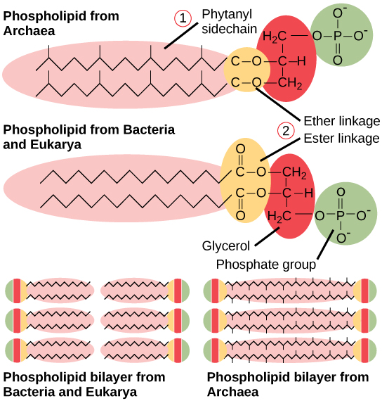

Figure 22.15図 22.15
Bacterial and archaeal phospholipids. Archaeal phospholipids differ from those found in Bacteria and Eukarya in two ways. First, they have branched phytanyl sidechains instead of linear ones. Second, an ether bond instead of an ester bond connects the lipid to the glycerol.
英語のヒント
細菌と古細菌のリン脂質。古細菌のリン脂質は、細菌や真核生物のものと二つの点で異なる。第一に、直鎖状ではなく、枝分かれしたフィタニル側鎖をもつ。第二に、脂質とグリセロールをつなぐのが、エステル結合ではなくエーテル結合である。
The Cell Wall of Prokaryotes原核生物の細胞壁
The cytoplasm of prokaryotic cells has a high concentration of dissolved solutes. Therefore, the osmotic pressure within the cell is relatively high. The cell wall is a protective layer that surrounds some cells and gives them shape and rigidity. It is located outside the cell membrane and prevents osmotic lysis (bursting due to increasing volume). The chemical composition of the cell wall varies between Archaea and Bacteria, and also varies between bacterial species.
英語のヒント
原核細胞の細胞質には、溶質が高濃度で溶けている。そのため、細胞内の浸透圧は比較的高い。細胞壁は、一部の細胞を囲む保護層で、形と硬さを与える。細胞膜の外側に位置し、浸透圧による溶解、すなわち体積の増加による破裂を防ぐ。細胞壁の化学組成は、古細菌と細菌の間で異なり、細菌の種同士でも異なる。
Bacterial cell walls contain peptidoglycan, composed of polysaccharide chains that are cross-linked by unusual peptides containing both L- and D-amino acids including D-glutamic acid and D-alanine. (Proteins normally have only L-amino acids; as a consequence, many of our antibiotics work by mimicking D-amino acids and therefore have specific effects on bacterial cell-wall development.) There are more than 100 different forms of peptidoglycan. S-layer (surface layer) proteins are also present on the outside of cell walls of both Archaea and Bacteria.
英語のヒント
細菌の細胞壁にはペプチドグリカンが含まれる。これは、特殊なペプチドで架橋された多糖鎖からなる。そのペプチドには、D-グルタミン酸やD-アラニンを含む、L型とD型の両方のアミノ酸が含まれる。通常、タンパク質にはL-アミノ酸しか含まれない。そのため、私たちが使う抗生物質の多くは、D-アミノ酸をまねることで作用し、細菌の細胞壁形成に特異的な効果を及ぼす。ペプチドグリカンには100種類以上の異なる形がある。古細菌と細菌の両方で、細胞壁の外側にはS層(表面層)タンパク質も存在する。
Bacteria are divided into two major groups: Gram positive and Gram negative, based on their reaction to Gram staining. Note that all Gram-positive bacteria belong to two phyla (Firmicutes and Actinobacteria); bacteria in the other phyla (Proteobacteria, Chlamydias, Spirochetes, Cyanobacteria, and others) are Gram-negative. The Gram staining method is named after its inventor, Danish scientist Hans Christian Gram (1853–1938). The different bacterial responses to the staining procedure are ultimately due to cell wall structure. Gram-positive organisms typically lack the outer membrane found in Gram-negative organisms (Figure 22.16). Up to 90 percent of the cell-wall in Gram-positive bacteria is composed of peptidoglycan, and most of the rest is composed of acidic substances called teichoic acids. Teichoic acids may be covalently linked to lipids in the plasma membrane to form lipoteichoic acids. Lipoteichoic acids anchor the cell wall to the cell membrane. Gram-negative bacteria have a relatively thin cell wall composed of a few layers of peptidoglycan (only 10 percent of the total cell wall), surrounded by an outer envelope containing lipopolysaccharides (LPS) and lipoproteins. This outer envelope is sometimes referred to as a second lipid bilayer. The chemistry of this outer envelope is very different, however, from that of the typical lipid bilayer that forms plasma membranes.
英語のヒント
細菌は、グラム染色への反応に基づき、グラム陽性とグラム陰性の二つの大きな群に分かれる。グラム陽性細菌はすべて、フィルミクテス門とアクチノバクテリア門という二つの門に属する。一方、プロテオバクテリア、クラミジア、スピロヘータ、シアノバクテリアなど、ほかの門の細菌はグラム陰性である。グラム染色法は、その考案者であるデンマークの科学者ハンス・クリスチャン・グラム(1853〜1938)にちなんで名づけられた。染色法への細菌の反応の違いは、最終的には細胞壁の構造による。グラム陽性生物には通常、グラム陰性生物にある外膜がない(図22.16)。グラム陽性細菌では、細胞壁の最大90%がペプチドグリカンでできており、残りの大部分は、テイコ酸と呼ばれる酸性物質からなる。テイコ酸は、細胞膜の脂質へ共有結合して、リポテイコ酸を作ることがある。リポテイコ酸は細胞壁を細胞膜につなぎ止める。グラム陰性細菌には、ペプチドグリカンの数層からなる比較的薄い細胞壁があり、ペプチドグリカンは細胞壁全体のわずか10%である。その周囲を、リポ多糖(LPS)とリポタンパク質を含む外側の被膜が囲む。この外側の被膜は、第二の脂質二重層と呼ばれることもある。しかし、その化学的性質は、細胞膜を作る典型的な脂質二重層とは大きく異なる。
Archaean cell walls do not have peptidoglycan. There are four different types of archaean cell walls. One type is composed of pseudopeptidoglycan, which is similar to peptidoglycan in morphology but contains different sugars in the polysaccharide chain. The other three types of cell walls are composed of polysaccharides, glycoproteins, or pure protein. Other differences between Bacteria and Archaea are seen in Table 22.2. Note that features related to DNA replication, transcription and translation in Archaea are similar to those seen in eukaryotes.
英語のヒント
古細菌の細胞壁には、ペプチドグリカンがない。古細菌の細胞壁には四つの異なる型がある。一つは、擬似ペプチドグリカンからなる。これは、形態はペプチドグリカンに似るが、多糖鎖に含まれる糖が異なる。残る三つの型は、それぞれ多糖、糖タンパク質、純粋なタンパク質からなる。細菌と古細菌のほかの違いを表22.2に示す。古細菌のDNA複製、転写、翻訳に関わる特徴は、真核生物のものと似ていることに注目してほしい。
Differences and Similarities between Bacteria and Archaea 細菌と古細菌の相違点と類似点 |
||
|---|---|---|
Structural Characteristic 構造上の特徴 |
Bacteria 細菌 |
Archaea 古細菌 |
Cell type 細胞の型 |
Prokaryotic 原核性 |
Prokaryotic 原核性 |
Cell morphology 細胞の形態 |
Variable 多様 |
Variable 多様 |
Cell wall 細胞壁 |
Contains peptidoglycan ペプチドグリカンを含む |
Does not contain peptidoglycan ペプチドグリカンを含まない |
Cell membrane type 細胞膜の型 |
Lipid bilayer 脂質二重層 |
Lipid bilayer or lipid monolayer 脂質二重層または脂質単分子層 |
Plasma membrane lipids 細胞膜の脂質 |
Fatty acids-glycerol ester 脂肪酸とグリセロールのエステル |
Phytanyl-glycerol ethers フィタニルとグリセロールのエーテル |
Chromosome 染色体 |
Typically circular 通常は環状 |
Typically circular 通常は環状 |
Replication origins 複製起点 |
Single 一つ |
Multiple 複数 |
RNA polymerase RNAポリメラーゼ |
Single 一つ |
Multiple 複数 |
Initiator tRNA 開始tRNA |
Formyl-methionine ホルミルメチオニン |
Methionine メチオニン |
Streptomycin inhibition ストレプトマイシンによる阻害 |
Sensitive 感受性 |
Resistant 耐性 |
Calvin cycle カルビン回路 |
Yes あり |
No なし |