第36章感覚系
36.5 Vision36.5 視覚
Learning Objectives学習目標
By the end of this section, you will be able to do the following:
この節を学び終えると、次のことができるようになる。
- Explain how electromagnetic waves differ from sound waves電磁波が音波とどう異なるか説明する。
- Trace the path of light through the eye to the point of the optic nerve眼を通り視神経へ至るまでの、光の経路をたどる。
- Explain tonic activity as it is manifested in photoreceptors in the retina網膜の視細胞にみられる持続性活動を説明する。
Vision is the ability to detect light patterns from the outside environment and interpret them into images. Animals are bombarded with sensory information, and the sheer volume of visual information can be problematic. Fortunately, the visual systems of species have evolved to attend to the most-important stimuli. The importance of vision to humans is further substantiated by the fact that about one-third of the human cerebral cortex is dedicated to analyzing and perceiving visual information.
英語のヒント
視覚は、外界からの光のパターンを検出し、像として解釈する能力である。動物には感覚情報が押し寄せ、視覚情報の量の多さ自体が問題になりうる。幸い、各種の視覚系は、最も重要な刺激に注意を向けるよう進化してきた。ヒトの大脳皮質のおよそ3分の1が、視覚情報の分析と知覚を担っている。この事実も、ヒトにとって視覚が重要であることを裏づける。
Light光
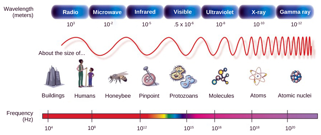
As with auditory stimuli, light travels in waves. The compression waves that compose sound must travel in a medium—a gas, a liquid, or a solid. In contrast, light is composed of electromagnetic waves and needs no medium; light can travel in a vacuum (Figure 36.17). The behavior of light can be discussed in terms of the behavior of waves and also in terms of the behavior of the fundamental unit of light—a packet of electromagnetic radiation called a photon. A glance at the electromagnetic spectrum shows that visible light for humans is just a small slice of the entire spectrum, which includes radiation that we cannot see as light because it is below the frequency of visible red light and above the frequency of visible violet light.
聴覚刺激と同じく、光も波として進む。音を構成する圧縮波は、気体、液体、固体などの媒質を伝わる必要がある。対して光は電磁波であり、媒質を必要とせず、真空も進める(図36.17)。光の振る舞いは、波の振る舞いとしても、光子と呼ばれる電磁放射のひとかたまり、すなわち光の基本単位の振る舞いとしても論じられる。電磁スペクトルを見ると、ヒトの可視光は全体のほんの一部でしかない。赤い可視光より周波数が低い放射や、紫の可視光より周波数が高い放射は、光として見ることができない。
Certain variables are important when discussing perception of light. Wavelength (which varies inversely with frequency) manifests itself as hue. Light at the red end of the visible spectrum has longer wavelengths (and is lower frequency), while light at the violet end has shorter wavelengths (and is higher frequency). The wavelength of light is expressed in nanometers (nm); one nanometer is one billionth of a meter. Humans perceive light that ranges between approximately 380 nm and 740 nm. Some other animals, though, can detect wavelengths outside of the human range. For example, bees see near-ultraviolet light in order to locate nectar guides on flowers, and some non-avian reptiles sense infrared light (heat that prey gives off).
英語のヒント
光の知覚を考える際には、いくつかの変数が重要になる。周波数に反比例する波長は、色相として現れる。可視スペクトルの赤い端の光は波長が長く、周波数が低い。紫の端では波長が短く、周波数が高い。光の波長はナノメートル(nm)で表す。1 nmは10億分の1メートルである。ヒトが知覚するのは、およそ380~740 nmの光である。ヒトの範囲外の波長を検出する動物もいる。たとえばハチは、花の蜜への案内模様を見つけるため近紫外線を見る。鳥類以外の一部の爬虫類は、獲物の放つ熱である赤外線を感知する。
OpenStax, Rice University; Biology 2e. (credit: modification of work by NASA) · CC BY 4.0 · Source出典

Figure 36.17図 36.17
In the electromagnetic spectrum, visible light lies between 380 nm and 740 nm. (credit: modification of work by NASA)
電磁スペクトルで、可視光は380~740 nmの範囲にある。(出典:NASAの作品を改変)
Wave amplitude is perceived as luminous intensity, or brightness. The standard unit of intensity of light is the candela, which is approximately the luminous intensity of one common candle.
波の振幅は、光度、すなわち明るさとして知覚される。光度の標準単位はカンデラで、普通のろうそく一本の光度におよそ相当する。
Light waves travel 299,792 km per second in a vacuum, (and somewhat slower in various media such as air and water), and those waves arrive at the eye as long (red), medium (green), and short (blue) waves. What is termed “white light” is light that is perceived as white by the human eye. This effect is produced by light that stimulates equally the color receptors in the human eye. The apparent color of an object is the color (or colors) that the object reflects. Thus a red object reflects the red wavelengths in mixed (white) light and absorbs all other wavelengths of light.
英語のヒント
光波は真空中を秒速299,792 kmで進み、空気や水などの媒質では少し遅くなる。長い波長(赤)、中間の波長(緑)、短い波長(青)の波として眼に届く。「白色光」と呼ばれるのは、ヒトの眼に白く知覚される光である。眼の色の受容器を等しく刺激することで、この効果が生じる。物体の見かけの色は、その物体が反射する色である。したがって赤い物体は、混ざった白色光のうち赤の波長を反射し、ほかのすべての波長を吸収する。
Anatomy of the Eye眼の解剖
The photoreceptive cells of the eye, where transduction of light to nervous impulses occurs, are located in the retina (shown in Figure 36.18) on the inner surface of the back of the eye. But light does not impinge on the retina unaltered. It passes through other layers that process it so that it can be interpreted by the retina (Figure 36.18b). The cornea, the front transparent layer of the eye, and the crystalline lens, a transparent convex structure behind the cornea, both refract (bend) light to focus the image on the retina. The iris, which is conspicuous as the colored part of the eye, is a circular muscular ring lying between the lens and cornea that regulates the amount of light entering the eye. In conditions of high ambient light, the iris contracts, reducing the size of the pupil at its center. In conditions of low light, the iris relaxes and the pupil enlarges.
英語のヒント
The main function of the lens is to focus light on the retina and fovea centralis. The lens is dynamic, focusing and re-focusing light as the eye rests on near and far objects in the visual field. The lens is operated by muscles that stretch it flat or allow it to thicken, changing the focal length of light coming through it to focus it sharply on the retina. With age comes the loss of the flexibility of the lens, and a form of farsightedness called presbyopia results. Presbyopia occurs because the image focuses behind the retina. Presbyopia is a deficit similar to a different type of farsightedness called hyperopia caused by an eyeball that is too short. For both defects, images in the distance are clear but images nearby are blurry. Myopia (nearsightedness) occurs when an eyeball is elongated and the image focus falls in front of the retina. In this case, images in the distance are blurry but images nearby are clear.
英語のヒント
水晶体の主な機能は、網膜と中心窩に光の焦点を合わせることである。視野内の近い物と遠い物へ眼を向けるたびに、焦点を調節し直す動的な構造である。筋肉が水晶体を平たく伸ばしたり、厚くなるようにしたりして、通る光の焦点距離を変え、網膜上にくっきり焦点を合わせる。加齢とともに水晶体の柔軟性が失われ、老視という、遠くが見やすく近くが見にくい状態が生じる。老視では網膜の後ろに焦点が合う。これは、眼球が短すぎるために起こる遠視と似た障害である。どちらでも、遠くの像は鮮明だが、近くの像はぼやける。近視は眼球が長く、網膜より前に焦点が合う場合に起こる。この場合、遠くはぼやけるが、近くは鮮明に見える。
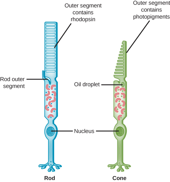
There are two types of photoreceptors in the retina: rods and cones, named for their general appearance as illustrated in Figure 36.19. Rods are strongly photosensitive and are located in the outer edges of the retina. They detect dim light and are used primarily for peripheral and nighttime vision. Cones are weakly photosensitive and are located near the center of the retina. They respond to bright light, and their primary role is in daytime, color vision.
網膜には桿体と錐体という二種類の視細胞があり、図36.19のような全体の形にちなんで名づけられた。桿体は光に非常に敏感で、網膜の周縁部にある。弱い光を検出し、主に周辺視と夜間の視覚に使われる。錐体は光への感受性が弱く、網膜の中心付近にある。明るい光に応答し、主に日中の色覚を担う。
OpenStax, Rice University; Biology 2e. (credit: modification of work by Piotr Sliwa) · CC BY 4.0 · Source出典

Figure 36.19図 36.19
Rods and cones are photoreceptors in the retina. Rods respond in low light and can detect only shades of gray. Cones respond in intense light and are responsible for color vision. (credit: modification of work by Piotr Sliwa)
桿体と錐体は網膜の視細胞である。桿体は弱い光に応答し、灰色の濃淡だけを検出する。錐体は強い光に応答し、色覚を担う。(出典:Piotr Sliwaの作品を改変)
The fovea is the region in the center back of the eye that is responsible for acute vision. The fovea has a high density of cones. When you bring your gaze to an object to examine it intently in bright light, the eyes orient so that the object’s image falls on the fovea. However, when looking at a star in the night sky or other object in dim light, the object can be better viewed by the peripheral vision because it is the rods at the edges of the retina, rather than the cones at the center, that operate better in low light. In humans, cones far outnumber rods in the fovea.
英語のヒント
中心窩は眼の後部中央にあり、鋭い視覚を担う。錐体の密度が高い。明るい場所で物をよく調べようと視線を向けると、像が中心窩に結ばれるように眼の向きが変わる。しかし夜空の星など、暗い中の物を見るときは、周辺視を使う方がよく見える。弱い光でよく働くのは中心の錐体ではなく、網膜の周縁部の桿体だからである。ヒトの中心窩では、錐体の数が桿体を大きく上回る。