第36章感覚系
36.2 Somatosensation36.2 体性感覚
Learning Objectives学習目標
By the end of this section, you will be able to do the following:
この節を学び終えると、次のことができるようになる。
- Describe four important mechanoreceptors in human skinヒトの皮膚の四つの重要な機械受容器を説明する。
- Describe the topographical distribution of somatosensory receptors between glabrous and hairy skin無毛皮膚と有毛皮膚で、体性感覚受容器がどこに分布するかを説明する。
- Explain why the perception of pain is subjective痛みの知覚が主観的である理由を説明する。
Somatosensation is a mixed sensory category and includes all sensation received from the skin and mucous membranes, as well from as the limbs and joints. Somatosensation is also known as tactile sense, or more familiarly, as the sense of touch. Somatosensation occurs all over the exterior of the body and at some interior locations as well. A variety of receptor types—embedded in the skin, mucous membranes, muscles, joints, internal organs, and cardiovascular system—play a role.
体性感覚は複数の感覚を含む分類で、皮膚と粘膜、手足、関節から受け取るすべての感覚を含む。触覚とも呼ばれ、より日常的には触れる感覚として知られる。体の外表全体と、一部の内部にも生じる。皮膚、粘膜、筋肉、関節、内臓、心血管系に埋め込まれた、さまざまな受容器が関与する。
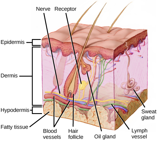
Recall that the epidermis is the outermost layer of skin in mammals. It is relatively thin, is composed of keratin-filled cells, and has no blood supply. The epidermis serves as a barrier to water and to invasion by pathogens. Below this, the much thicker dermis contains blood vessels, sweat glands, hair follicles, lymph vessels, and lipid-secreting sebaceous glands (Figure 36.4). Below the epidermis and dermis is the subcutaneous tissue, or hypodermis, the fatty layer that contains blood vessels, connective tissue, and the axons of sensory neurons. The hypodermis, which holds about 50 percent of the body’s fat, attaches the dermis to the bone and muscle, and supplies nerves and blood vessels to the dermis.
英語のヒント
哺乳類の皮膚の最外層は表皮である。比較的薄く、ケラチンで満たされた細胞からなり、血液は供給されない。水や病原体の侵入に対する障壁として働く。その下の真皮ははるかに厚く、血管、汗腺、毛包、リンパ管、脂質を分泌する皮脂腺を含む(図36.4)。表皮と真皮の下には皮下組織がある。これは血管、結合組織、感覚ニューロンの軸索を含む脂肪の層である。皮下組織は体脂肪のおよそ50%を含み、真皮を骨や筋肉につなぎ、真皮へ神経と血管を供給する。
OpenStax, Rice University; Biology 2e. (credit: modification of work by Don Bliss, National Cancer Institute) · CC BY 4.0 · Source出典

Figure 36.4図 36.4
Mammalian skin has three layers: an epidermis, a dermis, and a hypodermis. (credit: modification of work by Don Bliss, National Cancer Institute)
哺乳類の皮膚には、表皮、真皮、皮下組織の三層がある。(出典:米国国立がん研究所のDon Blissの作品を改変)
Somatosensory Receptors体性感覚受容器
Somatosensory receptors are classified into four categories: mechanoreceptors, thermoreceptors, pain receptors (nociceptors), and chemoreceptors. These categories are based on the nature of stimuli each receptor class transduces. What is commonly referred to as “touch” involves more than one kind of stimulus and more than one kind of receptor. Mechanoreceptors in the skin are described as encapsulated (that is, surrounded by a capsule) or unencapsulated (a group that includes free nerve endings). A free nerve ending, as its name implies, is an unencapsulated dendrite of a sensory neuron. Free nerve endings are the most common nerve endings in skin, and they extend into the middle of the epidermis. Free nerve endings are sensitive to painful stimuli, to hot and cold, and to light touch. They are slow to adjust to a stimulus and so are less sensitive to abrupt changes in stimulation.
英語のヒント
体性感覚受容器は、機械受容器、温度受容器、痛覚受容器(侵害受容器)、化学受容器の四つに分けられる。分類は、それぞれが変換する刺激の性質に基づく。一般に「触覚」と呼ぶものには、複数の種類の刺激と受容器が関わる。皮膚の機械受容器は、被膜性、すなわち被膜に囲まれたものと、自由神経終末などの非被膜性のものに分けられる。自由神経終末は、その名のとおり被膜のない感覚ニューロンの樹状突起である。皮膚で最も多い神経終末で、表皮の中ほどまで伸びる。自由神経終末は、痛みの刺激、熱さと冷たさ、軽い接触に反応する。刺激に順応するのが遅いため、刺激の急な変化には比較的鈍感である。
There are three classes of mechanoreceptors: tactile, proprioceptors, and baroreceptors. Mechanoreceptors sense stimuli due to physical deformation of their plasma membranes. They contain mechanically gated ion channels whose gates open or close in response to pressure, touch, stretching, and sound. There are four primary tactile mechanoreceptors in human skin: Merkel’s disks, Meissner’s corpuscles, Ruffini endings, and Pacinian corpuscles; two are located toward the surface of the skin and two are located deeper. A fifth type of mechanoreceptor, Krause end bulbs, are found only in specialized regions. Merkel’s disks (shown in Figure 36.5) are found in the upper layers of skin near the base of the epidermis, both in skin that has hair and on glabrous skin, that is, the hairless skin found on the palms and fingers, the soles of the feet, and the lips of humans and other primates. Merkel’s disks are densely distributed in the fingertips and lips. They are slow-adapting, unencapsulated nerve endings, and they respond to light touch. Light touch, also known as discriminative touch, is a light pressure that allows the location of a stimulus to be pinpointed. The receptive fields of Merkel’s disks are small with well-defined borders. That makes them finely sensitive to edges and they come into use in tasks such as typing on a keyboard.
英語のヒント
機械受容器には、触覚受容器、固有受容器、圧受容器の三種類がある。細胞膜の物理的な変形による刺激を感じ取る。機械的な力で開閉するイオンチャネルをもち、圧力、接触、伸張、音に応じてゲートが開閉する。ヒトの皮膚の主要な触覚性機械受容器は、メルケル盤、マイスナー小体、ルフィニ終末、パチニ小体の四つである。二つは皮膚の表面寄りに、二つはより深い位置にある。第五の種類であるクラウゼ終球は、特定の領域だけにみられる。メルケル盤(図36.5)は、表皮の基部に近い、皮膚の上層にある。有毛皮膚にも無毛皮膚にも存在する。無毛皮膚とは、ヒトなどの霊長類の手のひら、指、足の裏、唇にある毛のない皮膚である。メルケル盤は特に指先と唇に密集する。順応の遅い非被膜性の神経終末で、軽い接触に応答する。軽い接触は識別性触覚ともいい、刺激の場所を正確に特定できる軽い圧力のことである。メルケル盤の受容野は小さく、境界が明確である。そのため縁を精細に感じ取れ、キーボードの入力などで働く。
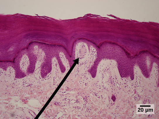
Meissner’s corpuscles, (shown in Figure 36.6) also known as tactile corpuscles, are found in the upper dermis, but they project into the epidermis. They, too, are found primarily in the glabrous skin on the fingertips and eyelids. They respond to fine touch and pressure, but they also respond to low-frequency vibration or flutter. They are rapidly adapting, fluid-filled, encapsulated neurons with small, well-defined borders and are responsive to fine details. Like Merkel’s disks, Meissner’s corpuscles are not as plentiful in the palms as they are in the fingertips.
マイスナー小体(図36.6)は触小体とも呼ばれ、真皮上層にあるが、表皮へと突出している。これも主に、指先やまぶたの無毛皮膚にみられる。微細な接触や圧力だけでなく、低周波の振動やはためくような刺激にも応答する。液体で満たされ、被膜に包まれた、順応の速いニューロンである。範囲は小さく境界が明確で、細部に応答する。メルケル盤と同様、手のひらより指先の方が多い。
OpenStax, Rice University; Biology 2e. (credit: modification of work by "Wbensmith"/Wikimedia Commons; scale-bar data from Matt Russell) · CC BY 4.0 · Source出典

Figure 36.6図 36.6
Meissner corpuscles in the fingertips, such as the one viewed here using bright field light microscopy, allow for touch discrimination of fine detail. (credit: modification of work by "Wbensmith"/Wikimedia Commons; scale-bar data from Matt Russell)
この明視野光学顕微鏡像に示すような指先のマイスナー小体により、細部を触覚で識別できる。(出典:「Wbensmith」/Wikimedia Commonsの作品を改変。スケールバーのデータ:Matt Russell)
Deeper in the epidermis, near the base, are Ruffini endings, which are also known as bulbous corpuscles. They are found in both glabrous and hairy skin. These are slow-adapting, encapsulated mechanoreceptors that detect skin stretch and deformations within joints, so they provide valuable feedback for gripping objects and controlling finger position and movement. Thus, they also contribute to proprioception and kinesthesia. Ruffini endings also detect warmth. Note that these warmth detectors are situated deeper in the skin than are the cold detectors. It is not surprising, then, that humans detect cold stimuli before they detect warm stimuli.
表皮のより深い、基部に近い位置には、球状小体とも呼ばれるルフィニ終末がある。無毛皮膚と有毛皮膚の両方に存在する。順応の遅い被膜性の機械受容器で、皮膚の伸張や関節内の変形を検出する。物を握り、指の位置や動きを制御するための重要なフィードバックを与え、固有感覚と運動感覚にも寄与する。ルフィニ終末は温かさも検出する。温かさの受容器は冷たさの受容器より皮膚の深い位置にある。このため、ヒトが温刺激より先に冷刺激を検出しても不思議ではない。
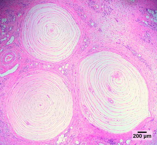
Pacinian corpuscles (seen in Figure 36.7) are located deep in the dermis of both glabrous and hairy skin and are structurally similar to Meissner’s corpuscles; they are found in the bone periosteum, joint capsules, pancreas and other viscera, breast, and genitals. They are rapidly adapting mechanoreceptors that sense deep transient (but not prolonged) pressure and high-frequency vibration. Pacinian receptors detect pressure and vibration by being compressed, stimulating their internal dendrites. There are fewer Pacinian corpuscles and Ruffini endings in skin than there are Merkel’s disks and Meissner’s corpuscles.
英語のヒント
パチニ小体(図36.7)は無毛皮膚と有毛皮膚の真皮深部にあり、構造はマイスナー小体に似ている。骨膜、関節包、膵臓などの内臓、乳房、生殖器にもみられる。順応の速い機械受容器で、深部の一過性の圧力と高周波の振動を感じるが、持続する圧力は感じない。圧縮されることで内部の樹状突起が刺激され、圧力と振動を検出する。皮膚にあるパチニ小体とルフィニ終末の数は、メルケル盤やマイスナー小体より少ない。
OpenStax, Rice University; Biology 2e. (credit: modification of work by Ed Uthman; scale-bar data from Matt Russell) · CC BY 4.0 · Source出典

Figure 36.7図 36.7
Pacinian corpuscles, such as these visualized using bright field light microscopy, detect pressure (touch) and high-frequency vibration. (credit: modification of work by Ed Uthman; scale-bar data from Matt Russell)
この明視野光学顕微鏡像に示すようなパチニ小体は、圧力(接触)と高周波の振動を検出する。(出典:Ed Uthmanの作品を改変。スケールバーのデータ:Matt Russell)
In proprioception, proprioceptive and kinesthetic signals travel through myelinated afferent neurons running from the spinal cord to the medulla. Neurons are not physically connected, but communicate via neurotransmitters secreted into synapses or “gaps” between communicating neurons. Once in the medulla, the neurons continue carrying the signals to the thalamus.
固有感覚では、固有感覚と運動感覚の信号が、脊髄から延髄へ走る有髄の求心性ニューロンを伝わる。ニューロン同士は物理的につながっておらず、シナプス、すなわち伝え合うニューロン間の「隙間」に分泌される神経伝達物質で情報をやり取りする。延髄に達した後、ニューロンは信号をさらに視床へ運ぶ。
Muscle spindles are stretch receptors that detect the amount of stretch, or lengthening of muscles. Related to these are Golgi tendon organs, which are tension receptors that detect the force of muscle contraction. Proprioceptive and kinesthetic signals come from limbs. Unconscious proprioceptive signals run from the spinal cord to the cerebellum, the brain region that coordinates muscle contraction, rather than to the thalamus, like most other sensory information.
英語のヒント
筋紡錘は、筋肉の伸張量、すなわち長さの増加を検出する伸張受容器である。関連するゴルジ腱器官は、筋収縮の力を検出する張力受容器である。固有感覚と運動感覚の信号は手足から来る。意識にのぼらない固有感覚の信号は、大部分のほかの感覚情報のように視床へ向かわず、脊髄から小脳へ伝わる。小脳は筋収縮を協調させる脳領域である。
Baroreceptors detect pressure changes in an organ. They are found in the walls of the carotid artery and the aorta where they monitor blood pressure, and in the lungs where they detect the degree of lung expansion. Stretch receptors are found at various sites in the digestive and urinary systems.
圧受容器は、器官内の圧力の変化を検出する。頸動脈と大動脈の壁にあって血圧を監視し、肺では膨張の程度を検出する。伸張受容器は、消化器系と泌尿器系の各所にもある。
In addition to these two types of deeper receptors, there are also rapidly adapting hair receptors, which are found on nerve endings that wrap around the base of hair follicles. There are a few types of hair receptors that detect slow and rapid hair movement, and they differ in their sensitivity to movement. Some hair receptors also detect skin deflection, and certain rapidly adapting hair receptors allow detection of stimuli that have not yet touched the skin.
深部の二種類の受容器に加え、順応の速い毛受容器もある。毛包の基部を取り巻く神経終末にみられる。毛受容器には数種類あり、ゆっくりした毛の動きと速い動きを検出し、動きに対する感受性が異なる。皮膚のたわみも検出するものや、まだ皮膚に触れていない刺激を検出できる順応の速いものもある。