第36章感覚系
Higher Processing高次の処理
The inner hair cells are most important for conveying auditory information to the brain. About 90 percent of the afferent neurons carry information from inner hair cells, with each hair cell synapsing with 10 or so neurons. Outer hair cells connect to only 10 percent of the afferent neurons, and each afferent neuron innervates many hair cells. The afferent, bipolar neurons that convey auditory information travel from the cochlea to the medulla, through the pons and midbrain in the brainstem, finally reaching the primary auditory cortex in the temporal lobe.
脳へ聴覚情報を伝えるうえで、最も重要なのは内有毛細胞である。求心性ニューロンの約90%が内有毛細胞からの情報を運び、一つの有毛細胞が約10個のニューロンとシナプスをつくる。外有毛細胞につながるのは求心性ニューロンの10%だけで、一つの求心性ニューロンが多数の有毛細胞を支配する。聴覚情報を運ぶ求心性の双極性ニューロンは、蝸牛から延髄へ進み、脳幹の橋と中脳を通って、最終的に側頭葉の一次聴覚野に達する。
Vestibular Information前庭情報
The stimuli associated with the vestibular system are linear acceleration (gravity) and angular acceleration and deceleration. Gravity, acceleration, and deceleration are detected by evaluating the inertia on receptive cells in the vestibular system. Gravity is detected through head position. Angular acceleration and deceleration are expressed through turning or tilting of the head.
前庭系に関わる刺激は、直線加速度(重力)と、回転の加速・減速である。前庭系の受容細胞にかかる慣性を評価することで、重力、加速、減速を検出する。重力は頭の位置によって、回転の加速・減速は頭の回転や傾きによって表される。
The vestibular system has some similarities with the auditory system. It utilizes hair cells just like the auditory system, but it excites them in different ways. There are five vestibular receptor organs in the inner ear: the utricle, the saccule, and three semicircular canals. Together, they make up what’s known as the vestibular labyrinth that is shown in Figure 36.16. The utricle and saccule respond to acceleration in a straight line, such as gravity. The roughly 30,000 hair cells in the utricle and 16,000 hair cells in the saccule lie below a gelatinous layer, with their stereocilia projecting into the gelatin. Embedded in this gelatin are calcium carbonate crystals—like tiny rocks. When the head is tilted, the crystals continue to be pulled straight down by gravity, but the new angle of the head causes the gelatin to shift, thereby bending the stereocilia. The bending of the stereocilia stimulates the neurons, and they signal to the brain that the head is tilted, allowing the maintenance of balance. It is the vestibular branch of the vestibulocochlear cranial nerve that deals with balance.
英語のヒント
前庭系は聴覚系と似た点がある。同じように有毛細胞を使うが、興奮させる方法が異なる。内耳には、卵形嚢、球形嚢、三つの半規管という五つの前庭受容器官がある。合わせて前庭迷路と呼ばれ、図36.16に示される。卵形嚢と球形嚢は、重力のような直線方向の加速度に応答する。卵形嚢には約30,000個、球形嚢には約16,000個の有毛細胞がある。ゼラチン状の層の下に位置し、不動毛がその層へ伸びる。層の中には、小さな石のような炭酸カルシウムの結晶が埋まっている。頭を傾けても結晶は重力で真下へ引かれ続けるが、頭の角度が変わるためゼラチン状の層がずれ、不動毛が曲がる。その曲がりがニューロンを刺激し、頭が傾いたことを脳へ知らせるので、平衡を保てる。平衡を担当するのは、内耳神経の前庭枝である。
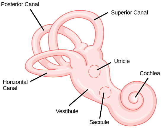

Figure 36.16図 36.16
The structure of the vestibular labyrinth is shown. (credit: modification of work by NIH)
前庭迷路の構造。(出典:米国国立衛生研究所〔NIH〕の作品を改変)
The fluid-filled semicircular canals are tubular loops set at oblique angles. They are arranged in three spatial planes. The base of each canal has a swelling that contains a cluster of hair cells. The hairs project into a gelatinous cap called the cupula and monitor angular acceleration and deceleration from rotation. They would be stimulated by driving your car around a corner, turning your head, or falling forward. One canal lies horizontally, while the other two lie at about 45 degree angles to the horizontal axis, as illustrated in Figure 36.16. When the brain processes input from all three canals together, it can detect angular acceleration or deceleration in three dimensions. When the head turns, the fluid in the canals shifts, thereby bending stereocilia and sending signals to the brain. Upon cessation of accelerating or decelerating—or just moving—the movement of the fluid within the canals slows or stops. For example, imagine holding a glass of water. When moving forward, water may splash backwards onto the hand, and when motion has stopped, water may splash forward onto the fingers. While in motion, the water settles in the glass and does not splash. Note that the canals are not sensitive to velocity itself, but to changes in velocity, so moving forward at 60mph with your eyes closed would not give the sensation of movement, but suddenly accelerating or braking would stimulate the receptors.
英語のヒント
液体で満たされた半規管は、互いに斜めの角度をなす管状の輪で、空間の三つの平面に配置されている。各管の基部には膨らみがあり、有毛細胞が集まる。毛はクプラというゼラチン状の帽子へ伸び、回転による加速と減速を監視する。車で角を曲がる、頭を回す、前へ倒れるといった動きで刺激される。図36.16のように、一つの管は水平で、残り二つは水平軸に約45度の角度をなす。脳は三つの管からの入力を一緒に処理し、三次元での回転の加速や減速を検出する。頭を回すと管内の液体がずれ、不動毛が曲がって脳へ信号を送る。加速、減速、あるいは単なる運動が止まると、管内の液体の動きも遅くなるか止まる。水の入ったコップを持つところを想像しよう。前へ動き始めると水は後ろへはねて手にかかり、動きを止めると前へはねて指にかかることがある。動いている間は、コップの中で水は落ち着き、はねない。半規管は速度そのものではなく、速度の変化に敏感である。そのため、目を閉じて時速60マイルで前進しても動きは感じないが、急に加速したりブレーキをかけたりすると受容器が刺激される。
Higher Processing高次の処理
Hair cells from the utricle, saccule, and semicircular canals also communicate through bipolar neurons to the cochlear nucleus in the medulla. Cochlear neurons send descending projections to the spinal cord and ascending projections to the pons, thalamus, and cerebellum. Connections to the cerebellum are important for coordinated movements. There are also projections to the temporal cortex, which account for feelings of dizziness; projections to autonomic nervous system areas in the brainstem, which account for motion sickness; and projections to the primary somatosensory cortex, which monitors subjective measurements of the external world and self-movement. People with lesions in the vestibular area of the somatosensory cortex see vertical objects in the world as being tilted. Finally, the vestibular signals project to certain optic muscles to coordinate eye and head movements.
卵形嚢、球形嚢、半規管の有毛細胞も、双極性ニューロンを介して延髄の蝸牛神経核に情報を伝える。蝸牛ニューロンは、脊髄へ下行性の投射を、橋、視床、小脳へ上行性の投射を送る。小脳とのつながりは、協調した運動に重要である。側頭皮質への投射はめまいの感覚を、脳幹の自律神経系の領域への投射は乗り物酔いをもたらす。一次体性感覚野への投射もあり、外界や自分の動きについての主観的な評価を監視する。体性感覚野の前庭領域に病変がある人には、垂直な物体が傾いて見える。最後に、前庭信号は特定の眼筋にも投射し、眼と頭の動きを協調させる。