第36章感覚系
Transduction of Light光の変換
The rods and cones are the site of transduction of light to a neural signal. Both rods and cones contain photopigments. In vertebrates, the main photopigment, rhodopsin, has two main parts (Figure 36.20): an opsin, which is a membrane protein (in the form of a cluster of α-helices that span the membrane), and retinal—a molecule that absorbs light. When light hits a photoreceptor, it causes a shape change in the retinal, altering its structure from a bent (cis) form of the molecule to its linear (trans) isomer. This isomerization of retinal activates the rhodopsin, starting a cascade of events that ends with the closing of Na+ channels in the membrane of the photoreceptor. Thus, unlike most other sensory neurons (which become depolarized by exposure to a stimulus) visual receptors become hyperpolarized and thus driven away from threshold (Figure 36.21).
英語のヒント
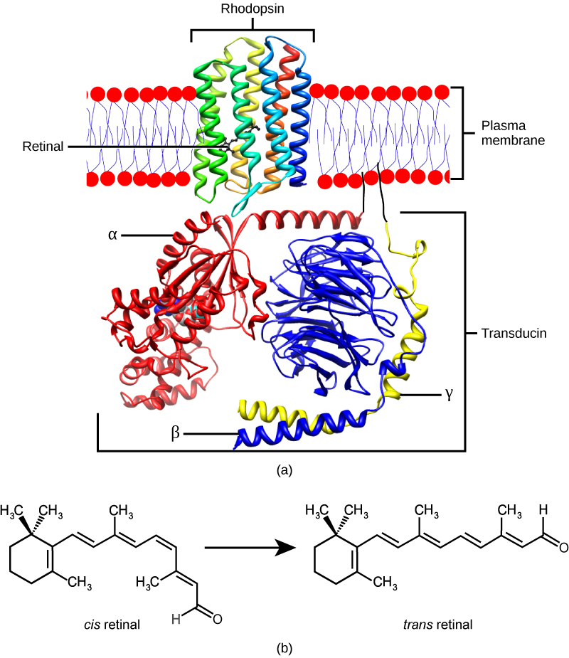
Figure 36.20図 36.20
(a) Rhodopsin, the photoreceptor in vertebrates, has two parts: the trans-membrane protein opsin, and retinal. When light strikes retinal, it changes shape from (b) a cis to a trans form. The signal is passed to a G-protein called transducin, triggering a series of downstream events.
(a)脊椎動物の光受容体ロドプシンは、膜貫通タンパク質オプシンとレチナールの二部分からなる。光がレチナールに当たると、(b)シス型からトランス型へ形が変わる。信号はトランスデューシンというGタンパク質に伝わり、下流の一連の反応を引き起こす。
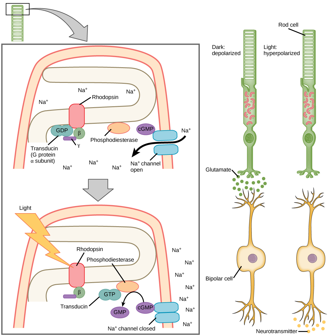
Figure 36.21図 36.21
When light strikes rhodopsin, the G-protein transducin is activated, which in turn activates phosphodiesterase. Phosphodiesterase converts cGMP to GMP, thereby closing sodium channels. As a result, the membrane becomes hyperpolarized. The hyperpolarized membrane does not release glutamate to the bipolar cell.
光がロドプシンに当たると、Gタンパク質のトランスデューシンが活性化され、続いてホスホジエステラーゼを活性化する。この酵素はcGMPをGMPへ変換し、ナトリウムチャネルを閉じる。その結果、膜が過分極する。過分極した膜からは、双極細胞へグルタミン酸が放出されない。
Trichromatic Coding三色による符号化
There are three types of cones (with different photopsins), and they differ in the wavelength to which they are most responsive, as shown in Figure 36.22. Some cones are maximally responsive to short light waves of 420 nm, so they are called S cones (“S” for “short”); others respond maximally to waves of 530 nm (M cones, for “medium”); a third group responds maximally to light of longer wavelengths, at 560 nm (L, or “long” cones). With only one type of cone, color vision would not be possible, and a two-cone (dichromatic) system has limitations. Primates use a three-cone (trichromatic) system, resulting in full color vision.
英語のヒント
錐体には、異なるフォトプシンをもつ三種類があり、最もよく応答する波長が異なる(図36.22)。420 nmの短波長に最大の応答を示すものを、shortのSからS錐体という。530 nmに最も応答するのがmediumのM錐体、より長い560 nmに最も応答するのがlongのL錐体である。錐体が一種類だけでは色覚は成り立たず、二種類による二色型の系にも限界がある。霊長類は三種類の錐体による三色型の系を使い、豊かな色覚を得ている。
The color we perceive is a result of the ratio of activity of our three types of cones. The colors of the visual spectrum, running from long-wavelength light to short, are red (700 nm), orange (600 nm), yellow (565 nm), green (497 nm), blue (470 nm), indigo (450 nm), and violet (425 nm). Humans have very sensitive perception of color and can distinguish about 500 levels of brightness, 200 different hues, and 20 steps of saturation, or about 2 million distinct colors.
英語のヒント
知覚する色は、三種類の錐体の活動の比率から生じる。可視スペクトルを長波長から短波長へ並べると、赤(700 nm)、橙(600 nm)、黄(565 nm)、緑(497 nm)、青(470 nm)、藍(450 nm)、紫(425 nm)である。ヒトの色の知覚は非常に敏感で、約500段階の明るさ、200種類の色相、20段階の彩度を区別できる。合計すると約200万種類の色になる。
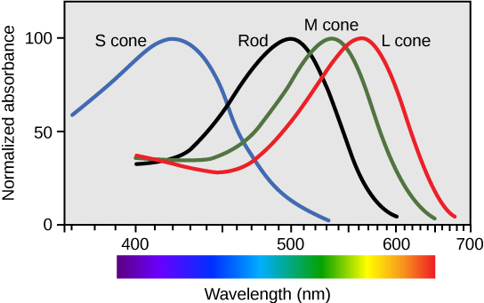
Figure 36.22図 36.22
Human rod cells and the different types of cone cells each have an optimal wavelength. However, there is considerable overlap in the wavelengths of light detected.
ヒトの桿体と各種類の錐体には、それぞれ最適な波長がある。ただし、検出する光の波長範囲には大きな重なりがある。
Retinal Processing網膜での処理
Visual signals leave the cones and rods, travel to the bipolar cells, and then to ganglion cells. A large degree of processing of visual information occurs in the retina itself, before visual information is sent to the brain.
視覚信号は錐体と桿体を出て、双極細胞へ、さらに神経節細胞へ伝わる。脳へ送られる前に、視覚情報の多くの処理が網膜自体で行われる。
Photoreceptors in the retina continuously undergo tonic activity. That is, they are always slightly active even when not stimulated by light. In neurons that exhibit tonic activity, the absence of stimuli maintains a firing rate at a baseline; while some stimuli increase firing rate from the baseline, and other stimuli decrease firing rate. In the absence of light, the bipolar neurons that connect rods and cones to ganglion cells are continuously and actively inhibited by the rods and cones. Exposure of the retina to light hyperpolarizes the rods and cones and removes their inhibition of bipolar cells. The now active bipolar cells in turn stimulate the ganglion cells, which send action potentials along their axons (which leave the eye as the optic nerve). Thus, the visual system relies on change in retinal activity, rather than the absence or presence of activity, to encode visual signals for the brain. Sometimes horizontal cells carry signals from one rod or cone to other photoreceptors and to several bipolar cells. When a rod or cone stimulates a horizontal cell, the horizontal cell inhibits more distant photoreceptors and bipolar cells, creating lateral inhibition. This inhibition sharpens edges and enhances contrast in the images by making regions receiving light appear lighter and dark surroundings appear darker. Amacrine cells can distribute information from one bipolar cell to many ganglion cells.
英語のヒント
網膜の視細胞は、持続的な活動を続けている。つまり、光で刺激されていないときでも、常にわずかに活動する。このようなニューロンでは、刺激がないときに基準となる発火頻度が保たれ、ある刺激は基準より発火を増やし、別の刺激は減らす。暗いとき、桿体と錐体を神経節細胞につなぐ双極ニューロンは、桿体と錐体から持続的、能動的に抑制される。網膜に光が当たると、桿体と錐体が過分極し、双極細胞への抑制が解除される。活動するようになった双極細胞は神経節細胞を刺激し、神経節細胞は軸索を通して活動電位を送る。この軸索が視神経として眼を出る。したがって視覚系は、活動の有無ではなく網膜活動の変化に基づいて、脳へ送る視覚信号を符号化する。水平細胞が、一つの桿体や錐体から、ほかの視細胞や複数の双極細胞へ信号を運ぶこともある。桿体や錐体が水平細胞を刺激すると、水平細胞が離れた視細胞と双極細胞を抑え、側方抑制を生む。光を受ける領域をより明るく、暗い周囲をより暗く見せるため、像の輪郭が鋭くなり、コントラストが高まる。アマクリン細胞は、一つの双極細胞から多くの神経節細胞へ情報を配れる。
You can demonstrate this using an easy demonstration to “trick” your retina and brain about the colors you are observing in your visual field. Look fixedly at Figure 36.23 for about 45 seconds. Then quickly shift your gaze to a sheet of blank white paper or a white wall. You should see an afterimage of the Norwegian flag in its correct colors. At this point, close your eyes for a moment, then reopen them, looking again at the white paper or wall; the afterimage of the flag should continue to appear as red, white, and blue. What causes this? According to an explanation called opponent process theory, as you gazed fixedly at the green, black, and yellow flag, your retinal ganglion cells that respond positively to green, black, and yellow increased their firing dramatically. When you shifted your gaze to the neutral white ground, these ganglion cells abruptly decreased their activity and the brain interpreted this abrupt downshift as if the ganglion cells were responding now to their “opponent” colors: red, white, and blue, respectively, in the visual field. Once the ganglion cells return to their baseline activity state, the false perception of color will disappear.
英語のヒント
視野の色について網膜と脳を「だます」簡単な実演で、この仕組みを確かめられる。図36.23を約45秒間じっと見てから、白紙や白い壁へ素早く視線を移そう。本来の色のノルウェー国旗の残像が見えるはずだ。少し目を閉じ、再び開いて白紙や壁を見ても、赤、白、青の残像が続いて見えるだろう。何が起こっているのだろうか。反対色過程説によれば、緑、黒、黄の旗を凝視している間、その色に正に応答する網膜神経節細胞の発火が大きく増える。中立な白い背景へ視線を移すと、これらの細胞の活動が急に低下する。脳はこの急低下を、それぞれの「反対」の色、すなわち赤、白、青に細胞が今応答しているかのように解釈する。神経節細胞が基準の活動状態へ戻ると、実際とは異なる色の知覚は消える。
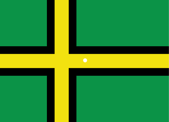


Figure 36.23図 36.23
View this flag to understand how retinal processing works. Stare at the center of the flag (indicated by the white dot) for 45 seconds, and then quickly look at a white background, noticing how colors appear.
この旗を見て、網膜での処理の働きを確かめよう。白点で示された中央を45秒間凝視した後、素早く白い背景を見て、どのような色が現れるか観察する。