第36章感覚系
Transduction of Sound音の変換
Vibrating objects, such as vocal cords, create sound waves or pressure waves in the air. When these pressure waves reach the ear, the ear transduces this mechanical stimulus (pressure wave) into a nerve impulse (electrical signal) that the brain perceives as sound. The pressure waves strike the tympanum, causing it to vibrate. The mechanical energy from the moving tympanum transmits the vibrations to the three bones of the middle ear. The stapes transmits the vibrations to a thin diaphragm called the oval window, which is the outermost structure of the inner ear. The structures of the inner ear are found in the labyrinth, a bony, hollow structure that is the most interior portion of the ear. Here, the energy from the sound wave is transferred from the stapes through the flexible oval window and to the fluid of the cochlea. The vibrations of the oval window create pressure waves in the fluid (perilymph) inside the cochlea. The cochlea is a whorled structure, like the shell of a snail, and it contains receptors for transduction of the mechanical wave into an electrical signal (as illustrated in Figure 36.14). Inside the cochlea, the basilar membrane is a mechanical analyzer that runs the length of the cochlea, curling toward the cochlea’s center.
英語のヒント
声帯などの振動する物体は、空気中に音波、すなわち圧力波をつくる。耳に届くと、耳は圧力波という機械的刺激を神経インパルス、すなわち電気信号に変え、脳がそれを音として知覚する。圧力波は鼓膜を振動させ、その機械的エネルギーが中耳の三つの骨へ振動を伝える。アブミ骨は、内耳の最外側の構造である卵円窓という薄い膜へ振動を伝える。内耳の構造は、耳の最も奥にある、骨性の空洞構造である迷路の中に収まる。音波のエネルギーはアブミ骨から柔軟な卵円窓を通り、蝸牛の液体へ伝わる。卵円窓の振動は、蝸牛内の外リンパ液に圧力波を生む。蝸牛はカタツムリの殻のように巻いた構造で、機械的な波を電気信号に変える受容器を含む(図36.14)。内部では、基底膜が蝸牛の全長を走り、中心へ向かって巻きながら、機械的な分析装置として働く。
The mechanical properties of the basilar membrane change along its length, such that it is thicker, tauter, and narrower at the outside of the whorl (where the cochlea is largest), and thinner, floppier, and broader toward the apex, or center, of the whorl (where the cochlea is smallest). Different regions of the basilar membrane vibrate according to the frequency of the sound wave conducted through the fluid in the cochlea. For these reasons, the fluid-filled cochlea detects different wave frequencies (pitches) at different regions of the membrane. When the sound waves in the cochlear fluid contact the basilar membrane, it flexes back and forth in a wave-like fashion. Above the basilar membrane is the tectorial membrane.
英語のヒント
基底膜の機械的性質は、長さに沿って変わる。蝸牛が最も大きい渦巻きの外側では、厚く、張りが強く、幅が狭い。蝸牛が最も小さい渦巻きの頂点、すなわち中心へ近づくほど、薄く、柔らかく、幅広くなる。蝸牛の液体を伝わる音波の周波数に応じて、基底膜の異なる領域が振動する。そのため、液体で満たされた蝸牛は、膜の異なる場所で異なる周波数、すなわち音の高さを検出する。液体中の音波が基底膜に接すると、膜は波のように前後にたわむ。基底膜の上には蓋膜がある。
The site of transduction is in the organ of Corti (spiral organ). It is composed of hair cells held in place above the basilar membrane like flowers projecting up from soil, with their exposed short, hair-like stereocilia contacting or embedded in the tectorial membrane above them. The inner hair cells are the primary auditory receptors and exist in a single row, numbering approximately 3,500. The stereocilia from inner hair cells extend into small dimples on the tectorial membrane’s lower surface. The outer hair cells are arranged in three or four rows. They number approximately 12,000, and they function to fine tune incoming sound waves. The longer stereocilia that project from the outer hair cells actually attach to the tectorial membrane. All of the stereocilia are mechanoreceptors, and when bent by vibrations they respond by opening a gated ion channel (refer to Figure 36.15). As a result, the hair cell membrane is depolarized, and a signal is transmitted to the chochlear nerve. Intensity (volume) of sound is determined by how many hair cells at a particular location are stimulated.
英語のヒント
変換が起こる場所は、コルチ器(ラセン器)である。基底膜の上に固定された有毛細胞からなり、土から花が伸びるように並ぶ。露出した短い毛状の不動毛は、上の蓋膜に接触するか、埋め込まれている。内有毛細胞は主要な聴覚受容器で、一列に約3,500個並ぶ。不動毛は、蓋膜下面の小さなくぼみへ伸びる。外有毛細胞は3~4列に約12,000個並び、入ってくる音波を微調整する。外有毛細胞のより長い不動毛は、蓋膜に実際に付着する。すべての不動毛は機械受容器で、振動で曲がると、開閉型イオンチャネルを開いて応答する(図36.15)。その結果、有毛細胞の膜が脱分極し、蝸牛神経へ信号が伝わる。音の強さ、すなわち音量は、ある場所で刺激される有毛細胞の数によって決まる。
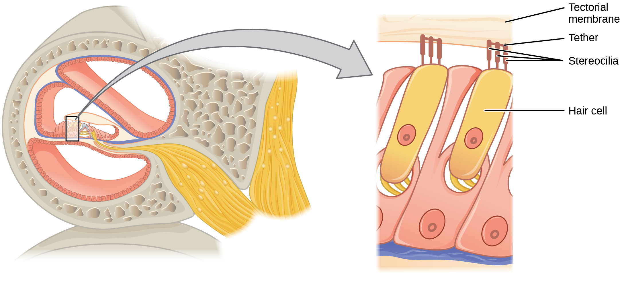

Figure 36.15図 36.15
The hair cell is a mechanoreceptor with an array of stereocilia emerging from its apical surface. The stereocilia are tethered together by proteins that open ion channels when the array is bent toward the tallest member of their array, and closed when the array is bent toward the shortest member of their array.
有毛細胞は、頂端面から一列の不動毛が伸びる機械受容器である。不動毛同士はタンパク質で結ばれる。列が最も長い不動毛の側へ曲がるとイオンチャネルが開き、最も短い側へ曲がると閉じる。
The hair cells are arranged on the basilar membrane in an orderly way. The basilar membrane vibrates in different regions, according to the frequency of the sound waves impinging on it. Likewise, the hair cells that lay above it are most sensitive to a specific frequency of sound waves. Hair cells can respond to a small range of similar frequencies, but they require stimulation of greater intensity to fire at frequencies outside of their optimal range. The difference in response frequency between adjacent inner hair cells is about 0.2 percent. Compare that to adjacent piano strings, which are about six percent different. Place theory, which is the model for how biologists think pitch detection works in the human ear, states that high frequency sounds selectively vibrate the basilar membrane of the inner ear near the entrance port (the oval window). Lower frequencies travel farther along the membrane before causing appreciable excitation of the membrane. The basic pitch-determining mechanism is based on the location along the membrane where the hair cells are stimulated. The place theory is the first step toward an understanding of pitch perception. Considering the extreme pitch sensitivity of the human ear, it is thought that there must be some auditory “sharpening” mechanism to enhance the pitch resolution.
英語のヒント
有毛細胞は基底膜上に規則正しく並ぶ。基底膜は、加わる音波の周波数によって異なる領域が振動する。その上の有毛細胞も同様に、特定の周波数の音波に最も敏感である。近い周波数の狭い範囲には応答できるが、最適な範囲を外れた周波数で発火するには、より強い刺激が必要になる。隣り合う内有毛細胞の応答周波数の違いは約0.2%である。隣り合うピアノの弦では約6%違うことと比べてみよう。場所説は、ヒトの耳が音の高さを検出する仕組みについて、生物学者が考えるモデルである。高周波の音は、内耳の入口である卵円窓の近くの基底膜を選択的に振動させる。低周波の音は膜をより遠くまで進んでから、十分な興奮を引き起こす。音の高さを決める基本的な仕組みは、膜上のどこで有毛細胞が刺激されるかに基づく。場所説は、音の高さの知覚を理解する第一歩である。ヒトの耳は高さの違いに非常に敏感なため、高さの分解能を高める、何らかの聴覚の「先鋭化」の仕組みがあるはずだと考えられる。
When sound waves produce fluid waves inside the cochlea, the basilar membrane flexes, bending the stereocilia that attach to the tectorial membrane. Their bending results in action potentials in the hair cells, and auditory information travels along the neural endings of the bipolar neurons of the hair cells (collectively, the auditory nerve) to the brain. When the hairs bend, they release an excitatory neurotransmitter at a synapse with a sensory neuron, which then conducts action potentials to the central nervous system. The cochlear branch of the vestibulocochlear cranial nerve sends information on hearing. The auditory system is very refined, and there is some modulation or “sharpening” built in. The brain can send signals back to the cochlea, resulting in a change of length in the outer hair cells, sharpening or dampening the hair cells’ response to certain frequencies.
音波が蝸牛内の液体に波を起こすと、基底膜がたわみ、蓋膜につながる不動毛を曲げる。曲がることで有毛細胞に活動電位が生じ、聴覚情報は有毛細胞の双極性ニューロンの神経終末を通って脳へ伝わる。これらを合わせて聴神経という。毛が曲がると、感覚ニューロンとのシナプスで興奮性の神経伝達物質を放出し、そのニューロンが中枢神経系へ活動電位を伝える。脳神経である内耳神経の蝸牛枝は、聴覚情報を送る。聴覚系は精細で、調節、すなわち「先鋭化」の仕組みが組み込まれている。脳は蝸牛へ信号を送り返して外有毛細胞の長さを変え、特定の周波数への応答を強めたり弱めたりできる。