第39章呼吸器系
39.3 Breathing39.3 呼吸運動
Learning Objectives学習目標
By the end of this section, you will be able to do the following:
この節を終えると、次のことができるようになる。
- Describe how the structures of the lungs and thoracic cavity control the mechanics of breathing肺と胸腔の構造が、呼吸の力学をどのように制御するかを説明する。
- Explain the importance of compliance and resistance in the lungs肺のコンプライアンスと抵抗の重要性を説明する。
- Discuss problems that may arise due to a V/Q mismatchV/Qの不均衡によって生じうる問題を論じる。
Mammalian lungs are located in the thoracic cavity where they are surrounded and protected by the rib cage, intercostal muscles, and bound by the chest wall. The bottom of the lungs is contained by the diaphragm, a skeletal muscle that facilitates breathing. Breathing requires the coordination of the lungs, the chest wall, and most importantly, the diaphragm.
英語のヒント
哺乳類の肺は胸腔内にあり、胸郭と肋間筋に囲まれて保護され、胸壁によって境界を定められている。肺の底部には、呼吸を助ける骨格筋である横隔膜がある。呼吸には、肺、胸壁、そしてとりわけ横隔膜の協調が必要である。
Types of Breathing呼吸の種類
Amphibians have evolved multiple ways of breathing. Young amphibians, like tadpoles, use gills to breathe, and they don’t leave the water. Some amphibians retain gills for life. As the tadpole grows, the gills disappear and lungs grow. These lungs are primitive and not as evolved as mammalian lungs. Adult amphibians are lacking or have a reduced diaphragm, so breathing via lungs is forced. The other means of breathing for amphibians is diffusion across the skin. To aid this diffusion, amphibian skin must remain moist.
英語のヒント
両生類は、複数の呼吸方法を進化させてきた。オタマジャクシなどの幼生は、えらで呼吸し、水中から出ない。一生えらをもつ両生類もいる。オタマジャクシが成長すると、えらがなくなり、肺が発達する。これらの肺は原始的で、哺乳類の肺ほど発達していない。成体の両生類では横隔膜がないか、縮小しているため、肺へ空気を押し込んで呼吸する。両生類のもう1つの呼吸方法は、皮膚を通した拡散である。この拡散を助けるため、皮膚は湿った状態を保たなければならない。
Birds face a unique challenge with respect to breathing: They fly. Flying consumes a great amount of energy; therefore, birds require a lot of oxygen to aid their metabolic processes. Birds have evolved a respiratory system that supplies them with the oxygen needed to enable flying. Similar to mammals, birds have lungs, which are organs specialized for gas exchange. Oxygenated air, taken in during inhalation, diffuses across the surface of the lungs into the bloodstream, and carbon dioxide diffuses from the blood into the lungs and expelled during exhalation. The details of breathing between birds and mammals differ substantially.
英語のヒント
鳥類は、呼吸に関して特有の課題に直面している。それは、空を飛ぶということである。飛行は大量のエネルギーを消費するため、鳥類は代謝を支えるために多くの酸素を必要とする。鳥類は、飛行を可能にするだけの酸素を供給できる呼吸器系を進化させてきた。鳥類も哺乳類と同じく、ガス交換に特化した器官である肺をもつ。吸気で取り込まれた酸素を含む空気は、肺の表面を通って血流へ拡散し、二酸化炭素は血液から肺へ拡散して、呼気で排出される。しかし、呼吸の細かな仕組みは、鳥類と哺乳類で大きく異なる。
In addition to lungs, birds have air sacs inside their body. Air flows in one direction from the posterior air sacs to the lungs and out of the anterior air sacs. The flow of air is in the opposite direction from blood flow, and gas exchange takes place much more efficiently. This type of breathing enables birds to obtain the requisite oxygen, even at higher altitudes where the oxygen concentration is low. This directionality of airflow requires two cycles of air intake and exhalation to completely get the air out of the lungs.
英語のヒント
鳥類は、肺に加えて、体内に気嚢をもつ。空気は後方の気嚢から肺を通り、前方の気嚢から出るという、一方向の流れをとる。空気の流れる方向は血流と逆であり、ガス交換ははるかに効率よく行われる。この呼吸様式により、鳥類は酸素濃度の低い高い標高でも、必要な酸素を得られる。このように空気が一方向へ流れる仕組みでは、空気を肺から完全に出すために、吸気と呼気の周期が2回必要になる。
All mammals have lungs that are the main organs for breathing. Lung capacity has evolved to support the animal’s activities. During inhalation, the lungs expand with air, and oxygen diffuses across the lung’s surface and enters the bloodstream. During exhalation, the lungs expel air and lung volume decreases. In the next few sections, the process of human breathing will be explained.
すべての哺乳類は、呼吸の主な器官である肺をもつ。肺容量は、それぞれの動物の活動を支えられるように進化してきた。吸気では肺が空気で膨らみ、酸素が肺の表面を通って拡散し、血流へ入る。呼気では肺から空気が排出され、肺気量が減る。以下では、ヒトの呼吸の過程を説明する。
The Mechanics of Human Breathingヒトの呼吸の力学
Boyle’s Law is the gas law that states that in a closed space, pressure and volume are inversely related. As volume decreases, pressure increases and vice versa (Figure 39.15). The relationship between gas pressure and volume helps to explain the mechanics of breathing.
英語のヒント
ボイルの法則は、閉じた空間では圧力と体積が反比例することを述べた気体の法則である。体積が小さくなると圧力は大きくなり、その逆も成り立つ(図39.15)。気体の圧力と体積の関係は、呼吸の力学を説明するのに役立つ。
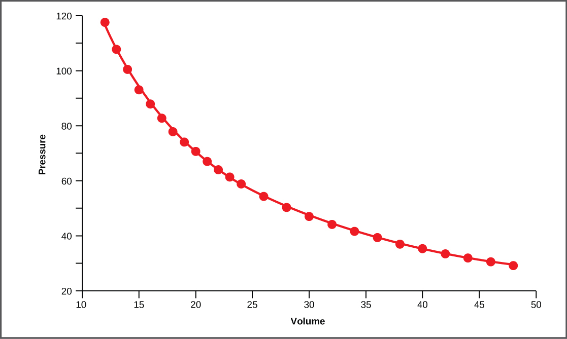
Figure 39.15図 39.15
This graph shows data from Boyle’s original 1662 experiment, which shows that pressure and volume are inversely related. No units are given as Boyle used arbitrary units in his experiments.
ボイルが1662年に行った実験のデータを示すグラフ。圧力と体積が反比例することを示している。ボイルは実験で任意の単位を使ったため、単位は記していない。
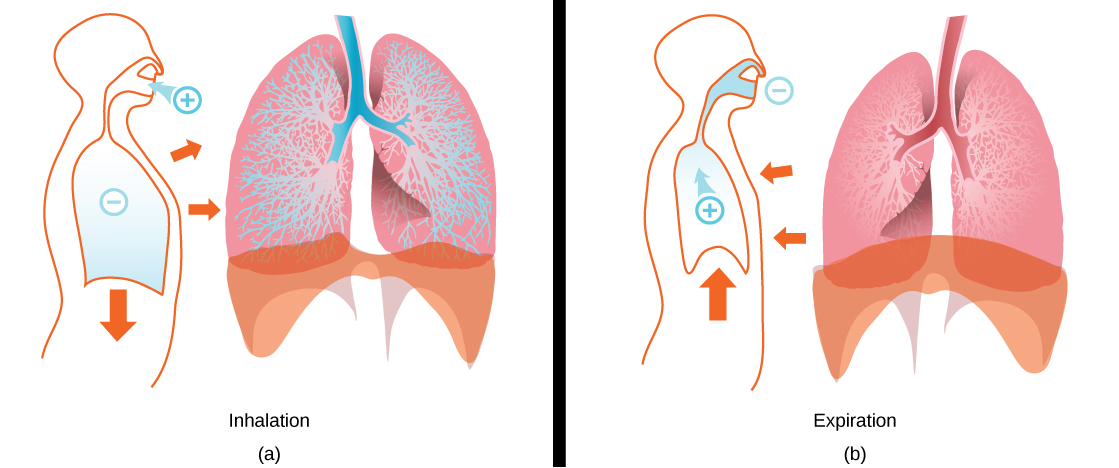
There is always a slightly negative pressure within the thoracic cavity, which aids in keeping the airways of the lungs open. During inhalation, volume increases as a result of contraction of the diaphragm, and pressure decreases (according to Boyle’s Law). This decrease of pressure in the thoracic cavity relative to the environment makes the cavity less than the atmosphere (Figure 39.16a). Because of this drop in pressure, air rushes into the respiratory passages. To increase the volume of the lungs, the chest wall expands. This results from the contraction of the intercostal muscles, the muscles that are connected to the rib cage. Lung volume expands because the diaphragm contracts and the intercostal muscles contract, thus expanding the thoracic cavity. This increase in the volume of the thoracic cavity lowers pressure compared to the atmosphere, so air rushes into the lungs, thus increasing its volume. The resulting increase in volume is largely attributed to an increase in alveolar space, because the bronchioles and bronchi are stiff structures that do not change in size.
英語のヒント
胸腔内には常にわずかな陰圧があり、肺の気道を開いた状態に保つのを助けている。吸気では、横隔膜が収縮して体積が増し、ボイルの法則に従って圧力が低下する。周囲に対して胸腔内の圧力が下がり、大気圧より低くなる(図39.16a)。この圧力低下によって、空気が呼吸の通路へ流れ込む。肺の体積を増やすために、胸壁は広がる。これは、胸郭につながる筋である肋間筋の収縮によって起こる。横隔膜と肋間筋が収縮して胸腔が広がるため、肺の体積が増える。胸腔の体積が増すと、その圧力が大気圧より低くなり、肺へ空気が流れ込んで、肺の体積がさらに増加する。この体積増加の大部分は、肺胞内の空間が広がることによる。細気管支や気管支は硬い構造で、大きさが変化しないためである。
OpenStax, Rice University; Biology 2e. (credit: modification of work by Mariana Ruiz Villareal) · CC BY 4.0 · Source出典
Figure 39.16図 39.16
The lungs, chest wall, and diaphragm are all involved in respiration, both (a) inhalation and (b) expiration. (credit: modification of work by Mariana Ruiz Villareal)
肺、胸壁、横隔膜は、(a)吸気と(b)呼気の両方に関わる。(出典:Mariana Ruiz Villarealの作品を改変)
The chest wall expands out and away from the lungs. The lungs are elastic; therefore, when air fills the lungs, the elastic recoil within the tissues of the lung exerts pressure back toward the interior of the lungs. These outward and inward forces compete to inflate and deflate the lung with every breath. Upon exhalation, the lungs recoil to force the air out of the lungs, and the intercostal muscles relax, returning the chest wall back to its original position (Figure 39.16b). The diaphragm also relaxes and moves higher into the thoracic cavity. This increases the pressure within the thoracic cavity relative to the environment, and air rushes out of the lungs. The movement of air out of the lungs is a passive event. No muscles are contracting to expel the air.
英語のヒント
胸壁は肺から離れるように外側へ広がる。肺には弾性があるため、空気で満たされると、肺組織の弾性反跳が肺の内側へ向かう圧力を生む。この外向きと内向きの力が拮抗し、呼吸のたびに肺が膨らんだりしぼんだりする。息を吐くときには、肺が弾性によって元に戻り、空気を押し出す。肋間筋は弛緩し、胸壁が元の位置へ戻る(図39.16b)。横隔膜も弛緩し、胸腔内で上方へ移動する。その結果、胸腔内の圧力が周囲より高くなり、空気が肺から流れ出る。肺から空気が出るのは受動的な現象であり、空気を排出するために筋が収縮するわけではない。
Each lung is surrounded by an invaginated sac. The layer of tissue that covers the lung and dips into spaces is called the visceral pleura. A second layer of parietal pleura lines the interior of the thorax (Figure 39.17). The space between these layers, the intrapleural space, contains a small amount of fluid that protects the tissue and reduces the friction generated from rubbing the tissue layers together as the lungs contract and relax. Pleurisy results when these layers of tissue become inflamed; it is painful because the inflammation increases the pressure within the thoracic cavity and reduces the volume of the lung.
英語のヒント
左右の肺は、それぞれ内側に折れ込んだ袋に包まれている。肺を覆い、隙間にも入り込む組織の層を、臓側胸膜という。もう1つの層である壁側胸膜は、胸部の内側を覆う(図39.17)。2層の間の胸膜腔には少量の液体があり、組織を保護するとともに、肺が縮んだり緩んだりするときに組織の層がこすれ合う摩擦を減らす。これらの組織層に炎症が起こると胸膜炎になる。炎症によって胸腔内の圧力が高まり、肺の体積が減るため、痛みを伴う。
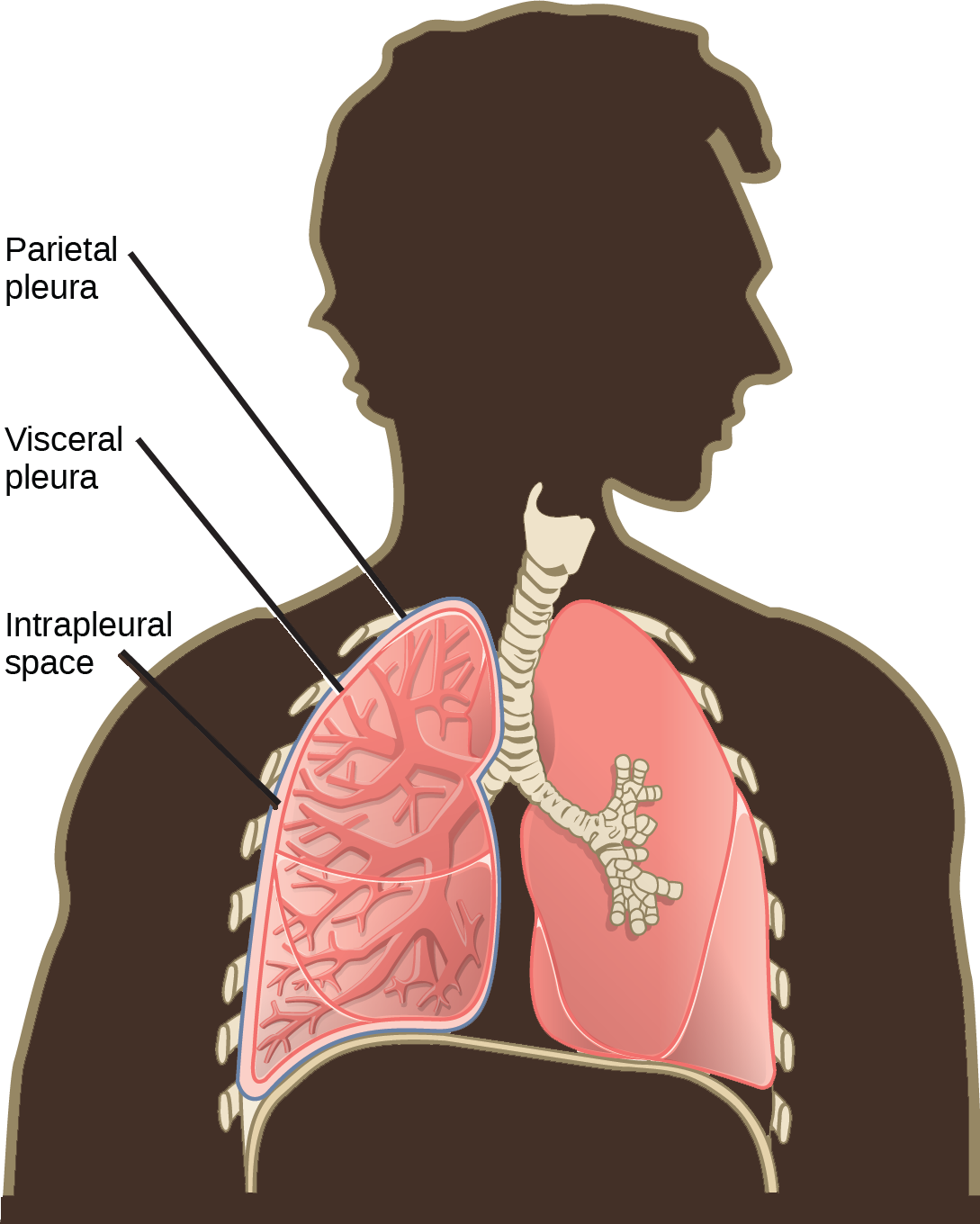


Figure 39.17図 39.17
A tissue layer called pleura surrounds the lung and interior of the thoracic cavity. (credit: modification of work by NCI)
胸膜という組織層が、肺と胸腔の内側を覆う。(出典:NCIの作品を改変)