第39章呼吸器系
Tracheal Systems気管系
Insect respiration is independent of its circulatory system; therefore, the blood does not play a direct role in oxygen transport. Insects have a highly specialized type of respiratory system called the tracheal system, which consists of a network of small tubes that carries oxygen to the entire body. The tracheal system is the most direct and efficient respiratory system in active animals. The tubes in the tracheal system are made of a polymeric material called chitin.
英語のヒント
昆虫の呼吸は循環器系から独立しているため、血液は酸素の輸送に直接関与しない。昆虫は気管系と呼ばれる高度に特殊化した呼吸器系をもち、細い管の網によって全身へ酸素を運ぶ。気管系は、活動的な動物の呼吸器系の中で最も直接的で効率がよい。気管系の管は、キチンと呼ばれる高分子材料でできている。
Insect bodies have openings, called spiracles, along the thorax and abdomen. These openings connect to the tubular network, allowing oxygen to pass into the body (Figure 39.6) and regulating the diffusion of CO2 and water vapor. Air enters and leaves the tracheal system through the spiracles. Some insects can ventilate the tracheal system with body movements.
英語のヒント
昆虫の体には、胸部と腹部に沿って、気門と呼ばれる開口部がある。気門は管の網につながり、酸素を体内へ通す(図39.6)とともに、CO2と水蒸気の拡散を調節する。空気は気門を通って気管系へ出入りする。一部の昆虫は、体を動かして気管系を換気できる。
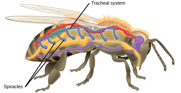
Figure 39.6図 39.6
Insects perform respiration via a tracheal system.
昆虫は気管系を介して呼吸する。
Mammalian Systems哺乳類の呼吸器系
In mammals, pulmonary ventilation occurs via inhalation (breathing). During inhalation, air enters the body through the nasal cavity located just inside the nose (Figure 39.7). As air passes through the nasal cavity, the air is warmed to body temperature and humidified. The respiratory tract is coated with mucus to seal the tissues from direct contact with air. Mucus is high in water. As air crosses these surfaces of the mucous membranes, it picks up water. These processes help equilibrate the air to the body conditions, reducing any damage that cold, dry air can cause. Particulate matter that is floating in the air is removed in the nasal passages via mucus and cilia. The processes of warming, humidifying, and removing particles are important protective mechanisms that prevent damage to the trachea and lungs. Thus, inhalation serves several purposes in addition to bringing oxygen into the respiratory system.
英語のヒント
哺乳類では、吸気(息を吸うこと)によって肺の換気が行われる。息を吸うと、鼻のすぐ内側にある鼻腔を通って空気が体内に入る(図39.7)。鼻腔を通る間に、空気は体温まで温められ、加湿される。呼吸の通り道は粘液で覆われ、組織が空気に直接触れるのを防いでいる。粘液は水分を多く含む。空気は粘膜の表面を通るときに水分を取り込む。これらの過程によって空気の状態が体内の条件に近づき、冷たく乾いた空気による損傷が抑えられる。空気中に浮遊する粒子状物質は、鼻の通り道で粘液と繊毛によって除去される。空気を温め、加湿し、粒子を取り除く過程は、気管や肺の損傷を防ぐ重要な防御機構である。このように、吸気は呼吸器系へ酸素を取り入れる以外にも、いくつかの役割を果たしている。
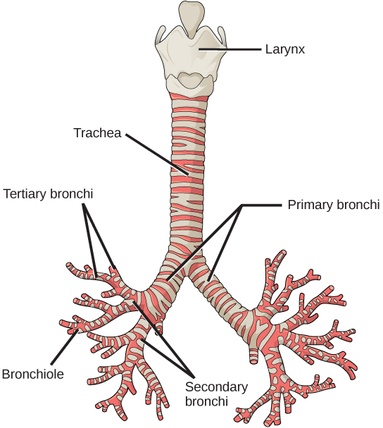
From the nasal cavity, air passes through the pharynx (throat) and the larynx (voice box), as it makes its way to the trachea (Figure 39.7). The main function of the trachea is to funnel the inhaled air to the lungs and the exhaled air back out of the body. The human trachea is a cylinder about 10 to 12 cm long and 2 cm in diameter that sits in front of the esophagus and extends from the larynx into the chest cavity where it divides into the two primary bronchi at the midthorax. It is made of incomplete rings of hyaline cartilage and smooth muscle (Figure 39.8). The trachea is lined with mucus-producing goblet cells and ciliated epithelia. The cilia propel foreign particles trapped in the mucus toward the pharynx. The cartilage provides strength and support to the trachea to keep the passage open. The smooth muscle can contract, decreasing the trachea’s diameter, which causes expired air to rush upwards from the lungs at a great force. The forced exhalation helps expel mucus when we cough. Smooth muscle can contract or relax, depending on stimuli from the external environment or the body’s nervous system.
英語のヒント
空気は鼻腔から咽頭(のど)と喉頭(声を出す器官)を通り、気管へ向かう(図39.7)。気管の主な機能は、吸い込んだ空気を肺へ導き、吐き出す空気を体外へ戻すことである。ヒトの気管は、長さ約10〜12cm、直径約2cmの円筒で、食道の前にある。喉頭から胸腔へ伸び、胸部の中央で2本の主気管支に分かれる。気管は、不完全な輪の形をした硝子軟骨と平滑筋からできている(図39.8)。内面には、粘液をつくる杯細胞と繊毛上皮が並ぶ。繊毛は、粘液に捕らえられた異物粒子を咽頭へ向けて運ぶ。軟骨は気管に強度と支えを与え、空気の通路を開いた状態に保つ。平滑筋が収縮すると気管の直径が小さくなり、肺からの呼気が強い勢いで上方へ押し出される。咳をするときには、この強い呼気が粘液の排出を助ける。平滑筋は、外部環境や体の神経系からの刺激に応じて収縮したり弛緩したりする。
OpenStax, Rice University; Biology 2e. (credit: modification of work by Gray's Anatomy) · CC BY 4.0 · Source出典
Figure 39.8図 39.8
The trachea and bronchi are made of incomplete rings of cartilage. (credit: modification of work by Gray's Anatomy)
気管と気管支は、不完全な輪の形をした軟骨からできている。(出典:Gray’s Anatomyの作品を改変)
Lungs: Bronchi and Alveoli肺:気管支と肺胞
The end of the trachea bifurcates (divides) to the right and left lungs. The lungs are not identical. The right lung is larger and contains three lobes, whereas the smaller left lung contains two lobes (Figure 39.9). The muscular diaphragm, which facilitates breathing, is inferior to (below) the lungs and marks the end of the thoracic cavity.
英語のヒント
気管の末端は二分し、左右の肺へ向かう。左右の肺は同じ形ではない。右肺は大きく、3つの葉をもつのに対し、小さい左肺には2つの葉がある(図39.9)。呼吸を助ける筋性の横隔膜は、肺の下方にあり、胸腔の下端を区切っている。
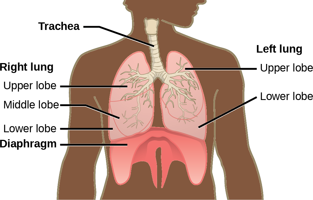


Figure 39.9図 39.9
The trachea bifurcates into the right and left bronchi in the lungs. The right lung is made of three lobes and is larger. To accommodate the heart, the left lung is smaller and has only two lobes.
気管は左右の気管支に分かれて肺へ入る。右肺は3葉からなり、左肺より大きい。左肺は心臓を収めるために小さく、2葉だけからなる。
In the lungs, air is diverted into smaller and smaller passages, or bronchi. Air enters the lungs through the two primary (main) bronchi (singular: bronchus). Each bronchus divides into secondary bronchi, then into tertiary bronchi, which in turn divide, creating smaller and smaller diameter bronchioles as they split and spread through the lung. Like the trachea, the bronchi are made of cartilage and smooth muscle. At the bronchioles, the cartilage is replaced with elastic fibers. Bronchi are innervated by nerves of both the parasympathetic and sympathetic nervous systems that control muscle contraction (parasympathetic) or relaxation (sympathetic) in the bronchi and bronchioles, depending on the nervous system’s cues. In humans, bronchioles with a diameter smaller than 0.5 mm are the respiratory bronchioles. They lack cartilage and therefore rely on inhaled air to support their shape. As the passageways decrease in diameter, the relative amount of smooth muscle increases.
英語のヒント
肺の中では、空気は気管支という、次第に細くなる通路へ分かれて流れる。空気は左右2本の主気管支を通って肺に入る。英語では複数形をbronchi、単数形をbronchusという。各気管支は第二次気管支、次いで第三次気管支へと分かれ、さらに肺内で枝分かれして広がるにつれ、直径の小さな細気管支になる。気管と同じく、気管支は軟骨と平滑筋からできている。細気管支では、軟骨が弾性線維に置き換わる。気管支には、副交感神経と交感神経の両方が分布する。神経系からの信号に応じて、副交感神経は気管支や細気管支の筋の収縮を、交感神経は弛緩を制御する。ヒトでは、直径0.5mm未満の細気管支が呼吸細気管支である。これらには軟骨がないため、吸い込んだ空気によって形が支えられている。通路の直径が小さくなるにつれ、平滑筋の相対的な量は増加する。
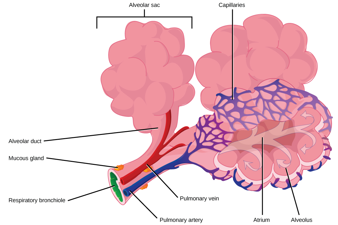
The terminal bronchioles subdivide into microscopic branches called respiratory bronchioles. The respiratory bronchioles subdivide into several alveolar ducts. Numerous alveoli and alveolar sacs surround the alveolar ducts. The alveolar sacs resemble bunches of grapes tethered to the end of the bronchioles (Figure 39.10). In the acinar region, the alveolar ducts are attached to the end of each bronchiole. At the end of each duct are approximately 100 alveolar sacs, each containing 20 to 30 alveoli that are 200 to 300 microns in diameter. Gas exchange occurs only in alveoli. Alveoli are made of thin-walled parenchymal cells, typically one-cell thick, that look like tiny bubbles within the sacs. Alveoli are in direct contact with capillaries (one-cell thick) of the circulatory system. Such intimate contact ensures that oxygen will diffuse from alveoli into the blood and be distributed to the cells of the body. In addition, the carbon dioxide that was produced by cells as a waste product will diffuse from the blood into alveoli to be exhaled. The anatomical arrangement of capillaries and alveoli emphasizes the structural and functional relationship of the respiratory and circulatory systems. Because there are so many alveoli (~300 million per lung) within each alveolar sac and so many sacs at the end of each alveolar duct, the lungs have a sponge-like consistency. This organization produces a very large surface area that is available for gas exchange. The surface area of alveoli in the lungs is approximately 75 m2. This large surface area, combined with the thin-walled nature of the alveolar parenchymal cells, allows gases to easily diffuse across the cells.
英語のヒント
終末細気管支は、呼吸細気管支と呼ばれる微細な枝に分かれる。呼吸細気管支は、さらに数本の肺胞管に分かれる。肺胞管の周りには、多数の肺胞と肺胞嚢がある。肺胞嚢は、細気管支の末端につながれたブドウの房に似ている(図39.10)。腺房領域では、各細気管支の末端に肺胞管がつながる。各肺胞管の末端には約100個の肺胞嚢があり、それぞれが、直径200〜300マイクロメートルの肺胞を20〜30個含む。ガス交換は肺胞だけで起こる。肺胞は、通常は細胞1層分の厚さの、壁の薄い実質細胞でできており、嚢の中の小さな泡のように見える。肺胞は、循環器系の毛細血管(厚さは細胞1層分)と直接接している。この密接な接触によって、酸素は肺胞から血液へ拡散し、全身の細胞へ行き渡る。また、細胞が老廃物としてつくった二酸化炭素は、血液から肺胞へ拡散し、吐き出される。毛細血管と肺胞の解剖学的な配置は、呼吸器系と循環器系の構造と機能のつながりをよく示している。それぞれの肺胞嚢の中には非常に多数の肺胞(片肺あたり約3億個)があり、各肺胞管の末端にも多数の肺胞嚢があるため、肺はスポンジのような性状をもつ。この構造によって、ガス交換に利用できる非常に大きな表面積が生まれる。肺の肺胞の表面積は約75m2である。この広い表面積と、肺胞の実質細胞の壁が薄いことによって、気体は細胞を容易に通り抜けて拡散できる。
OpenStax, Rice University; Biology 2e. (credit: modification of work by Mariana Ruiz Villareal) · CC BY 4.0 · Source出典

Figure 39.10図 39.10
Terminal bronchioles are connected by respiratory bronchioles to alveolar ducts and alveolar sacs. Each alveolar sac contains 20 to 30 spherical alveoli and has the appearance of a bunch of grapes. Air flows into the atrium of the alveolar sac, then circulates into alveoli where gas exchange occurs with the capillaries. Mucous glands secrete mucous into the airways, keeping them moist and flexible. (credit: modification of work by Mariana Ruiz Villareal)
終末細気管支は、呼吸細気管支を介して肺胞管と肺胞嚢につながる。各肺胞嚢には20〜30個の球形の肺胞があり、ブドウの房のように見える。空気は肺胞嚢腔に入り、次いで肺胞へ流れ、そこで毛細血管とのガス交換が起こる。粘液腺は気道に粘液を分泌し、湿り気と柔軟性を保つ。(出典:Mariana Ruiz Villarealの作品を改変)