第33章動物の体:基本的な形態と機能
33.2 Animal Primary Tissues33.2 動物の基本組織
Learning Objectives学習目標
By the end of this section, you will be able to do the following:
この節を学ぶと、次のことができるようになる。
- Describe epithelial tissues上皮組織を説明する。
- Discuss the different types of connective tissues in animals動物の結合組織のさまざまな種類を論じる。
- Describe three types of muscle tissues三種類の筋組織を説明する。
- Describe nervous tissue神経組織を説明する。
The tissues of multicellular, complex animals are four primary types: epithelial, connective, muscle, and nervous. Recall that tissues are groups of similar cells (cells carrying out related functions). These tissues combine to form organs—like the skin or kidney—that have specific, specialized functions within the body. Organs are organized into organ systems to perform functions; examples include the circulatory system, which consists of the heart and blood vessels, and the digestive system, consisting of several organs, including the stomach, intestines, liver, and pancreas. Organ systems come together to create an entire organism.
英語のヒント
複雑な多細胞動物の組織には、上皮組織、結合組織、筋組織、神経組織という四つの基本型がある。組織とは、関連した機能を行う、似た細胞の集まりであることを思い出そう。これらの組織が組み合わさり、皮膚や腎臓のように、体内で特定の専門的な機能を担う器官を作る。器官は器官系へと組織化され、機能を果たす。たとえば、心臓と血管からなる循環系や、胃、腸、肝臓、膵臓など複数の器官からなる消化系がある。器官系がまとまって、一つの生物全体を構成する。
Epithelial Tissues上皮組織
Epithelial tissues cover the outside of organs and structures in the body and line the lumens of organs in a single layer or multiple layers of cells. The types of epithelia are classified by the shapes of cells present and the number of layers of cells. Epithelia composed of a single layer of cells is called simple epithelia; epithelial tissue composed of multiple layers is called stratified epithelia. Table 33.2 summarizes the different types of epithelial tissues.
英語のヒント
上皮組織は、単層または複数層の細胞によって、体内の器官や構造の外側を覆い、器官の内腔を裏打ちする。上皮は、構成する細胞の形と細胞層の数によって分類される。1層の細胞からなるものを単層上皮、複数層からなるものを重層上皮という。表33.2は、さまざまな上皮組織の型をまとめたものである。
Different Types of Epithelial Tissues 上皮組織のさまざまな型 |
||
|---|---|---|
Cell shape 細胞の形 |
Description 特徴 |
Location 存在する場所 |
squamous 扁平 |
flat, irregular round shape 平たく、不規則な丸い形 |
simple: lung alveoli, capillaries; stratified: skin, mouth, vagina 単層:肺胞、毛細血管。重層:皮膚、口腔、膣。 |
cuboidal 立方 |
cube shaped, central nucleus 立方体状で、中央に核がある |
glands, renal tubules 腺、尿細管 |
columnar 円柱 |
tall, narrow, nucleus toward base; tall, narrow, nucleus along cell 背が高く細く、核が基部寄りにあるものと、核が細胞のさまざまな高さにあるもの |
simple: digestive tract; pseudostratified: respiratory tract 単層:消化管。偽重層:気道。 |
transitional 移行 |
round, simple but appear stratified 丸く、単層だが重層に見える |
urinary bladder 膀胱 |
Squamous Epithelia扁平上皮
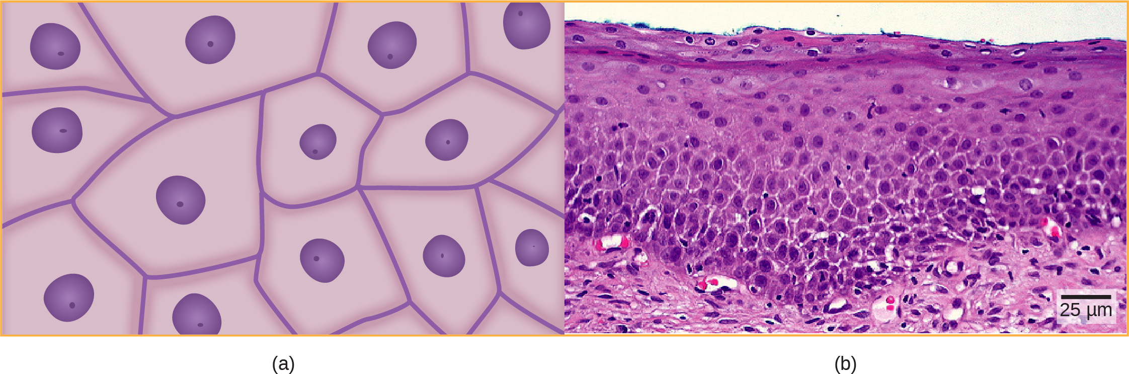
Squamous epithelial cells are generally round, flat, and have a small, centrally located nucleus. The cell outline is slightly irregular, and cells fit together to form a covering or lining. When the cells are arranged in a single layer (simple epithelia), they facilitate diffusion in tissues, such as the areas of gas exchange in the lungs and the exchange of nutrients and waste at blood capillaries.
英語のヒント
扁平上皮細胞は一般に丸く平たく、中央に小さな核をもつ。輪郭はやや不規則で、細胞同士が組み合わさって外側の覆いや内側の裏打ちを作る。単層に並ぶ場合は、肺でガス交換を行う領域や、毛細血管で栄養分と老廃物を交換する場所などで、組織内の拡散を促す。
OpenStax, Rice University; Biology 2e. (credit b: modification of work by Ed Uthman; scale-bar data from Matt Russell) · CC BY 4.0 · Source出典
Figure 33.7図 33.7
Squamous epithelia cells (a) have a slightly irregular shape, and a small, centrally located nucleus. These cells can be stratified into layers, as in (b) this human cervix specimen. (credit b: modification of work by Ed Uthman; scale-bar data from Matt Russell)
(a)扁平上皮細胞は、やや不規則な形と、中央にある小さな核をもつ。(b)このヒト子宮頸部の標本のように、何層にも重なって並ぶことがある。(クレジットb:Ed Uthmanの作品を改変;スケールバーのデータ:Matt Russell)
Figure 33.7a illustrates a layer of squamous cells with their membranes joined together to form an epithelium. Image Figure 33.7b illustrates squamous epithelial cells arranged in stratified layers, where protection is needed on the body from outside abrasion and damage. This is called a stratified squamous epithelium and occurs in the skin and in tissues lining the mouth and vagina.
Cuboidal Epithelia立方上皮
Cuboidal epithelial cells, shown in Figure 33.8, are cube-shaped with a single, central nucleus. They are most commonly found in a single layer representing a simple epithelia in glandular tissues throughout the body where they prepare and secrete glandular material. They are also found in the walls of tubules and in the ducts of the kidney and liver.
図33.8の立方上皮細胞は、立方体の形をし、中央に核を一つもつ。もっともよく見られるのは、全身の腺組織で単層上皮を作る場合であり、そこで腺の分泌物を作って分泌する。小管の壁や、腎臓・肝臓の管にも見られる。
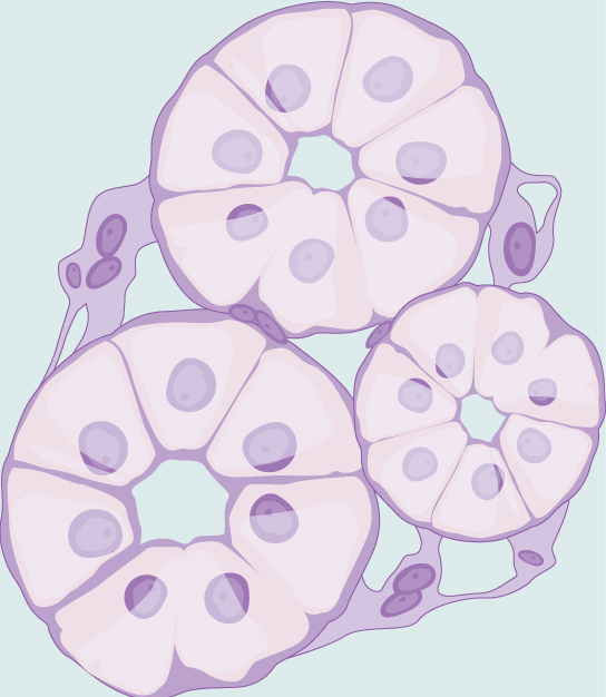
Figure 33.8図 33.8
Simple cuboidal epithelial cells line tubules in the mammalian kidney, where they are involved in filtering the blood.
単層立方上皮細胞は、哺乳類の腎臓の尿細管の内側を覆い、血液の濾過に関わる。
Columnar Epithelia円柱上皮
Columnar epithelial cells are taller than they are wide: they resemble a stack of columns in an epithelial layer, and are most commonly found in a single-layer arrangement. The nuclei of columnar epithelial cells in the digestive tract appear to be lined up at the base of the cells, as illustrated in Figure 33.9. These cells absorb material from the lumen of the digestive tract and prepare it for entry into the body through the circulatory and lymphatic systems.
英語のヒント
円柱上皮細胞は幅より高さが大きく、上皮層の中で柱が並んだように見える。もっともよく見られるのは単層の配列である。消化管の円柱上皮細胞の核は、図33.9のように、細胞の基部に並んで見える。これらの細胞は消化管の内腔から物質を吸収し、循環系やリンパ系を通して体内へ入るための準備をする。
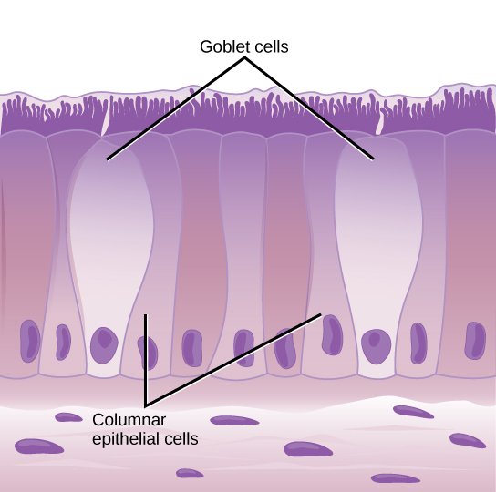
Figure 33.9図 33.9
Simple columnar epithelial cells absorb material from the digestive tract. Goblet cells secrete mucus into the digestive tract lumen.
単層円柱上皮細胞は、消化管から物質を吸収する。杯細胞は、消化管の内腔へ粘液を分泌する。
Columnar epithelial cells lining the respiratory tract appear to be stratified. However, each cell is attached to the base membrane of the tissue and, therefore, they are simple tissues. The nuclei are arranged at different levels in the layer of cells, making it appear as though there is more than one layer, as seen in Figure 33.10. This is called pseudostratified, columnar epithelia. This cellular covering has cilia at the apical, or free, surface of the cells. The cilia enhance the movement of mucus and trapped particles out of the respiratory tract, helping to protect the system from invasive microorganisms and harmful material that has been breathed into the body. Goblet cells are interspersed in some tissues (such as the lining of the trachea). The goblet cells contain mucus that traps irritants, which in the case of the trachea keep these irritants from getting into the lungs.
英語のヒント
気道の内側を覆う円柱上皮細胞は、重層しているように見える。しかし各細胞は組織の基底膜に付着しており、そのため単層の組織である。図33.10のように、核が細胞層のさまざまな高さに並ぶので、複数の層があるように見える。これを偽重層円柱上皮、すなわち多列円柱上皮という。この細胞の覆いには、頂端面、すなわち自由表面に繊毛がある。繊毛は、粘液や捕らえた粒子を気道の外へ動かすのを助け、侵入する微生物や吸い込まれた有害物質から呼吸器系を守る。気管の内面など、一部の組織では杯細胞が散在する。杯細胞には刺激物を捕らえる粘液が含まれ、気管では、これにより刺激物が肺へ入るのを防ぐ。
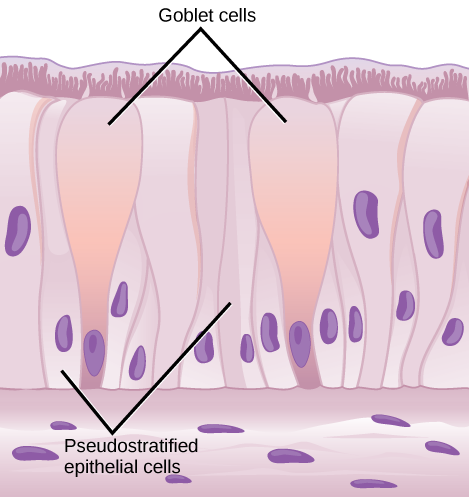


Figure 33.10図 33.10
Pseudostratified columnar epithelia line the respiratory tract. They exist in one layer, but the arrangement of nuclei at different levels makes it appear that there is more than one layer. Goblet cells interspersed between the columnar epithelial cells secrete mucus into the respiratory tract.
偽重層円柱上皮は気道の内側を覆う。実際には一層だが、核が異なる高さに並ぶため、複数層があるように見える。円柱上皮細胞の間に散在する杯細胞は、気道へ粘液を分泌する。