第33章動物の体:基本的な形態と機能
Transitional Epithelia移行上皮
Transitional or uroepithelial cells appear only in the urinary system, primarily in the bladder and ureter. These cells are arranged in a stratified layer, but they have the capability of appearing to pile up on top of each other in a relaxed, empty bladder, as illustrated in Figure 33.11. As the urinary bladder fills, the epithelial layer unfolds and expands to hold the volume of urine introduced into it. As the bladder fills, it expands and the lining becomes thinner. In other words, the tissue transitions from thick to thin.
移行上皮細胞、または尿路上皮細胞は、泌尿器系、とくに膀胱と尿管にだけ現れる。これらの細胞は重層に並ぶが、図33.11に示すように、弛緩した空の膀胱では、互いの上に積み重なったように見える。膀胱に尿がたまると、入ってきた尿の体積を収めるため、上皮層が伸び広がる。膀胱が満たされて拡大するにつれ、内側の覆いは薄くなる。言い換えると、組織が厚い状態から薄い状態へ移行する。
Connective Tissues結合組織
Connective tissues are made up of a matrix consisting of living cells and a nonliving substance, called the ground substance. The ground substance is made of an organic substance (usually a protein) and an inorganic substance (usually a mineral or water). The principal cell of connective tissues is the fibroblast. This cell makes the fibers found in nearly all of the connective tissues. Fibroblasts are motile, able to carry out mitosis, and can synthesize whichever connective tissue is needed. Macrophages, lymphocytes, and, occasionally, leukocytes can be found in some of the tissues. Some tissues have specialized cells that are not found in the others. Connective tissues are made up of living cells and a nonliving matrix of organic substances (usually proteins) and inorganic substances (usually a mineral or water).
英語のヒント
結合組織は、生きた細胞と、基質と呼ばれる非生体物質からなる細胞間の構成物でできている。基質は、有機物(通常はタンパク質)と無機物(通常は無機成分または水)からなる。結合組織の主な細胞は線維芽細胞であり、ほぼすべての結合組織に見られる線維を作る。線維芽細胞は移動でき、有糸分裂を行い、必要とされる結合組織を合成できる。一部の組織には、マクロファージ、リンパ球、ときに白血球も見られる。他の組織にはない特殊な細胞をもつ組織もある。結合組織は、生きた細胞と、有機物(通常はタンパク質)および無機物(通常は無機成分または水)からなる非生体の細胞外基質でできている。
The organic portion or protein fibers found in connective tissues are either collagen, elastic, or reticular fibers. Collagen fibers provide strength to the tissue, preventing it from being torn or separated from the surrounding tissues. Elastic fibers are made of the protein elastin; this fiber can stretch to one and one half of its length and return to its original size and shape. Elastic fibers provide flexibility to the tissues. Reticular fibers are the third type of protein fiber found in connective tissues. This fiber consists of thin strands of collagen that form a network of fibers to support the tissue and other organs to which it is connected. The various types of connective tissues, the types of cells and fibers they are made of, and sample locations of the tissues is summarized in Table 33.3.
英語のヒント
結合組織の有機成分、すなわちタンパク質線維には、膠原線維、弾性線維、細網線維がある。膠原線維は組織に強度を与え、裂けたり、周囲の組織から離れたりするのを防ぐ。弾性線維はエラスチンというタンパク質からなり、長さを1.5倍に伸ばした後でも、もとの大きさと形へ戻れる。組織に柔軟性を与える線維である。細網線維は、結合組織で見られる第3のタンパク質線維である。細いコラーゲンの束からなり、線維の網目を作って、組織やそれにつながるほかの器官を支える。結合組織のさまざまな型、それぞれを構成する細胞と線維の種類、存在する場所の例を表33.3にまとめる。
Connective Tissues 結合組織 |
|||
|---|---|---|---|
Tissue 組織 |
Cells 細胞 |
Fibers 線維 |
Location 存在する場所 |
loose/areolar 疎性/疎性結合組織 |
fibroblasts, macrophages, some lymphocytes, some neutrophils 線維芽細胞、マクロファージ、少数のリンパ球と好中球 |
few: collagen, elastic, reticular 少量の膠原線維、弾性線維、細網線維 |
around blood vessels; anchors epithelia 血管の周囲。上皮を固定する。 |
dense, fibrous connective tissue 密性の線維性結合組織 |
fibroblasts, macrophages 線維芽細胞、マクロファージ |
mostly collagen 主に膠原線維 |
irregular: skin; regular: tendons, ligaments 不規則:皮膚。規則的:腱、靱帯。 |
cartilage 軟骨 |
chondrocytes, chondroblasts 軟骨細胞、軟骨芽細胞 |
hyaline: few: collagen fibrocartilage: large amount of collagen 硝子軟骨:少量の膠原線維。線維軟骨:多量の膠原線維。 |
shark skeleton, fetal bones, human ears, intervertebral discs サメの骨格、胎児の骨、ヒトの耳、椎間板 |
bone 骨 |
osteoblasts, osteocytes, osteoclasts 骨芽細胞、骨細胞、破骨細胞 |
some: collagen, elastic 少量の膠原線維、弾性線維 |
vertebrate skeletons 脊椎動物の骨格 |
adipose 脂肪組織 |
adipocytes 脂肪細胞 |
few 少量 |
adipose (fat) 脂肪組織(脂肪) |
blood 血液 |
red blood cells, white blood cells 赤血球、白血球 |
none なし |
blood 血液 |
Loose/Areolar Connective Tissue疎性結合組織
Loose connective tissue, also called areolar connective tissue, has a sampling of all of the components of a connective tissue. As illustrated in Figure 33.12, loose connective tissue has some fibroblasts; macrophages are present as well. Collagen fibers are relatively wide and stain a light pink, while elastic fibers are thin and stain dark blue to black. The material in the connective tissue gives it a loose consistency similar to a cotton ball that has been pulled apart. Loose connective tissue is found around every blood vessel and helps to keep the vessel in place. The tissue is also found around and between most body organs. In summary, areolar tissue is tough, yet flexible, and comprises membranes.
英語のヒント
疎性結合組織には、結合組織のあらゆる構成要素が少しずつ含まれる。図33.12のように、線維芽細胞がいくつかあり、マクロファージも存在する。膠原線維は比較的太く、淡いピンク色に染まる。一方、弾性線維は細く、濃い青から黒に染まる。組織の構成物によって、引きほぐした綿球のような緩い質感になる。疎性結合組織はすべての血管の周囲にあり、血管を所定の位置に保つ助けとなる。また、大部分の器官の周囲や器官同士の間にもある。つまり、疎性結合組織は丈夫でありながら柔軟で、膜を構成する。
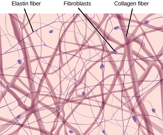

Figure 33.12図 33.12
Loose connective tissue is composed of loosely woven collagen and elastic fibers. The fibers and other components of the connective tissue matrix are secreted by fibroblasts.
疎性結合組織は、緩く織り合わさった膠原線維と弾性線維からなる。線維と、結合組織の細胞外基質のほかの成分は、線維芽細胞によって分泌される。